NICOTINE REPLACEMENT THERAPY

Cigarettes contain nicotine and by smoking regularly and for long hours, your body becomes dependent on it. Giving up smoking can cause nicotine withdrawal symptoms which are :

  • Restlessness ,impatience
  • Eating more than usual
  • Anxiety / tension
  • Headaches,irritability / anger
  • Difficulty in concentration + Depression
  • Loss of energy,dizziness
  • Sweating
  • Insomnia
  • Stomach or bowel problems
  • Heart palpitations
  • Tremors
  • Craving for tobacco

Stop smoking medicines along with nicotine replacement therapy are effective aid to tobacco cessation & can help control these symptoms.

What are these?

3 types –

  • Champix tablets (varenicline)
  • Zyban tablets (bupropion)
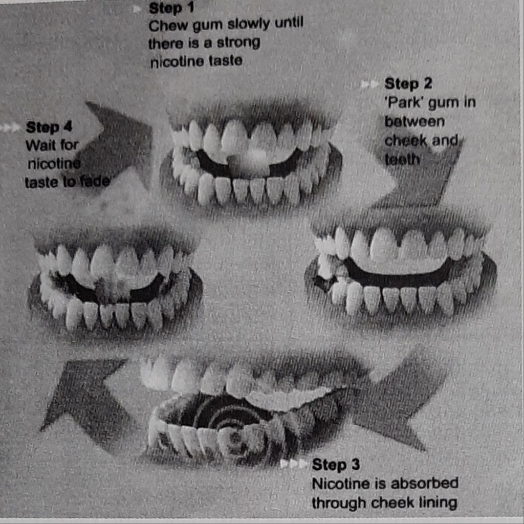
  • Nicotine replacement therapies (gums,patches,lozenges,microtabs,sublingual tablets,insulators,cigs,nasal sprays)

How does it work?

Tobacco intakers who are motivated to quit the habit & are dependent on nicotine should be offered NRT.

  • Prescribed for 6 to 8 weeks,in blocks of up to 2 weeks,contingent on continued abstinence.
  • If one type of NRT is not working for the patient ,the health professional is advised to prescribe a suitable type informing about the dosage & maximum amount to take a day.

Is it safe?

NRT is safe because of the facts that the nicotine levels are low and it’s less addictive delivery mechanism (unlike smoking tobacco where the nicotine reaches the brain quickly) and also because most of the harmful problems are caused by the other components of tobacco smoke ,not by the nicotine.

NRT is safe for most adults and in people with stable cardiac diseases, but caution needed in unstable,acute cardiovascular disease,pregnancy,or breastfeeding or in those aged under 18 years.

Brownie points-reduces the constant urge to munch on food,thus reducing weight gain.😊

When should one stop using NRT?

Most courses of NRT recommend use for about 12 weeks.This is because it takes this much time for the brain to adjust to working without the high doses of nicotine that the cigarettes supply.However there is no hard and fast rule.After starting the therapy,most people mistake the lack of discomfort for the belief that the addiction is over,leading to stop using the product too soon. This can result only in reappearing of the symptoms.

The best method is to take the help of the health professional when you start the therapy and keep them updated about the progress.

Lastly ,we all are not the same….each tobacco smoker’s tendency & pattern to quit may vary & it depends on different factors like age,gender,environment, general physical and mental health.

Sources:S.S Hiremath textbook of preventive and community dentistry, http://www.healthunlocked.com(Quit Support)

TOBACCO CESSATION MOTIVATION

Tobacco is the leading preventable cause of death in the world and is the only consumer product that kills when used as intended by its manufacturers ;which may become deadly for non-smokers also.

FACTS & FIGURES:

  • Tobacco causes 1 in 10 adult deaths worldwide,nearly 5 million deaths a year or one death every 6.5 seconds,killing 50% of regular users.
  • Total global smoking prevalence is 29%;47.5% men & 10.3% women.
  • By 2030,70% of deaths in the world is attributable to tobacco.
  • It’s a known or probable cause of about 25 diseases.
  • Smokeless tobacco causes oral cancer,especially in lips,tongue,mouth and throat area.
  • Breathing Environmental Tobacco Smoke(ETS) (i.e .side stream,exhaled smoke from cigarettes,cigars and pipes)causes serious health problems & aggravates allergies and increase the severity of symptoms in children & adolescents, with asthma and heart diseases.
  • People who start using tobacco early have more difficulty in quitting,are more likely to be heavy smokers and if young people donot begin to use tobacco before the age of 20,they are unlikely to start the habit.
  • The World Bank estimated that smoking prevention is among the most effective of all health interventions.
Deadlier than ever-how cigarettes have evolved over last 50 years.

EFFECTS OF NICOTINE :

  • Electroencephalographic desynchronisation.
  • Increased circulating levels of catecholamines, vasopressin, growth hormone,adenocorticotropic hormone,cortisol,prolactin,and beta-endorphin.
  • Increased metabolic rate
  • Lipolysis,increased free fatty acids.
  • Heart rate acceleration, nicotine can increase the heart rate by 10-15 beats/min.
  • Cutaneous & coronary vasoconstriction
  • Increased cardiac output & blood pressure by 5-10 mm Hg
  • Skeletal muscle relaxation
  • Nicotine can induce pathogenic changes to the endothelium associated with atherosclerotic process.
  • Halitosis,staining of teeth and soft tissues(smokers melanosis),drying of mouth.

‘Nicotine itself is not carcinogenic unless it undergoes nitrosation to form nitrosamines(during tobacco curing & combustion).’

HOW TO ASSESS TOBACCO DEPENDENCE?

A question-answer session with the individual would be very helpful ….

The total score can be calculated to know the dependence.

THE 5 A’S :

  • ASK- health care professionals / dentists should ask the patient about his or her tobacco intake habits which includes the questions discussed above,during every visit.
  • ADVICE-health care professional / dentists should continually advice patient to quit the habit thereby emphasizing the importance of the issue.
  • ASSESS- patients readiness & motivation to quit the habit must be assessed- cause this is a ‘major lifestyle change & requires preparation, readiness & several failed attempts’.
  • ASSIST-health care professionals/ dentists should assist those individuals who are motivated- by informing,suggesting and prescribing a pharmacological cessation aids ( nicotine replacement therapy ) and providing or referring the patient to counseling ( individual, group or over telephone ) and behavioral therapies and support services where available.
  • Lastly,ARRANGE-follow up services are often critical & the dentists can help the patient be tobacco free by providing services like advising availability of national hotlines,support from non-smoking friends or colleagues,or community based support groups.
The best time to quit smoking was the day you started,the second best time is today.

Lastly ,dentists play a major role in helping a patient quit smoking because we might be the first to detect an abnormality( be it a small stain or an abnormal mass ) in the oral cavity during routine examination. Do your part ,every small step counts……

Sources- S.S Hiremath textbook of preventive and community dentistry ,www.alhambraesd.org ,www.tobaccofreekids.org

NRT DRUGS

Written by : Dr. Urusa I Inamdar

Nicotine gum

Nicotine polacrilex .

  • Buccal absorption
  • 4 mg of nicotine ( India )
  • Two varieties in India : gutkha and mint flavour
  • Duration of treatment is 4-6 weeks ; start weaning after 2-3 months .
  • Brand name : nicorette , nulife , nicotex

Nicotine patch ( transdermal )

  • Ready absorption of nicotine across the skin .
  • 3 of the patches are for 24 hr use and one is for 16 hr ( waking ) use .
  • Starting doses are 21-22 mg/ 24 hr patch and 15 mg/ 16 hr patch .
  • The recommended total duration of treatment is usually 6-12 weeks .
  • Brand name : habitrol , nicodern – cq , nicotrol

Patient instruction for nicotine patch:

  • Do not smoke while using the patch .
  • Rotate the patch site to minimize skin irritation .
  • If insomnia occurs , remove patch before going to bed or use 16 hr patch .
  • Apply a new patch every day ( remove old patch ) in a location between the neck and waist that is relatively hairless and where the skin is not broken . Apply to a different location each day .

Nicotine nasal spray

  • 8-40 dosage per day .
  • Nasal irritation may occur .
  • Treatment time : 3-6 months .
  • Brand name : nicotrol nasal spray

Nicotine inhaler

  • 6-16 cartridges /day
  • Mouth and throat irritation may occur .
  • Treatment time : upto 6 months .
  • Brand name : nicotrol inhaler

References

  • Dental notes
  • Google.com

FLUORIDE TOXICITY


• Fluoride can be toxic, if administered in excess quantity.
• The toxic effects of fluoride can be either acute or chronic.
• Acute toxicity occurs due to a single ingestion of large amount of fluoride.
• Chronic toxicity occurs dụe to long-term ingestion of small amounts of fluoride excessive than the required optimal level.

Acute Fluoride Toxicity:
• Acute fluoride poisoning can occur either as a result of accidental ingestion or from deliberate attempts like suicide
• This can be from the use of fluoride containing products such as toothpaste, mouth rinse, tablets or household insecticides.
• Acute fluoride poisoning can also occur from overfeeds in community and school water fluoridation systems.
• Most of the time overfeeds occur in small water supplies.

Terms used in relation to fluoride toxicity:
Safely Tolerated Dose (STD): Dose below which symptoms of toxicity are unlikely to occur
(1 mg/kg of body weight).

Potentially Lethal Dose (PLD): Lowest dose associated with a fatality.

(5 mg/kg of body weight).

Certainly Lethal Dose (CLD): Survival after consuming this amount of fluoride is unlikely (32-64 mg/kg of body weight).

Features of acute toxicity of fluorides are:

  1. Nausea
  2. Vomiting
  3. Diarrhoea
  4. Abdominal pain
  5. Hypocalcaemia
  6. Acidosis
  7. Progressive hypotension
  8. Ventricular tachycardia and fibrillation.

The ingested fluoride combines with hydrochloric acid in the stomach to form hydrofluoric acid which exhibits a corrosive effect on the gastric mucosa causing nausea, vomiting, diarrhoea and abdominal pain.


*Hypocalcemia is caused by the affinity of fluoride to cations in the serum.
**Hypocalcemia is associated with paresthesia, paresis, muscle fibrillation, tetany, convulsions, decreased myocardial contractility and cardiovascular collapse.

Death usually occurs due to:

  1. Convulsions
  2. Cardiac arrhythmias
  3. Coma.

Chronic Fluoride Toxicity:
• Long-term ingestion of small amounts of excessive fluoride will lead to chronic fluoride toxicity which is often referred to as fluorosis.
• Fluorosis usually affects the bones and the teeth.
• Fluorosis of the bone is called as osteofluorosis and the fluorosis of the teeth is called as dental fluorosis.

MANAGEMENT:

REFERENCES:

  • Pediatric Dentistry: Principles & Practice, MS Muthu(2nd Edition)
  • downtoearth.org.in

PREVENTIVE RESIN RESTORATIONS

:
Preventive resin restoration (PRR) is a thin, resin coating applied to the chewing surface of molars, premolars and any deep grooves of the teeth.
• They are a natural extension of the use of occlusal sealants.
• It consists of an enamel sealant with a resin filling.
• If caries is present in one area or parts of pits and fissures, that particular area of caries is restored and fissures are protected with sealants.
• It integrates the preventive approach of the sealant therapy for caries susceptible pit & fissure with therapeutic restoration incipient caries with composite resin that occur on the same occlusal surface.
• They are the conservative answer to conventional extension for prevention” philosophy of Class I amalgam cavity preparation.

Preventive resin restoration(PRR) consists of a thin, resin coating applied to the occlusal surface of molars, premolars and deep grooves.
Most decays begin in the deep grooves. Thus, teeth with this condition are difficult to clean and becomes more susceptible to caries.
PRR protects the tooth by sealing the deep grooves and creating a smooth, easy to clean surface.
By this way, the teeth is protected from decay for several years, provided they are checked for thinning and wear at regular dental check-ups.

  • Dental caries is due to the imbalance between loss & gain of
  • minerals from a tooth surface.
  • The loss of minerals from our teeth occurs from the bacteria from the foods and producing acids, whereas the tooth gains minerals from our saliva and fluoride that is present within our mouth which over a period of time becomes a tooth decay.
  • Fissure sealants are a preventive treatment that is part of the minimal intervention dentistry approach to dental care.
  • This approach facilitates prevention and early intervention, in order to prevent or stop the dental caries process before it reaches the ends stage of the disease.

CLASSIFICATION:

There are three types of preventive resin restoration based on the extent & depth of carious lesion as determined by exploratory preparation.

Simonsen (1978) has classified them:

TYPE A:

• Suspicious pits & fissures where caries removal is limited to enamel
• Local anesthesia is not required.
• A slow speed 4 or 2 round burs. used to remove decalcified enamel
• Sealant is placed

TYPE B:

• Incipient lesion in dentin that is small & confined.
• No local anesthesia is needed.
• An appropriate base is placed in areas of dentin exposure, composite resin is placed & the remaining pit & fissure are covered with a sealant.

TYPE C:
• More extensive dentinal involvement & requires restorations with posterior composite material
• Appropriate base is placed over the dentin.
• Pits & fissures are covered with sealant.
• Local anesthesia is required.

PROCEDURE:

CONCLUSION:
• Regular maintenance and sealant addition when necessary is important in long-term caries protection after sealant placement.
• Much better effectiveness data will result if sealants are used on teeth with a true predilection to caries.
• Better materials and better use of bonding agents with sealants will improve overall effectiveness on all teeth,particularly on those teeth now thought of as difficult to seal.
• Use of sealants has proved to have good results.
• For prevention of dental caries in pit and fissure, sealants were introduced.
• There is evidence suggesting effectiveness of sealants.
• Sealants prevent bacteria growth which causes caries.
• Biomaterials to seal pit and fissure should present with the simple application method, biocompatibility, low viscosity and good surface retention and low solubility.
• To improve this biomaterial, more laboratory should be developed.

REFERENCES:

  • Essentials of Public Health Dentistry, Soben Peter (6th Edition).
  • FENESTRA 2002-18, Dr. Bruno Jacquot(Bruno.Jacqout@odonto.u-nancy.fr)

Halitosis

Written by - Dr.Urusa I Inamdar

Also called as oral malodor.

” Halitosis may rank only behind dental caries and periodontal disease as the cause of the patients visit to the dentist.”

Origin

Oral

  • Poor oral hygiene
  1. Retention of odoriferous food particles on and between the teeth.
  2. Coated tongue.
  3. Artificial dentures.
  • Acute Necrotizing Ulcerative Gingivitis.
  • Pericoronitis.
  • Abscess.
  • Dehydration states.
  • Ulceration in the oral cavity.
  • Hyposalivation/Xerostomia.
  • Bone disease ( dry socket , Osteomyelitis , osteonecrosis and malignancy )
  • Smoker’s breath.
  • Healing oral wounds.
  • Chronic periodontitis with pocket formation.

Extraoral ( Conditions that can contribute to presence of oral malodor )

  • Sinusitis and other bacterial infections.
  • Dry nasal mucosa.
  • Blocked nose ( which can cause mouth breathing )
  • Tonsillitis/ tonsil stones.
  • Various carcinomas.
  • Infections of the respiratory tract ( bronchitis , pneumonia , bronchiectasis )
  • Alcoholic breath.
  • Uremic breath of kidney dysfunction.
  • Acetone odor of Diabetes.

It is important for the dental professional to eliminate systemic conditions that may be contributing to the presence of oral malodor.

The clinical assessment of oral malodor is either subjective or objective . Subjective assessment is based on smelling the exhaled air of the mouth and nose and comparing the two ( organoleptic assessment ).

Organoleptic scoring scale

  • Absence of odor.
  • Questionable to slight malodor. Odor is deemed to exceed the threshold of malodor detection.
  • Moderate malodor. Odor is definitely detected.
  • Strong malodor. Malodor is objectionable but examiner can tolerate.
  • Severe malodor. Overwhelming malodor. Examiner cannot tolerate.

Various scoring systems, such as 0 to 5 point scale and a 0 to 10 point scale can be used to estimate the intensity of exhaled oral odor, tongue odor and nasal odor , among others.

Methods for objective measurement of the breath include :

  • Detection of sulphides with an appropriate monitor- simple but may fail to detect oral malodor caused by nonsulphide components. Halimeter is a instrument that can be used chair side to measure volatile sulfur compounds in the exhaled air.
  • Gas chromatography- not applicable for routine clinical practice.
  • Bacterial detection ( such as benzoylarginine – naphthylamide test – BANA test ) , polymerase chain reaction, dark field microscopy ) – not applicable for routine clinical practice.

References

  • Dental notes.
  • A practical manual of Public Health Dentistry – C M Marya.

Bad breath/ Halitosis

August 1st,NATIONAL ORAL HYGIENE DAY 🦷
A reminder that oral health is the door to overall health and reinforcing the fact that “Brush and floss ,before the loss.”
Amidst the Covid era,where wearing a mask
has become a part and parcel,many of us face the problem of “BAD BREATH” or halitosis and people often end up in temporary solutions without knowing the root cause.
Here are some of the causes & remedy which might prove helpful.

Sources:instagram @team_dentistree @theunicorndentist @drsana_daruwala

Minimal Invasive Dentistry

– current approach in dentistry in Covid state Amidst the pandemic state with the high transmissibility of the disease through air & droplets and considering that routine dental procedures usually generate aerosols; alterations to dental treatment is of prime concern to maintain a healthy environment for patient & dental team.Here is where the approach of […]

Minimal Invasive Dentistry

Sources: Slideshare-Minimal invasive dentistry by Nabeela Basha , Minimal intervention dentistry by Dr.Nagamaheswari, Sturdvent’s South Asian edition,Clinical operative dentistry principles & practice by Ramya Raghu,textbook of preventive and community dentistry by SS Hiremath

Sources: