Bioprogressive therapy was developed to correct the limitations of conventional edgewise treatment, especially anchorage loss, unwanted incisor flaring, occlusal plane dumping, and overreliance on heavy mechanics. Ricketts emphasizes that treatment should be built around biologic force levels, cortical bone considerations, and prefabricated appliance components rather than endless chairside wire bending.
Historical progression
Stage
Main feature
Main drawback
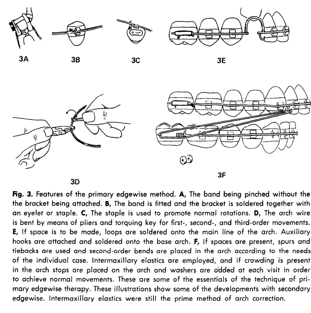
Primary edgewise
Fully banded, custom bends, gold bands, heavy manual finishing
Time-consuming, rigid, depends on full eruption, difficult finishing
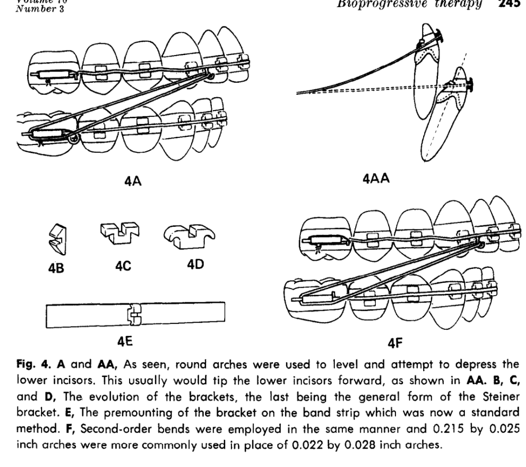
Secondary edgewise
Round wire used more often, later rectangular finishing
Flaring, anchorage loss, protrusion in nonextraction cases
Requires thoughtful planning and case-specific customization
Why bioprogressive emerged
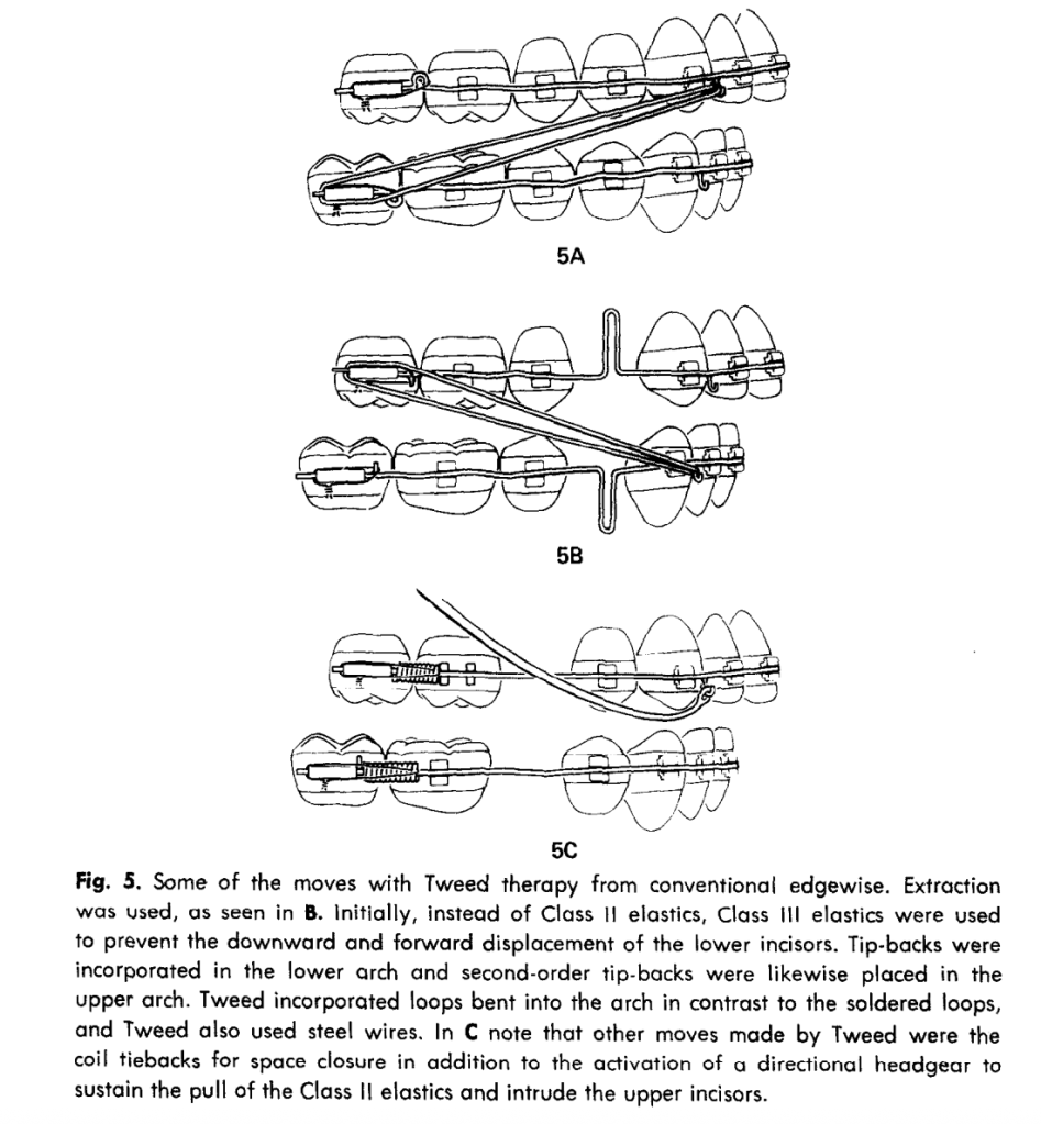
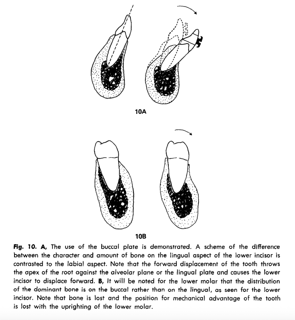
The author’s main criticism of older systems is that round-wire leveling and heavy intermaxillary mechanics often caused unwanted tooth movement, especially forward tipping of lower incisors and extrusion of molars. He links these problems to cortical bone resistance and shows that orthodontic movement is not just about moving teeth through cancellous bone; the compact bone plates matter greatly
Force and biology
Concept
Exam point
Light forces
Favored for biologic efficiency and reduced tissue damage
Optimal force range
Storey and Smith’s canine retraction work is cited as supporting 150–300 g for translatory retraction
Pressure concept
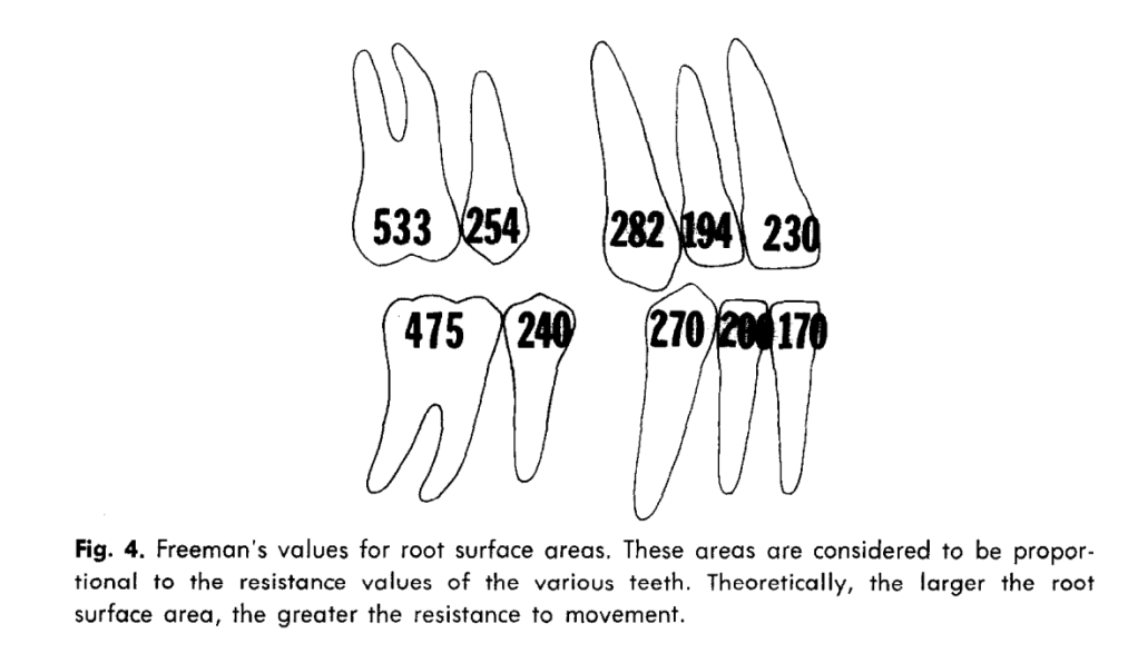
Force should be considered relative to root surface area and tissue response
Cortical bone
Lower incisors and molars behave differently because bone support differs by site
Appliance logic
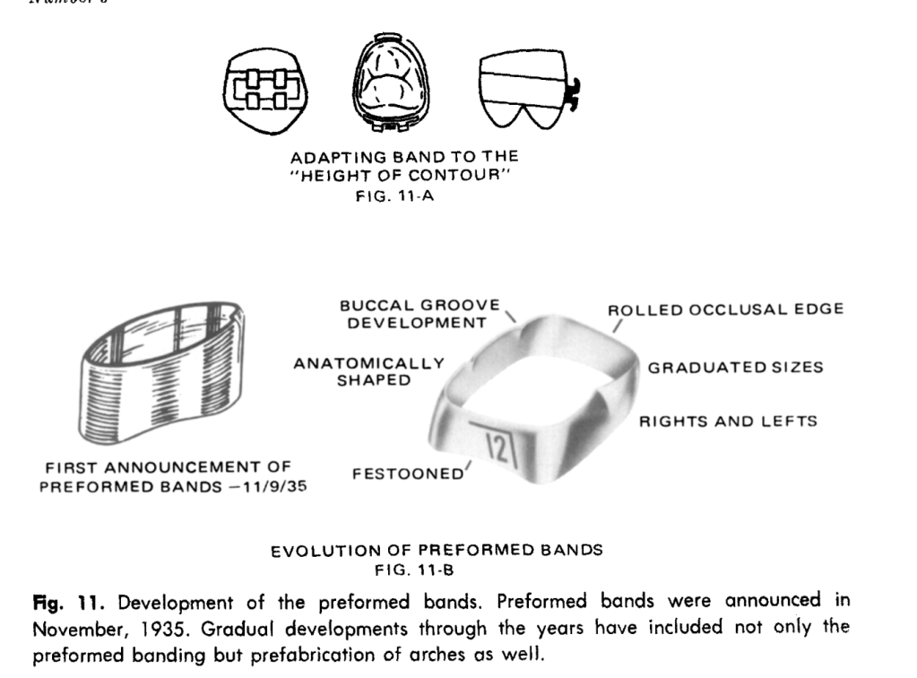
Bioprogressive therapy uses a prefabricated system with bands, brackets, and arch forms designed in advance, reducing chairside bending and standardizing control. Ricketts’ philosophy is that the appliance should do more of the work, while the clinician still retains control through selective adjustments and overtreatment where needed.
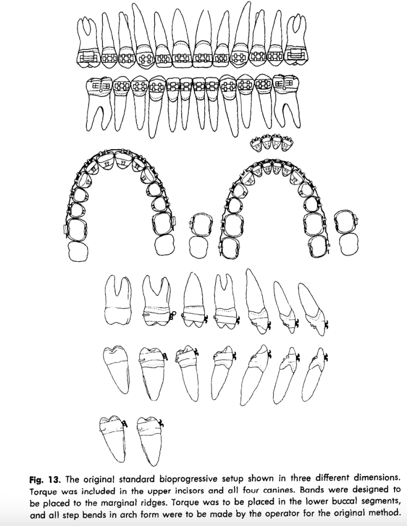
Three bioprogressive setups
Setup
Main design idea
Best use
Standard bioprogressive
Torque built into upper incisors and canines; lower torque largely managed in wire
General cases and balanced control
Full-torque bioprogressive
Adds lower posterior torque to the standard setup
Cases needing more complete torque control
Triple-control bioprogressive
Adds step-outs/step-ins and more overtreatment of rotations and posterior segments
Cases needing maximal built-in control and less wire bending
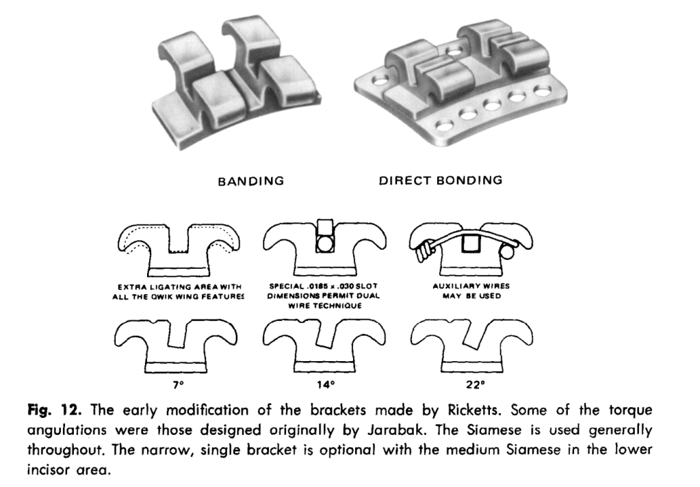
Bracket prescriptions
Tooth group
Prescription emphasized in the paper
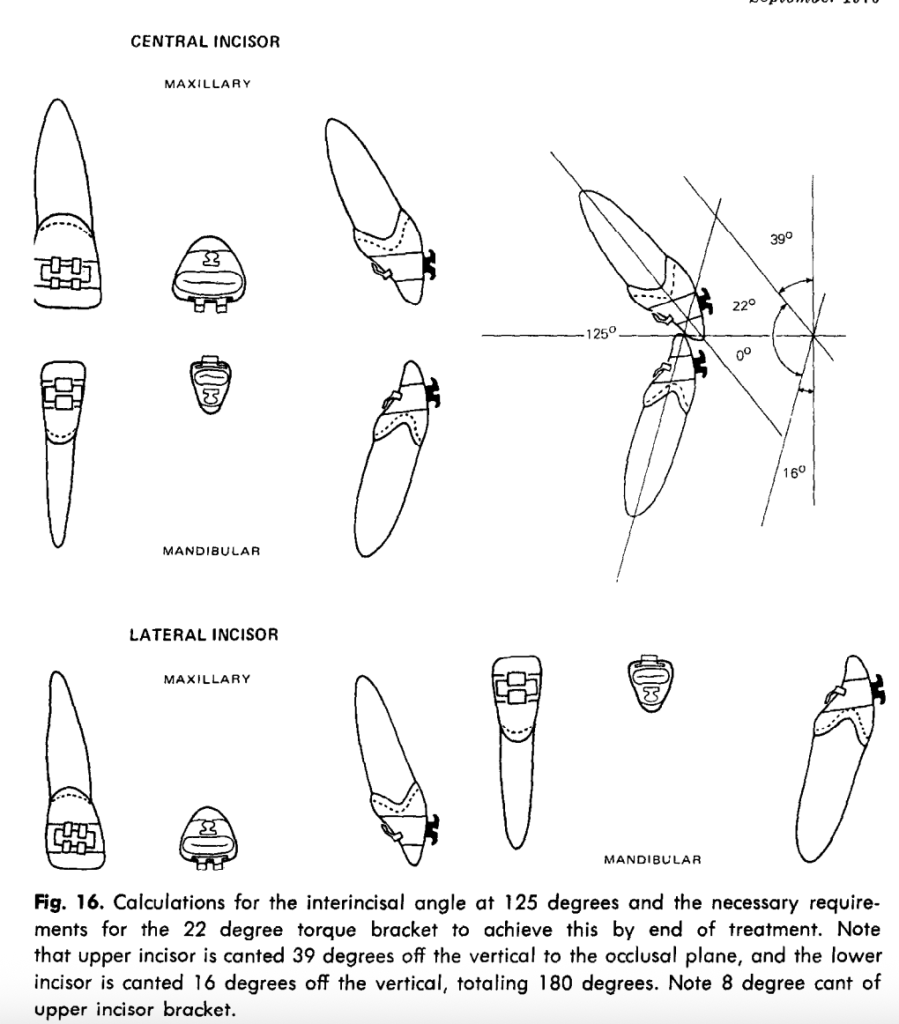
Upper central incisors
22 degrees root to palatal
Upper lateral incisors
14 degrees root to palatal, 8 degrees tip down distally
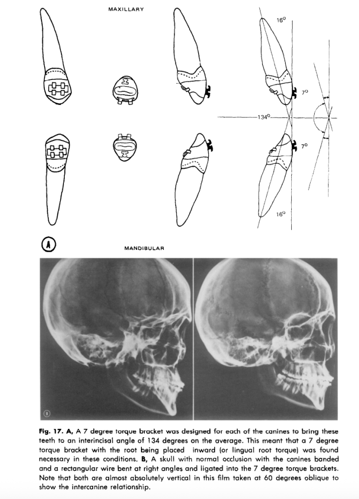
Upper canines
7 degrees root to palatal, 5 degrees tip down distally
Lower canines
7 degrees root to lingual, 5 degrees tip down distally
Lower second premolars
14 degrees root to buccal in full-torque and triple-control setups
Lower first molars
22 degrees root to buccal in full-torque and triple-control setups
Rotation and step control
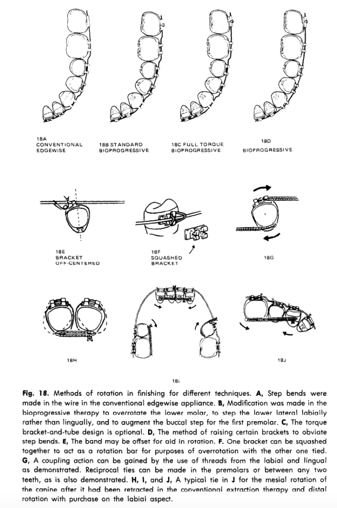
Ricketts treats rotation as an essential part of first-order control and provides multiple methods to overcorrect or maintain rotation without excessive archwire complexity. These include off-centering the band, squashing a bracket, reciprocal ties, and lingual cleats, showing that the system is flexible rather than purely “straight wire.”
Second molars and anchorage
Second molars are not automatically banded in every case, especially upper second molars, because they may erupt into better function later and can complicate treatment if included too early. Lower second molars, however, are usually important for anchorage and proper arch development, and the system is designed to accommodate them without full rebanding.
Viva-style one-liners
Bioprogressive therapy is a biologically oriented evolution of edgewise mechanics.
Its philosophy is light force, preformed components, and anchorage preservation.
Ricketts strongly emphasizes cortical bone and regional tooth behavior.
The system reduces chairside bending by using standardized bands, brackets, and arches.
Standard, full-torque, and triple-control are the three major setups described in Part I
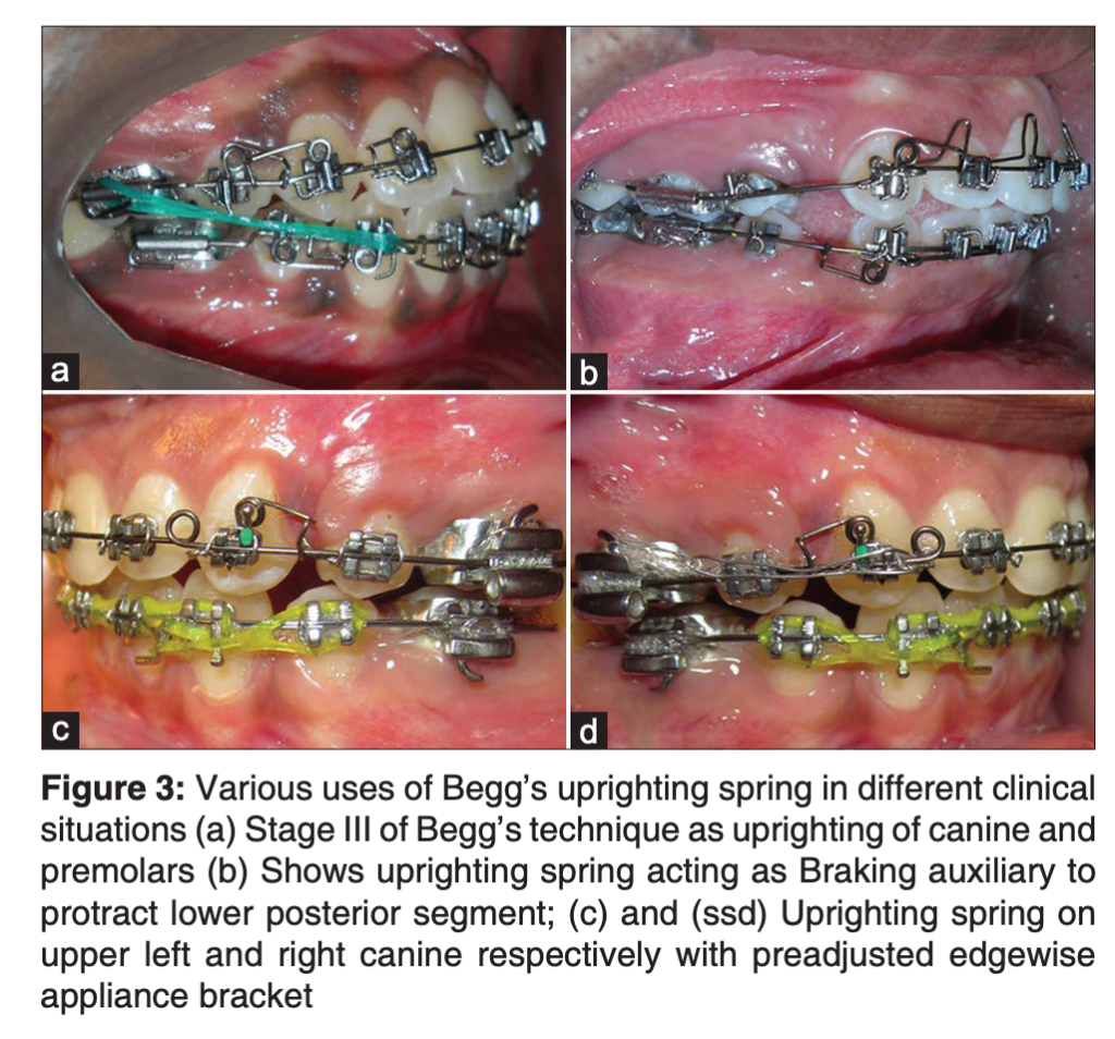
Somewhere in every orthodontic department, there’s a forgotten drawer. Inside? Old Begg pliers, Australian wire, random elastomeric chains—and one underrated genius of biomechanics: the Begg’s uprighting spring.
Modern orthodontics loves sleek prescriptions, digital setups, and aligner simulations. But when anchorage falters or teeth tip uncontrollably, this vintage auxiliary stages a silent comeback.
What Is It?
A light-wire auxiliary for mesiodistal root uprighting, anchorage reinforcement, controlled movement, and braking during space closure. Born in Percy Raymond Begg’s differential force technique, it now aids preadjusted edgewise systems with vertical slots.
Core Principle: Moments Matter
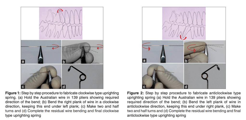
Decide root movement first—clockwise or anticlockwise. This sets coil direction for precise moments.
Desired Movement
Spring Type
Clockwise uprighting
Clockwise coil
Anticlockwise uprighting
Anticlockwise coil
Fabrication Essentials
Wire: 0.009″–0.018″ Australian for resilience and activation range.
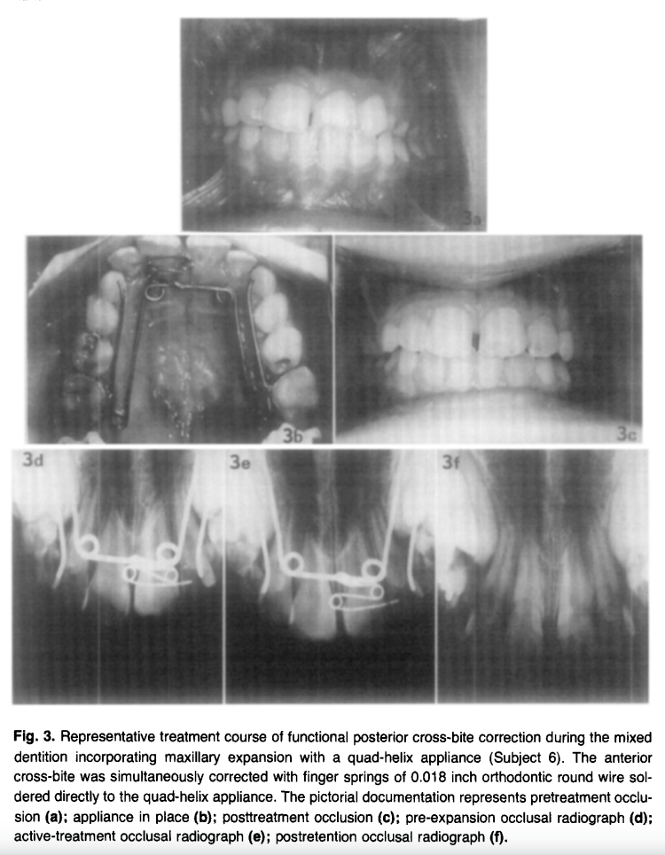
One-line takeaway: The quad-helix produces significant, stable maxillary expansion (mean +5.3 mm intermolar, +4.1 mm intercanine) with midpalatal suture opening in both deciduous and mixed dentitions — with no significant difference between the two groups.
Why Early Maxillary Expansion Matters
Functional posterior crossbite is commonly associated with a transverse maxillary deficiency. In such cases, the mandible often shifts laterally during closure to avoid occlusal interference. This functional shift can lead to several secondary problems, including:
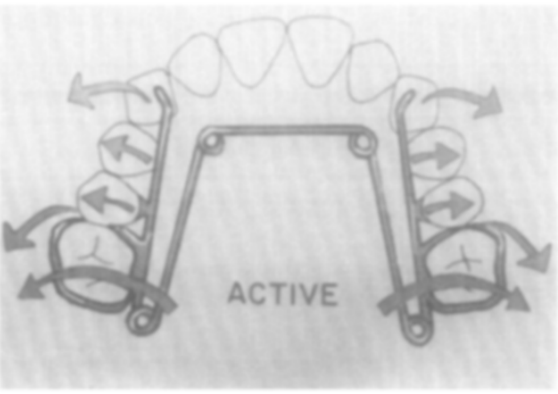
The quad-helix appliance is essentially a modification of the W-arch appliance, with the addition of four helices. These helices provide greater flexibility and allow a wider range of activation compared with traditional lingual arch expansion appliances.
Typically, the appliance is fabricated from 0.036-inch stainless steel wire and soldered to bands placed on the maxillary molars. The helices act as force modulators, delivering low, continuous expansion forces to the maxillary arch.
The quad-helix is a W-arch modification with 4 helical loops incorporated. These loops deliver four specific advantages over a standard W-arch:
Refined adjustment capability — fine-tune forces without full removal
💡 Exam Distinction: Quad-helix = slow/continuous expansion vs. jackscrew = rapid expansion. Both open the midpalatal suture, but quad-helix produces more physiologic bone remodeling with less relapse risk.
↑ Range of force application — stores energy over greater activation distances
↑ Flexibility — lighter, continuous, physiologic force
The typical treatment protocol involves an initial activation that produces a modest transverse expansion force. The patient is then monitored periodically, and adjustments are made only when expansion progress slows.
General clinical steps include:
Cementing the appliance onto molar bands.
Activating the appliance to produce expansion equivalent to approximately half the buccolingual width of the molars.
Monitoring the patient weekly or periodically during the active expansion phase.
Achieving slight overexpansion so that the lingual cusp of the maxillary molar contacts the buccal cusp slope of the mandibular molar in centric relation.
Maintaining the appliance in a passive state for a retention period.
The entire active phase of expansion typically lasts about one month, followed by a retention period of approximately six weeks.
📊 Treatment Course Data
Variable
Deciduous (x̄ 5y 3m)
Mixed (x̄ 8y 2m)
Correction time (days)
28.8 ± 4.9
31.8 ± 5.9
Retention time (days)
44.2 ± 1.8
45.2 ± 1.7
Total appliance time (days)
73.0 ± 5.9
77.0 ± 6.0
No. of adjustments
1.2 ± 0.4
1.0 ± 0.3
Midpalatal suture opening
✅ All subjects
✅ All subjects
Between-group significance
NS (p > 0.05)
← same
Memory hook: “30-45-75” — ~30 days active, ~45 days retention, ~75 days total.
📐 Transverse Dimensional Changes (The Core Data)
Measurement
Deciduous — Intercanine
Deciduous — Intermolar
Mixed — Intercanine
Mixed — Intermolar
Before treatment (mm)
27.5 ± 0.4
31.0 ± 0.4
29.3 ± 0.9
35.3 ± 2.0
Post-retention (mm)
31.4 ± 0.9
36.7 ± 0.6
33.7 ± 1.1
40.2 ± 1.2
3-month recall (mm)
29.8 ± 0.4
34.8 ± 0.4
31.5 ± 1.0
38.9 ± 1.5
Expansion increase
+3.9 ± 0.8
+5.7 ± 0.5
+4.4 ± 0.7
+4.8 ± 1.3
Relapse
−1.6 ± 0.9
−1.9 ± 0.3
−2.2 ± 0.3
−1.2 ± 0.4
Net gain
+2.3 ± 0.4
+3.9 ± 0.5
+2.2 ± 0.6
+3.6 ± 1.1
Significance (p)
< 0.01
< 0.001
< 0.01
< 0.02
Overall pooled means (both groups combined):
Intermolar expansion: +5.3 mm → net gain after relapse: ~+3.75 mm
Intercanine expansion: +4.1 mm → net gain after relapse: ~+2.25 mm
🔬 Sutural Opening — The Radiographic Finding
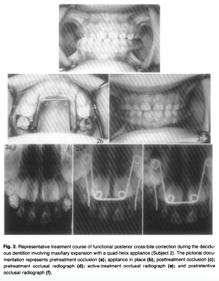
Every single subject (10/10) showed radiographic evidence of midpalatal suture opening on occlusal radiographs taken at 2 weeks of active treatment. The separation pattern was greatest anteriorly with a progressive posterior decrease — a classic sutural opening pattern. By end of retention, suture widening was no longer detectable radiographically, confirming bone fill-in.
📌 Exam alert: This finding proved the quad-helix produces orthopedic effects, not purely orthodontic tooth tipping — especially relevant in younger patients. This was the key debate this study addressed (W arch/Porter arch were thought to be purely orthodontic appliances).
↩️ Relapse & Overexpansion Protocol
Relapse averaged ~2 mm in both intercanine and intermolar dimensions after the 3-month post-retention period. The protocol to handle this:
Overexpand by 2–3 mm during active phase — lingual cusp tip contacts buccal cusp slope of mandibular molars bilaterally in centric relation
This slight overcorrection compensates for tooth uprighting relapse once appliance is removed
Slow expansion → more physiologic sutural remodeling → less relapse than rapid palatal expansion
⚡ Rapid vs. Slow Expansion
Feature
Quad-Helix (Slow)
RPE/Jackscrew (Rapid)
Force type
Low, continuous
High, intermittent
Suture opening
✅ Yes (both dentitions)
✅ Yes
Orthopedic effect
Present (especially young)
Dominant
Orthodontic effect
Present (tooth tipping)
Present
Relapse
Lower
Higher
Adjustments needed
~1.1 (minimal)
Multiple activations daily
Patient compliance
Not required
Device-dependent
Total treatment time
~75 days
3–6 months incl. retention
Berlocher et al. (RPE comparison): intermolar +4.2 mm, intercanine +3.8 mm using RPE — comparable to quad-helix results here.
❗ Key Conclusions — Write These in Your Answer
Functional posterior cross-bites are mandibular shift-related, causing midline deviation, condylar asymmetry, and arch constriction — early correction is essential
Quad-helix produces significant transverse increases in all subjects (p < 0.001 for intermolar)
No significant difference between deciduous and mixed dentition groups in expansion magnitude, rate, or relapse
Midpalatal suture opens in both dentitions — confirming orthopedic, not just orthodontic, mechanism
~2 mm overexpansion effectively compensates for expected relapse
Mandibular arch dimensions showed no significant change — no predictable expansion effect on the lower arch
Appliance had excellent patient tolerance — no pain, speech difficulty, or significant soft tissue issues
The pterygoid response manifests in a sequential timeline beginning the moment a functional appliance is placed, with the full clinical response becoming evident within approximately 2 weeks, though some sources cite 6–8 weeks for it to be clearly obvious.
Timeline of Manifestation
The sequence unfolds in stages:
Immediately upon appliance placement — The neuromuscular balance is altered; lateral pterygoid muscle activity increases significantly right after insertion as the mandible is held in a protruded positionmeridian.allenpress+1
Within ~2 weeks — The mandible adapts to its new protruded position; retraction back to the original position becomes effortful and painful — this is the classic pterygoid response as described by Clark (1988) [pmc.ncbi.nlm.nih]
6–8 weeks — The successful clinical pterygoid response becomes clearly obvious and is used as a clinical checkpoint to confirm Twin Block therapy is working [pmc.ncbi.nlm.nih]
4–6 months — Lateral pterygoid muscle activity gradually decreases as neuromuscular adaptation stabilizes, preceding the longer-term skeletal and condylar morphological changesjdat+1
Mechanism Behind It
When the mandible postures downward and forward (as directed by the Twin Block inclined planes), a tension zone forms above and behind the condyle. This area is rapidly invaded by proliferating blood vessels and connective tissue. A new pattern of muscle behavior is established, making it difficult — and ultimately painful — for the patient to retract the mandible to its former retruded position. McNamara and Petrovic (1980) attributed this to altered muscular activity of the lateral pterygoid and retractor muscles, followed by condylar adaptation. [journalijar]
Clinical Significance
The pterygoid response serves as a key clinical indicator that the Twin Block appliance is functioning correctly. If a patient can still comfortably retract their mandible after 6–8 weeks, it suggests the bite registration may not have adequately engaged the functional inclined planes or the appliance wear compliance is poor. [pmc.ncbi.nlm.nih]
Reference: Clark WJ. The twin block technique. A functional orthopedic appliance system. Am J Orthod Dentofacial Orthop. 1988 Jan;93(1):1–18.
🎬 WHY THIS PAPER EXISTS (The “So What” in 30 Seconds)
Orthodontics always taught: “Functional crossbite = symmetric mandible, just positioned wrong. Fix the maxilla, mandible self-corrects.” Clean. Simple. Reassuring.
Santos Pinto said: Not so fast.
In growing children, a mandible that’s been displaced for months to years actually remodels and becomes structurally asymmetric — especially at the ramus. This paper is the first to prove both morphological AND positional asymmetry exist simultaneously, and that early RPE can reverse both.
🔴 Examiner hook: “Functional crossbite means symmetric mandible.” — TRUE for adults, NOT fully true for growing children. This paper is your evidence.
LAYER 1 — POSITIONAL (Where is the mandible sitting?)
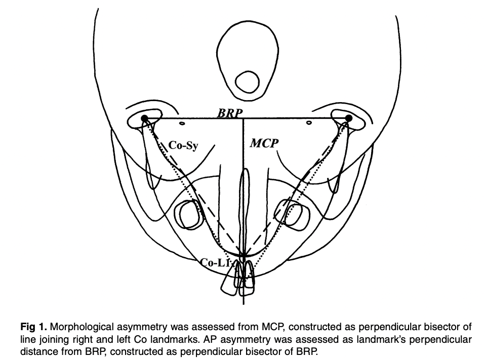
→ Whole mandible shifted LATERALLY + POSTERIORLY to crossbite side
→ Midline deviation = 1.6 mm toward crossbite side
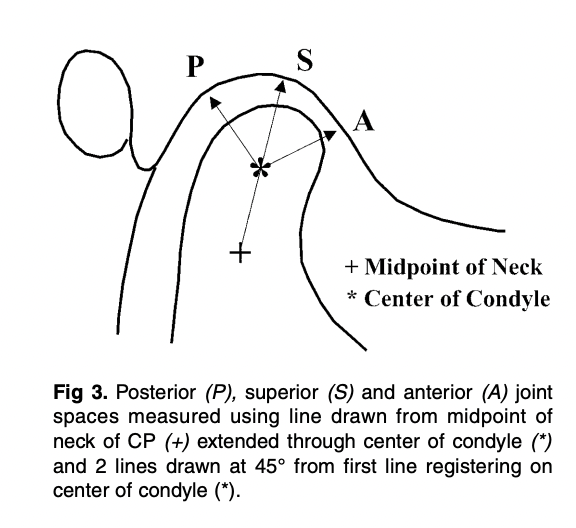
LAYER 2 — JOINT SPACE (Where is the condyle in the fossa?)
→ Noncrossbite condyle = more anterior on articular eminence
→ Superior joint space: 4.0 mm (non-XB) vs 3.2 mm (XB) ← SIGNIFICANT
→ Posterior: larger on non-XB (not significant)
→ Anterior: EQUAL on both sides ← MCQ TRAP
LAYER 3 — MORPHOLOGICAL (Has the bone actually changed shape?)
→ Yes! Ramus is SHORTER on crossbite side
→ Co–Sy: 75.5 mm (non-XB) vs 73.9 mm (XB) — 1.6 mm difference
→ Asymmetry in RAMUS (condyle + coronoid) but NOT in body (L6–L1 equal)
⚡ THE NUMBERS BANK — Memorise These 10 Numbers
Value
What It Represents
5.9–9.4%
Incidence of UPXB
67–79%
Proportion of UPXB that are functional
1.6 mm
Midline deviation + Co–Sy difference
~3 mm
L6 and coronoid horizontal offset (crossbite side more lateral)
4.0 vs 3.2 mm
Superior joint space (non-XB vs XB) — only SIGNIFICANT TMJ finding
8.8 yrs
Mean age of subjects
15
Sample size
1 month
RPE activation phase
6 months
Retention phase
11
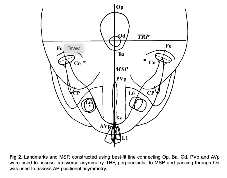
Total SMV landmarks digitized
🔥 MECHANISM CHAIN — Viva Storytelling Version
Examiner: “Walk me through how FUPXB causes skeletal asymmetry.”
YOUR ANSWER:
Narrow maxilla creates a dental interference → mandible must shift from CR to ICP, deviating laterally and anteroposteriorly toward the crossbite side → this asymmetric posture alters condylar loading: noncrossbite condyle rides higher on the articular eminence → muscle compensation: anterior temporalis fires more on the noncrossbite side; posterior temporalis fires more on the crossbite side → sustained asymmetric forces trigger adaptive bone remodeling → the ramus on the crossbite side becomes shorter (both condylar and coronoid processes affected) → result: a functionally crossbited child now has a morphologically asymmetric mandible
🎯 EXAMINER TRAPS — Don’t Fall For These
Trap Statement
The Truth
“All joint spaces are asymmetric in FUPXB”
❌ Only superior space is significant; anterior joint spaces are EQUAL
“RPE doesn’t affect morphological asymmetry”
❌ RPE + retention eliminated morphological asymmetry — crossbite side grew MORE
“The mandibular body is asymmetric”
❌ L6–L1 distance is EQUAL — asymmetry is in the ramus only
“Glenoid fossa is asymmetric too”
❌ Fossa position showed little/no transverse or AP asymmetry
“Chewing patterns normalize after RPE”
❌ Reverse chewing sequencing persists even after correction
“Functional crossbite = symmetric mandible”
⚠️ Only in adults — in growing children, morphological change occurs
🧠 MUSCLE MNEMONIC — Never Mix This Up
“At the PARTY, Non-Cross goes FORWARD, Cross goes BACK”
ANTERIOR temporalis (forward-pulling) → fires more on NON-crossbite side
POSTERIOR temporalis (backward-pulling) → fires more on CROSSBITE side
📊 Pre vs. Post Treatment — What Changed?
Measurement
Pre-Treatment
Post-Retention
Verdict
Co–Sy side difference
Significant
Not significant
✅ Resolved
L6 lateral offset
~3 mm
~0 mm
✅ Resolved
Midline deviation (L1)
1.6 mm
Not significant
✅ Resolved
Superior joint space gap
Significant
Not significant
✅ Resolved
Glenoid fossa position
Not significant
Not significant
➡️ Unchanged (already symmetric)
Chewing pattern (reverse)
Abnormal
Still abnormal
❌ NOT resolved
🔑 Key insight on growth: Crossbite-side ramus grew MORE than noncrossbite side during treatment — compensatory catch-up growth. The mandible also rotated forward and medially on the crossbite side, and backward and laterally on the noncrossbite side.
🏛️ LANDMARKS MNEMONIC (All 11 SMV Landmarks)
“Old Baboons Often Play Violins — Conducting Fine Concerts, Like Symphony”
❓ SELF-TEST — Rapid Fire (Cover answers, test yourself)
Question
Answer
Which ramus is LONGER in FUPXB?
Noncrossbite side
Which joint space is EQUAL on both sides?
Anterior
Which is the ONLY significantly different joint space?
Superior
Where does the asymmetry occur in the mandible?
Ramus (not body)
What persists even after successful RPE?
Reverse chewing sequencing
What muscle is more active on the noncrossbite side?
Anterior temporalis
What does the MCP stand for and how is it constructed?
Midcondylar reference plane — ⊥ bisector of Co-Co line
What was the midline deviation pre-treatment?
1.6 mm toward crossbite side
Which radiograph assessed TMJ spaces?
Zonograms (4-turn spiral tomography)
What is the key conclusion that overturns classic teaching?
Functional crossbites cause morphological (structural) mandibular asymmetry in growing children
🩺 VIVA CLINCHER — The One Paragraph Examiners Love
“Santos Pinto et al. demonstrated that the classic view of functional crossbite as purely a positional problem is incomplete in growing children. Their prospective study showed the mandible is both positionally displaced and morphologically asymmetric — with the ramus shorter on the crossbite side due to adaptive remodeling. Crucially, the asymmetry is ramus-specific; the mandibular body remains symmetric. Early bonded RPE successfully resolved both layers of asymmetry through compensatory growth, though abnormal chewing patterns persisted, highlighting the need for functional rehabilitation post-treatment.”
Picture this exam scenario: A 7-year-old child sits in your chair. Her mom says “her jaw looks crooked.” You notice her teeth bite on the right side but her chin shifts left. Is this dental? Skeletal? Functional? Do you treat now or wait?
Every answer in this review solves THAT case.
⚡ The “Know This Or Fail” Numbers
Stat
Value
Why It Matters
Posterior crossbite prevalence
7–23%
Most common transverse malocclusion
FXB = unilateral with shift
80–97% of all PXB
Nearly all UPXBs are functional!
Self-correction rate
0–9%
Never justify waiting
Deciduous dentition prevalence
8.4% → 7.2% mixed
Slight spontaneous decrease
Spontaneous new crossbite development
7%
Equals self-correction rate — net zero
Equilibration success (< 5 yrs)
27–64%
Only in very young, limited use
Arch perimeter gain
4 mm (85% stable long-term)
Bonus benefit of expansion
🔥 EXAM TRAP: “Posterior crossbite is self-correcting” → FALSE. Only 0–9%. Never a valid clinical justification.
🧩 Etiology: The BIG Picture First
Think in 3 layers — Genetic → Environmental → Habit
 NARROWED MAXILLA
      ↑
┌─────────────────────────────────┐
│  SKELETAL: Small Max/Mand ratio │ ← Genetic + mouth breathing
│  + Increased lower face height  │
└─────────────────────────────────┘
      ↑
┌───────────────────────────────────────┐
│  AIRWAY: Adenoids / Tonsils / Rhinitis│ → Mouth breathing → narrow maxilla
│  + Neonatal intubation                │ → Direct palatal deformation
└───────────────────────────────────────┘
      ↑
┌─────────────────────────────────────────────┐
│  HABIT: Pacifier / Digit sucking >4 yrs age │ → ↓ Max intercanine + ↑ Mand intercanine
└─────────────────────────────────────────────┘
🧠 Mnemonic: “GANH”(say it like “Gain” — because early treatment = gain!)
🔍 Differential Diagnosis — The Most Examined Section
The 3-Type Framework
Feature
✅ FXB (Functional)
Single Tooth XB
True Skeletal Bilateral XB
CO vs CR
Discrepancy (mandatory finding)
Coincident
Coincident
Mandibular midline
Deviated to crossbite side
Midline OK
Midline OK
Maxillary arch shape
Symmetrical (key!)
Asymmetrical
Symmetrical
Crowding pattern
More in maxilla (not mandible)
Localized
—-
Crossbite side molar
Class II (partial/full)
Varies
Bilateral Class II
Non-crossbite side molar
Class I
Normal
Bilateral Class II
Condyle position (tomogram)
Non-XB side: down & forward in fossa
Symmetric
Symmetric
Cause
Transverse maxillary deficiency
Overretained teeth / arch length
Severe skeletal discrepancy
🧠 Mnemonic: “SMACK-D” (What FXB gives you clinically)
Shift of mandible → toward crossbite side Midline mandibular deviation → toward crossbite side Arch — maxillary is symmetrical (despite appearing unilateral!) CO–CR discrepancy — the defining diagnostic feature Klass II on crossbite side / Class I on non-crossbite side (K for klass 😄) Deficiency maxillary arch → more crowding in upper than lower
🔥 EXAM TRAP: The maxillary arch in FXB is SYMMETRICAL. The unilateral appearance is caused by the mandibular shift — not by asymmetric maxillary constriction. Examiners love asking this!
⏰ Treatment Timing — The Golden Window
Think of the Midpalatal Suture as a WINDOW that closes with age:
Age:        2–5 yrs        6–8 yrs           9–11 yrs       12+ yrs         Adult
Suture:     Wide open      [BEST WINDOW]     Narrowing      Almost fused    Fused
Force:      Minimal        Small forces      Moderate       RME needed      SURGERY
Recommend:  Equilibration  ✅ IDEAL       ⚠️ Difficult   ⚠️ RME only     ❌ Ortho+Surg
Late deciduous / early mixed dentition = IDEAL → small forces open suture, permanent incisors get space before eruption
🏆 Gold standard — 1/3 cost of removable, 1/5 treatment time
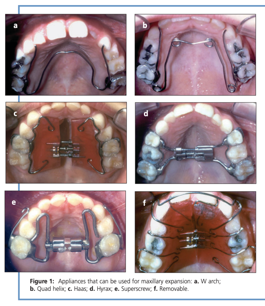
W-Arch
Slow
¼ turn / 2–3 days
6–12 wks
✅ Good alternative
Haas
RME
1–2 × ¼ turn/day
2–6 wks
✅ Tissue-borne, most skeletal effect
Hyrax
RME
1–2 × ¼ turn/day
2–6 wks
✅ Tooth-borne, hygienic
Superscrew
RME
1–2 × ¼ turn/day
2–6 wks
✅ Comparable to Haas/Hyrax
Removable plate
Slow
¼ turn / 5–7 days
Longest
❌ NOT recommended — compliance failure, relapse, lost appliances
📋 Retention Protocol — “SOLAR”
Stabilize screw with ligature wire or composite Overexpand — lingual cusps of upper contact buccal cusps of lower Leave appliance in place for retention OR make removable retainer At least 4–6 months retention minimum Rule: Retention period ≥ active treatment duration
⚠️ Side Effects of RME — “DEMO”
Diastema (midline maxillary — transient! closes via transeptal fibers) → warn patient/parent Expansion of mandibular intercanine width (spontaneous — actually a bonus in crowded cases) Maxillary protraction (forward movement of maxilla — useful in Class III patients!) Open bite (anterior) — especially if 2nd permanent molars present; control molar eruption carefully
🧬 The Adaptation Argument (Why You MUST Treat Early)
This is the biological rationale section — examiners love conceptual questions here.
If left untreated, 3 irreversible adaptations occur:
Condylar asymmetry → glenoid fossa and condyle remodel asymmetrically during growth
Mandibular rotation → mandible rotates relative to cranial base (submentovertex X-ray shows this in adults)
Muscle adaptation → masticatory cycle becomes asymmetric (Throckmorton et al.)
After early treatment: condyle symmetry restored, mandibular rotation corrected, masticatory symmetry re-established
Adult with untreated FXB: mandible is rotated relative to cranial base but symmetric within the fossa — adaptation has already “locked in” the asymmetry
🔥 EXAM TRAP: In adults with untreated posterior crossbite, condyles ARE symmetric within the fossa (adaptation is complete) but mandible IS asymmetric relative to the cranial base. Don’t confuse this!
AGAINST causation: Sari et al., Keeling et al. found no causal link
Safe conclusion:“Crossbite may be a cofactor in TMD identification, but its role should not be overstated”
🎯 The Selective Grinding Rule (< 5 Years Only)
Age limit: strictly < 5 years
Success rate: 27–64% (Lindner: 50% in 4-year-olds)
The magic number: maxillary intercanine width must be ≥ 3.3 mm greater than mandibular for best results
Beyond age 5 → expansion appliances required, not grinding
🏁 Master Flash Summary — “FEED-SOLAR”
(Treatment protocol in one phrase)
Functional shift eliminated by symmetric maxillary expansion Early treatment — late deciduous / early mixed dentition Expand symmetrically (even for unilateral presentation!) Don’t use removable appliances — Stabilize screw, Overexpand, Leave appliance in, At least 4–6 months, Retention ≥ treatment time
🔥 5 Rapid-Fire Viva Questions
Q1. Why do we expand the maxilla symmetrically for a unilateral crossbite? → Because the maxilla is bilaterally constricted; the unilateral appearance is due to the mandibular shift
Q2. What is the single most important diagnostic feature of FXB? → CO–CR discrepancy (centric occlusion ≠ centric relation)
Q3. Why is the Quad Helix preferred over removable plates? → 1/3 cost, 1/5 treatment time, no compliance issues
Q4. What happens to the midpalatal diastema created during RME? → Closes spontaneously via transeptal fiber pull and dental tipping
Q5. Name two studies supporting crossbite–TMD correlation. → Alamoudi (2000) and Egermark-Eriksson et al. (1990)
WHAT IS A ORTHODONTIC STUDY MODEL? It is an essential diagnostic record which help to study the occlusion and dentition from all 3 dimensions. The models are used as pre-treatment models, stage models and post treatment models. Purposes of Orthodontic Study Models Represent the case prior to orthodontic treatment. Aid the dentist in diagnosing […]
The angulation of the Twin Block appliance’s inclined planes underwent three distinct stages of development, each driven by clinical observations and biomechanical reasoning.
Stage 1 — 90° (Initial Design)
The earliest Twin Block appliances, developed by W.J. Clark, featured bite blocks angulated at 90° to the occlusal plane. Patients were required to consciously posture the mandible forward to occlude the blocks. However, many patients struggled to maintain this forward position and habitually returned to their original distal occlusal position, causing the flat-surfaced blocks to stack on top of each other. This resulted in a significant posterior open bite, a complication seen in approximately 30% of early Twin Block cases.
Stage 2 — 45° (Functional Correction)
To resolve the compliance problem, the angulation was modified to 45° to the occlusal plane. This immediately guided the mandible forward more passively, eliminating the stacking issue. A 45° angle provides an equal downward and forward force component to the lower dentition, promoting both vertical and sagittal growth stimuli. Clark continued using this angulation clinically for approximately 8 years before the next modification.
Stage 3 — 70° (Current Standard)
After the prolonged use of the 45° design, the angulation was increased to 70° to the occlusal plane — the current standard configuration. This steeper angle introduces a more horizontal force component, theoretically encouraging greater forward (sagittal) mandibular growth rather than a combined downward-forward stimulus. The 70° angle is now incorporated into the standard Twin Block design with maxillary and mandibular acrylic base plates.
Angulation at a Glance
Angulation
Rationale
Limitation
Angulation
Rationale
Limitation
90°
Original design; edge-to-edge block contact
~30% posterior open bite; poor compliance
45°
Equal forward + downward force vector
Used for 8 years; less horizontal growth stimulus
70°
More horizontal force; greater forward mandibular growth
May reduce mandibular postural guidance
Clinically, if a patient struggles to maintain the forward mandibular posture with a 70° design, it is advisable to revert to a 45° angulation to facilitate easier maintenance of the protruded position.
Posterior crossbite is one of the most common transverse discrepancies encountered in orthodontic practice. A transpalatal arch (TPA) is a deceptively simple appliance — but when activated using Burstone biomechanics, it becomes a powerful tool capable of producing controlled symmetric or asymmetric molar expansion.
Understanding force systems, moment-to-force ratios, and side effects is essential if one wants to use this appliance predictably.
This article walks through the biomechanics, clinical application, and outcomes of a Burstone-type TMA transpalatal arch.
1. Why Molar Transverse Position Matters
Correct positioning of maxillary first molars is critical for:
Functional occlusion
Arch coordination
Midline stability
TMJ health
Untreated transverse maxillary deficiency may cause:
Posterior crossbite
Functional mandibular shift
Midline deviation
TMJ strain
Posterior crossbite prevalence:
Unilateral: ~9%
Bilateral: ~4%
Quick Viva Pause
Q: Why is unilateral crossbite more problematic than bilateral crossbite?
A: Because it frequently causes functional mandibular shift, leading to asymmetry and midline deviation.
2. What is a Burstone-Type Transpalatal Arch?
A transpalatal arch (TPA) connects the maxillary first molars across the palate.
It can be used in two modes:
Mode
Purpose
Passive
Anchorage reinforcement / stabilization
Active
Tooth movement
The Burstone system differs from traditional TPA systems.
Key Differences
Feature
Burstone TPA
Goshgarian TPA
Attachment
Lingual bracket
Lingual sheath
Wire material
TMA
Stainless steel
Force magnitude
Lower
Higher
Control
High precision
Less controlled
TMA wires produce ~60% lower force compared to stainless steel, improving control and reducing unwanted side effects.
Viva Pause
Q: Why is TMA preferred over stainless steel in Burstone TPA?
Answer
Lower load-deflection rate
Greater formability
More controlled force delivery
Reduced risk of excessive tipping
3. Recommended Activation
Typical parameters reported:
Parameter
Value
Activation
3–10 mm
Expansive force
1.5–4 N
Wire dimension
0.032 × 0.032 TMA
A 10 mm activation produces approximately 4 N expansion force.
However, force depends on:
Wire length
Loop configuration
Height of arch
Patient anatomy
Viva Pause
Q: What happens if the TPA height increases?
Answer
The moment-to-force ratio changes, altering the type of tooth movement.
4. Types of Expansion Using TPA
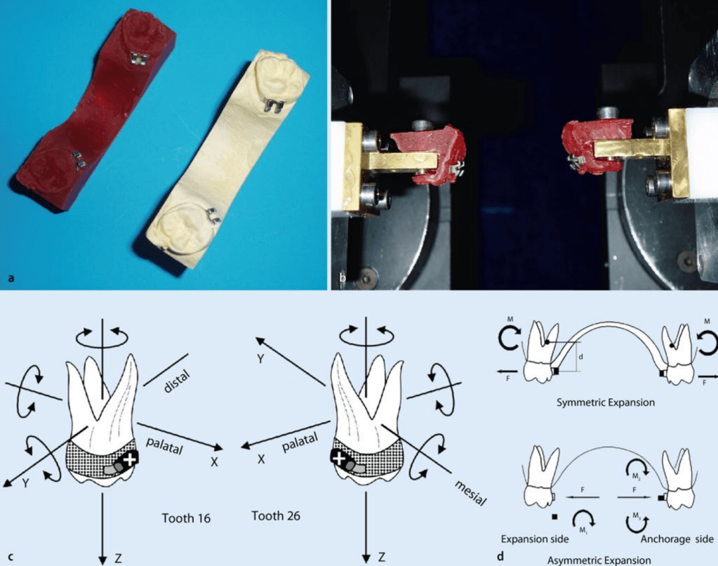
1. Symmetric Expansion
Both molars move buccally.
Used for:
Bilateral posterior crossbite
Narrow maxilla
2. Asymmetric Expansion
One side expands more than the other.
Used for:
Unilateral crossbite
This is achieved by creating moment differential between molars.
Biomechanical Principle
Side
Force System
Crossbite side
Force → tipping movement
Anchorage side
Force + counter-torque
This allows unilateral expansion without significant movement of the anchorage molar.
Viva Pause
Q: Why is tipping used on the crossbite side?
Answer
Because tipping requires less force than bodily movement, making unilateral correction easier.
5. Biomechanics of Burstone TPA
The appliance generates:
Force component
Effect
Expansive force
Buccal movement
Moment
Crown tipping
Vertical force
Minor extrusion/intrusion
The center of resistance of molars lies approximately:
7 mm apical to the bracket level in the furcation region.
Viva Pause
Q: Why does TPA cause buccal crown tipping?
Answer
Because the force is applied away from the center of resistance, creating a moment that tips the crown buccally.
6. Clinical Outcomes (Study Findings)
Symmetric Expansion
Parameter
Result
Mean expansion
~4.5 mm
Buccal tipping
~10°
Treatment time
12 weeks
Vertical side effects
Minimal
Expansion occurred primarily due to buccally directed forces acting at the crown level.
Viva Pause
Q: What is the main disadvantage of symmetric TPA expansion?
Answer
Buccal crown tipping of molars, which may require later torque correction.
7. Asymmetric Expansion Outcomes
For unilateral crossbite:
Parameter
Crossbite Side
Anchorage Side
Tooth movement
~2.5 mm
~0.8 mm
Torque
Higher
Lower
Vertical movement
Minimal
Minimal
Thus effective unilateral expansion was achieved in all patients.
Viva Pause
Q: Why does the anchorage side show less movement?
Answer
Because counter-torque increases moment-to-force ratio, resisting tipping.
8. Side Effects
Vertical Effects
Movement
Magnitude
Intrusion
~0.6 mm
Extrusion
~0.8 mm
These are considered clinically insignificant.
Sagittal Effects
Minor:
Mesial rotation of molars
Minimal sagittal displacement
Viva Pause
Q: What is the most common rotational side effect?
Answer
Mesial rotation of molars
9. Why Simulation Systems Were Used
The study used Orthodontic Measurement and Simulation System (OMSS).
Purpose:
Measure force systems
Predict tooth movement
Compare simulation vs clinical outcomes
Findings:
Simulated movements were highly consistent with clinical results.
Viva Pause
Q: Why can’t simulation fully replicate real orthodontic tooth movement?
Answer
Because it cannot account for:
Mastication
Occlusal contacts
Soft tissue forces
Material fatigue
Biological variability
10. Clinical Pearls for Orthodontists
1. TPA is not just an anchorage appliance
It can produce controlled molar movement.
2. Shape matters
Force depends on:
Height
Length
Configuration
3. Perfect force systems are difficult
Even identical activation may produce different forces due to anatomical variation.
4. Tipping is expected
Crossbite correction usually occurs by molar tipping rather than bodily movement.
5. Torque correction may be needed later
After expansion, clinicians may need to:
Add counter-torque
Use archwire adjustments
Rapid Revision Table
Feature
Symmetric Expansion
Asymmetric Expansion
Indication
Bilateral crossbite
Unilateral crossbite
Force system
Equal bilateral forces
Differential moment
Mean expansion
~4.5 mm
~2.5 mm on affected side
Crown tipping
Present
Controlled
Side effects
Minimal
Minimal
Ultimate Viva Questions (PG Level)
Basic
1. What is the function of a transpalatal arch?
Anchorage control
Molar rotation control
Transverse expansion
Intermediate
2. Why is TMA preferred in Burstone TPA?
Lower load-deflection rate
Better formability
More controlled forces
Advanced
3. How does asymmetric TPA correct unilateral crossbite?
By generating different moment-to-force ratios on each molar.
Clinical
4. What is the most common side effect of TPA expansion?
Buccal crown tipping.
Biomechanics
5. Why does tipping occur with TPA?
Force acts away from center of resistance, generating a moment.
Final Takeaway
The Burstone-type TPA is a biomechanically sophisticated appliance capable of producing:
Controlled symmetric molar expansion
Targeted asymmetric correction of unilateral crossbite
Minimal side effects
When understood biomechanically, it transforms from a simple wire into a precise orthodontic force delivery system.
 Dentowesome