ADAMS CLASP

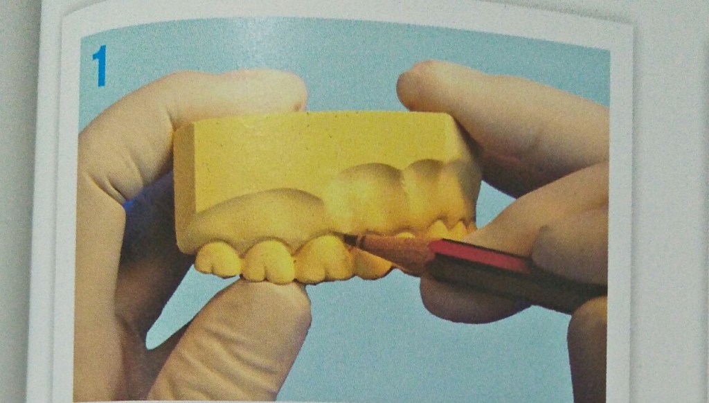
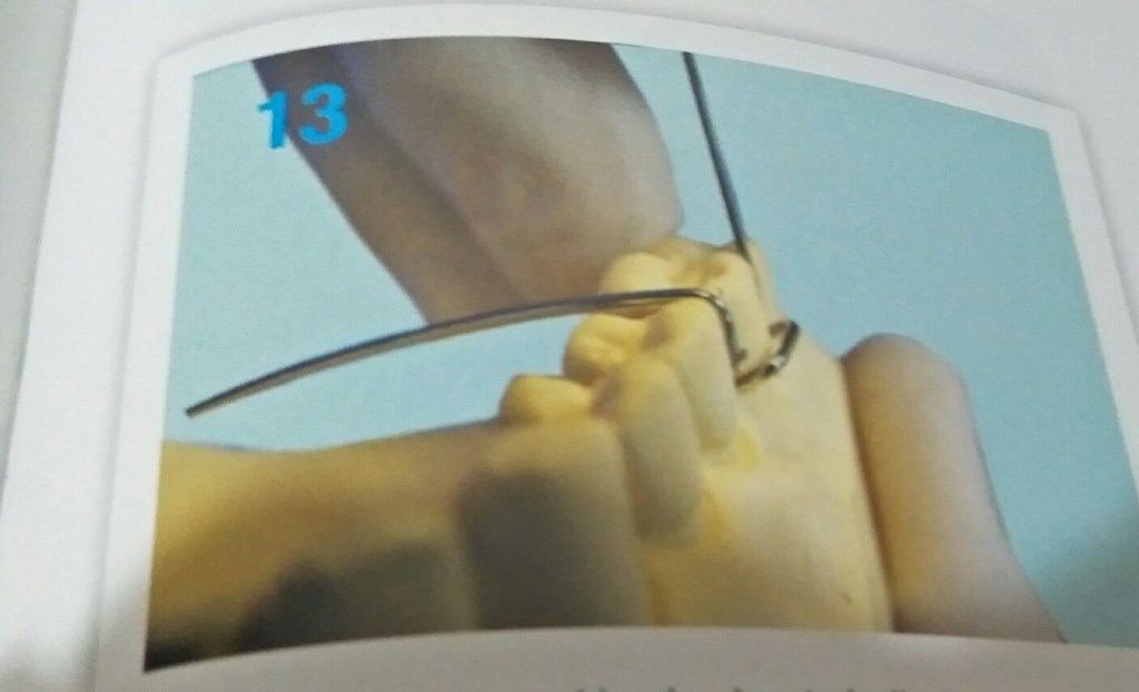
Clasps are the retentive components of the removable appliances.

Mode of action-

  • Clasps act by engaging certain areas of teeth called the undercuts.
  • Two types of undercuts are found in natural dentition
  • Buccal and lingual cervical
  • Mesial and distal proximal
  • Adams clasp engages the mesial and distal proximal undercuts.

Adams clasp also called as universal clasp, liverpool clasp and modified arrowhead clasp.

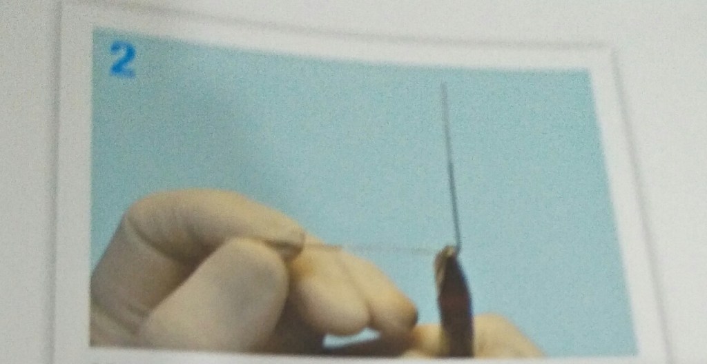
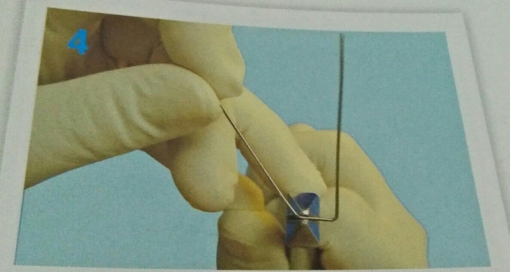
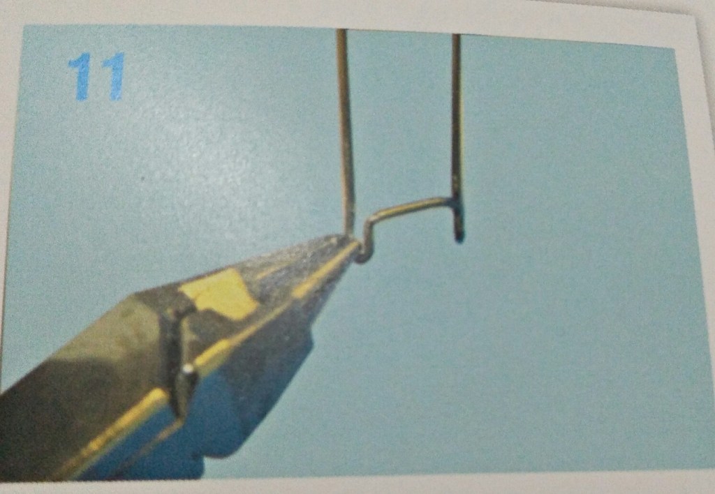
Parts of adams clasp-

  • Two arrowheads
  • Bridge
  • Two retentive arms

Advantages of adams clasp-

  • Rigid and offers excellent retention
  • Fabricated on deciduous and permanent dentition
  • Can be fabricated on fully or partially erupted tooth
  • Can be used on molars, premolars and incisors.
  • Small and occupies minimum space
  • Can be modified in many ways.
  • Universal pliers can be used for fabricating.

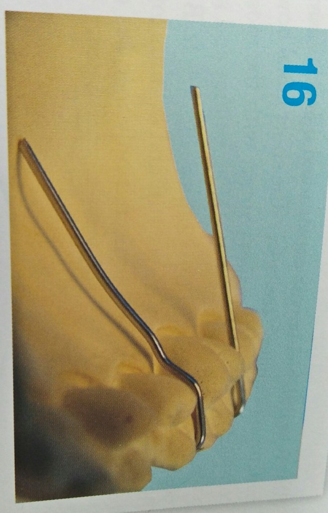
Modifications of adams clasp-

  • Adams with single arrowhead
  • Adams with J hook
  • Adams with incorporated helix
  • Adams with additional arrowhead
  • Adams on incisor and premolars
  • Adams with distal extension

Reference- Bhalajhi 7th edition

STEPS IN FABRICATION OF ADAMS CLASP

Mesial and distal undercuts of the molar are marked on the cast. This distance between these 2 marks would form the length of bridge of Adams.
22 gauge hard round stainless steel wire is used. 90 degree bend is made.
Wire is placed on the model and distance between mesial and distal undercuts is marked on wire.
The other 90 degree is made thus forming bridge of adams clasp
Round beak of universal plier is placed on outer side close to bridge of adams
Wire bent around to form a “U” thus forming the arrowhead
Arrow head is squeezed between the two beaks of pliers to make it as narrow and pointed as possible.
Arrow head is given a 45 degree twist so that arrow head at angle of 45 degree to bridge of adams
Outer arm of arrow head is given a 90 degree bend at a height that is half of arrow head. Done by placing round beak inside arrow head.
Other arrow head is also bent similarly so that the free end of wire rests in embrasure. Wire is bent down and adapted between teeth.
Place a mark beyond palatal side of contact area.
Palatal tag is bent down and is slightly kinked to form step over gingiva. Palatal wire is adapted to be parallel to the plaster.

3 things to be noted after the clasp is placed on tooth:-

  • When viewed from occlusal aspect bridge is parallel to buccal aspect.
  • Bridge to be at 45 degree to long axis of tooth.
  • When viewed from buccal aspect parallel to occlusal surface.

Reference- bhalajhi ortho

Ortho Case 3.3

A 14-year-old male presented with a class I malocclusion on a skeletal class I base with average vertical dimensions complicated by an ectopic and mesially-angulated UR3, crowding of both arches and centre line discrepancies.

What will be the treatment for creating space for canine and correcting central line discrepancies? Which teeth will be extracted? What appliances will be used and what force level should be used during space closure?

To understand this and more, read the pdf attached to the link. I have also tried to explain the case via digital images. I have attached 5 images. Do check it out 🙂

Happy Reading Friends!
LINK: https://drive.google.com/file/d/1cfWNTyZVX7IISDGJvJIZ8rjuXZ3fR2Hd/view?usp=sharing

Ortho Case 3.2

Link: https://drive.google.com/file/d/1htUo39ZpGIdf5XGRcSmcn3RAsYzJEY6k/view?usp=sharing

A 13-year-old female presented with a class I malocclusion on a mild skeletal class II pattern with average vertical dimensions complicated by impaction of the LR5 and an invaginated UL2

What is treatment plan? Which tooth will be extracted? What factors need to be considered when substituting a maxillary canine for a lateral incisor?

I have attached following images for easy read! Happy Reading Friends 🙂

Ortho Case 3.1

A 13-year-old female presented with a class I malocclusion on a mild skeletal class II pattern with average vertical dimensions complicated by moderate upper arch crowding, palatal displacement of the UL2 and a localized crossbite

Initially, Headgear was used but compliance was poor. It was therefore decided to remove both maxillary second premolars to recreate space for maxillary arch alignment.

Therefore, Self-ligating brackets (SLBs) were used in this case after extractions. What are SLBs and what are the advantages of these systems? Why has glass ionomer cement been placed on the maxillary first molars? What is the final buccal segment relationship

link: https://drive.google.com/file/d/1YaucTBr47omt-lD1Ok5kM-BMXc3R3c72/view?usp=sharing