Diagnosis Demystified – Case 8/255

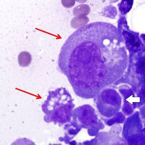
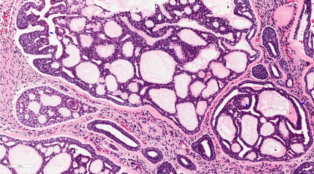
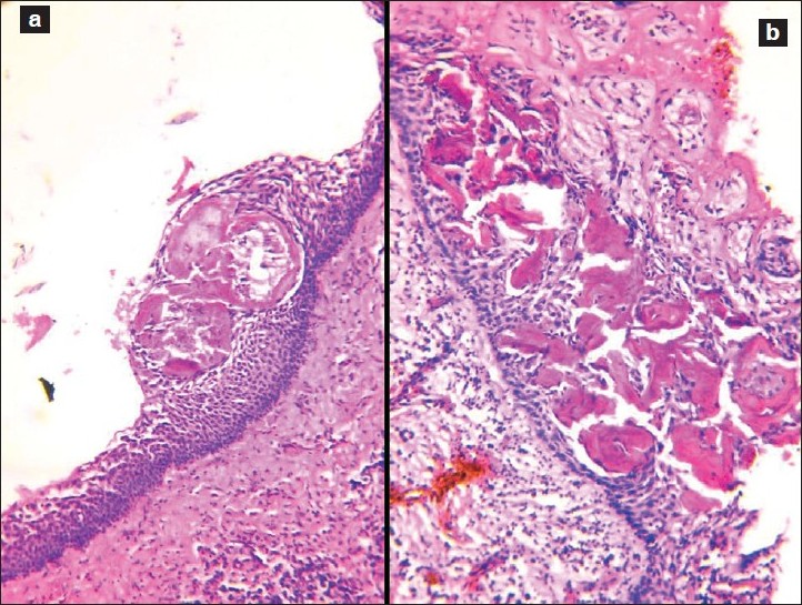
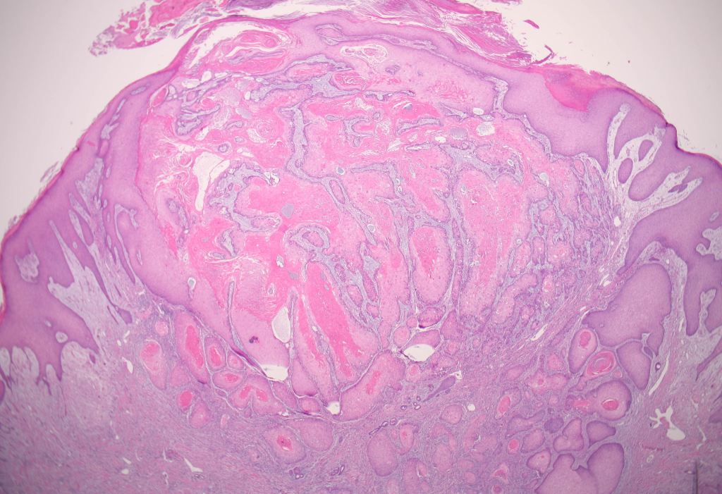
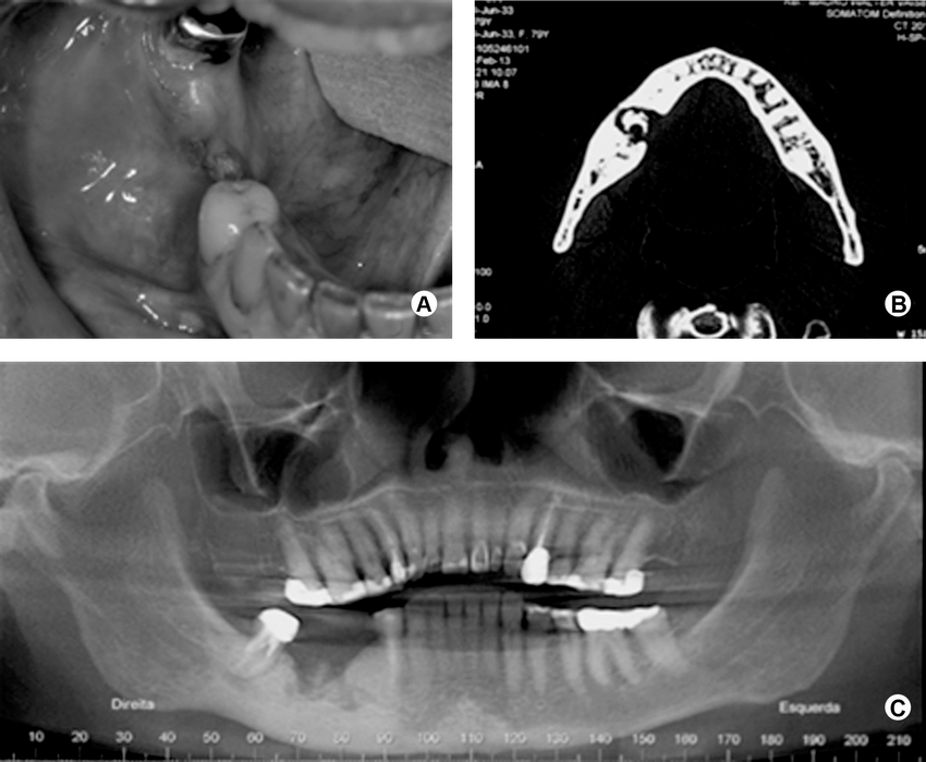
A cyst was removed along with an unerupted third molar tooth. The pathologist reported a fibrous capsule that was myxoid in places and that was lined by a thin layer of squamous and cuboidal epithelium. The cyst originated from the amelocemental junction.

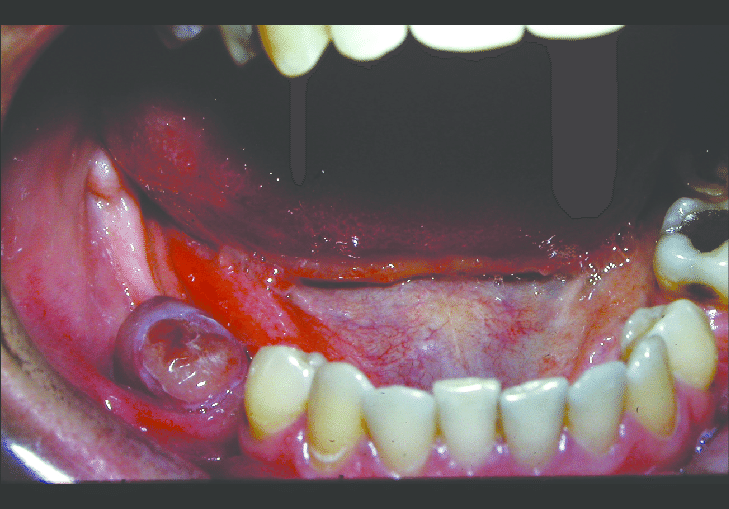
As a dental student, it’s important to have a good understanding of dentigerous cysts. Dentigerous cysts are a type of cyst that form around unerupted or impacted teeth. They develop from a structure called the dental follicle, which surrounds the tooth germ or the developing tooth.

When a tooth fails to erupt properly, sometimes a cyst can form around it. This cyst is called a dentigerous cyst. It is characterized by a fibrous capsule, which is like a protective layer, and it is lined by stratified squamous epithelium, which is a type of tissue that forms the outer layer of the cyst.

Histologically, dentigerous cysts may show various features such as the thickness of the epithelial lining or the presence of inflammation or keratinization. However, these features are not unique to dentigerous cysts and can also be seen in other types of cysts.

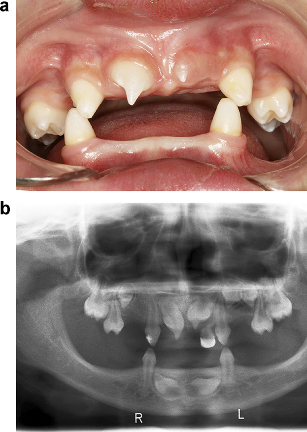
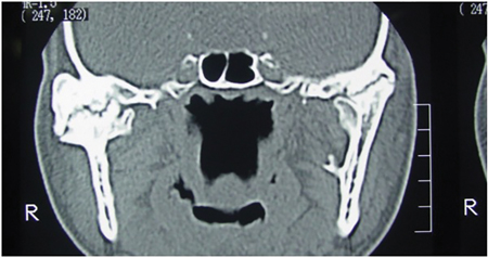
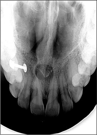
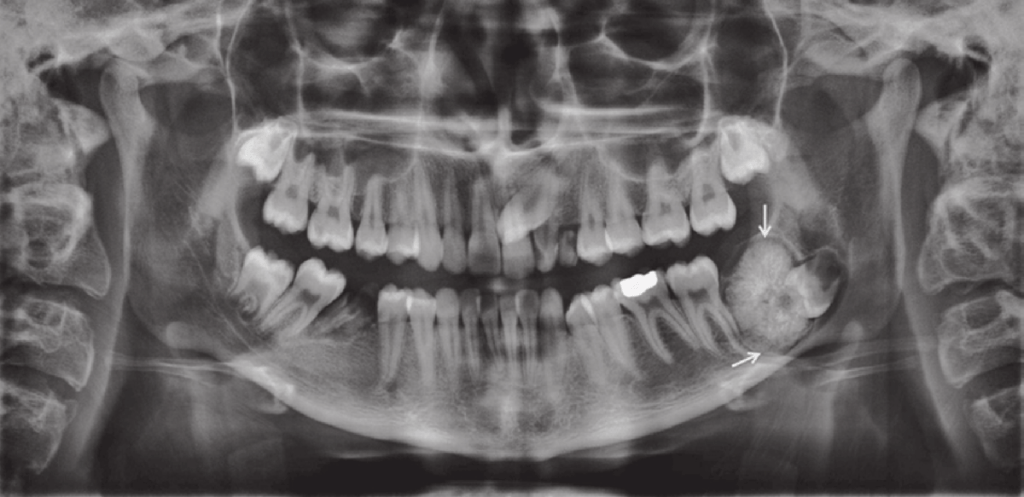
In terms of diagnosis, clinical features are important. Dentigerous cysts are usually discovered during routine dental exams or through radiographic evaluations. One of the key clinical features is that the cyst originates from the amelocemental junction, which is the junction between the enamel (the hard outer layer of the tooth) and the cementum (the specialized tissue covering the root of the tooth).

On an X-ray, the dentigerous cyst appears as a well-defined radiolucent area around the crown of the impacted tooth. It’s important to note that dentigerous cysts often don’t cause any symptoms and are found incidentally. However, if they grow in size or become infected, they can lead to swelling, pain, or displacement of nearby teeth.

Treatment for dentigerous cysts usually involves surgical removal or marsupialization, which is a procedure that creates a surgical opening to allow drainage and shrinkage of the cyst. This is done to prevent complications and facilitate the eruption of the impacted tooth.

Overall, understanding the clinical and histological features of dentigerous cysts is crucial for diagnosing and managing these conditions effectively as a dental student.

Diagnosis Demystified – Case 7/255

A cyst was enucleated from the posterior mandible of a 38-year-old man. The pathologist reported that the lining was composed of stratified squamous epithelium that showed parakeratosis and basal-cell palisading. Some areas were inflamed and cholesterol nodules were noted.

As a dental student, it’s important to understand the histological features of odontogenic keratocyst (OKCs) in order to recognize and diagnose them accurately. Here’s a breakdown of the key features:

  1. Basal-cell palisading: Odontogenic keratocysts have a specific arrangement of cells in their lining. The cells at the base of the epithelium align in a palisade-like fashion, with their nuclei positioned away from the central cavity of the cyst. This is a distinctive characteristic often seen in OKCs.
  2. Keratinisation: Odontogenic keratocysts exhibit a high degree of keratinization. This means that the cells within the lining of the cyst produce a protein called keratin, resulting in the formation of a layer of keratinized cells. This keratinization gives the lining a unique appearance when observed under a microscope.
  3. Uniform thickness: The epithelial lining of an odontogenic keratocyst tends to have a relatively consistent thickness throughout. This means that when examining a tissue sample of an OKC, you will notice that the lining is of similar thickness in different areas. However, it’s important to remember that this feature alone is not sufficient for making a definitive diagnosis.
  4. Lumen filled with keratinous material: The central space or lumen within an odontogenic keratocyst is typically filled with keratinous material. This material is composed of dead cells that have undergone keratinization. Essentially, the lining of the cyst sheds these keratinized cells, and over time, they accumulate within the cystic space, filling it with this keratin debris.

Remember, these histological features are indicative of an odontogenic keratocyst, but a comprehensive diagnosis also requires consideration of other clinical and radiographic findings. It’s crucial to consult with an experienced oral and maxillofacial pathologist for a definitive diagnosis and to determine the appropriate treatment plan for the patient.

Diagnosis Demystified – Case 6/255

A pear-shaped and well-circumscribed radiolucent lesion with a corticated outline was found on a radiograph related to the root of an upper central incisor. The tooth was not restored and proved vital on testing.

A nasopalatine duct cyst is a specific type of cyst that develops from remnants of the nasopalatine duct. During embryonic development, this duct is involved in the formation of the nasal and oral cavities. Sometimes, remnants of this duct can persist and give rise to a cystic lesion later in life.

The nasopalatine duct extends from a structure called the incisive canal, which is located in the midline of the maxillary bone (the bone that comprises the upper jaw). The cyst can develop anywhere along this tract.

When we examine the lining of a nasopalatine duct cyst under a microscope, we typically observe the presence of both respiratory and squamous epithelium. This finding is significant because it indicates the origin of the cyst from the nasopalatine duct. The respiratory epithelium represents the lining of the nasal portion of the duct, while the squamous epithelium represents the lining of the oral portion.

Furthermore, a nasopalatine duct cyst often contains a neurovascular bundle within its capsule. This bundle consists of nerve fibers and blood vessels that supply the nasopalatine area. Its presence within the cyst is a characteristic feature.

In clinical practice, nasopalatine duct cysts are usually asymptomatic and are often discovered incidentally during routine dental examinations. However, if the cyst becomes large or infected, it can cause pain, swelling, and discomfort in the affected area.

As a dental student, it’s important to be familiar with the clinical presentation, radiographic appearance, and management of nasopalatine duct cysts. Treatment typically involves surgical removal of the cyst to relieve symptoms and prevent potential complications.

Diagnosis Demystified- Case 6/255

A 26-year-old African patient presented with a rapidly growing lesion that expanded the mandible. There was bone destruction on the radiograph and therefore malignancy was suspected. On biopsy, the lesion was intraosseous and was cavitated. There was profuse bleeding and a small biopsy of the lining was taken. The pathologist reported osteoclast-like giant cells and granulation tissue with blood clots.

An aneurysmal bone cyst is a type of benign bone tumor that can occur in the jawbone. It is called an “aneurysmal” because it contains blood-filled spaces that can resemble the sac-like structure of an aneurysm. These cysts are relatively rare, accounting for less than 1% of all bone tumors.

Clinically, an aneurysmal bone cyst can be dramatic because it can grow rapidly and attain a large size. This can cause symptoms such as pain, swelling, and displacement of nearby teeth. When a biopsy is performed to confirm the diagnosis, bleeding can be profuse due to the presence of fragile blood vessels within the cyst. In some cases, a blood transfusion may be necessary to manage the bleeding.

It is important to note that aneurysmal bone cysts often form around an underlying primary lesion, such as a vascular malformation or bone tumor. Therefore, it is crucial to thoroughly evaluate any aneurysmal bone cyst to determine if there is an underlying primary lesion that needs to be addressed. Treatment options for aneurysmal bone cysts may include surgical excision or minimally invasive procedures such as sclerotherapy or curettage.

As a dental student, it is important to be aware of aneurysmal bone cysts as a potential differential diagnosis when evaluating patients with jaw pain, swelling, or other symptoms. If you suspect a patient may have an aneurysmal bone cyst, referring them to an oral and maxillofacial surgeon for further evaluation and management is important.

Diagnosis Demystified – Case 4/255

A 22-year-old man attended for treatment. He had a history of Gardnerʼs syndrome and noticed a bony hard lump on the ascending ramus of the mandible.

Gardner’s syndrome, also known as familial adenomatous polyposis (FAP), is a rare genetic disorder that affects the colon and rectum. It is caused by mutations in the APC gene and is inherited in an autosomal dominant pattern.

Individuals with Gardner’s syndrome develop numerous polyps in the colon and rectum, which can eventually lead to colorectal cancer if left untreated. Additionally, they may also develop polyps in the stomach and small intestine. The condition is typically diagnosed in the teenage years or early adulthood.

Gardner’s syndrome can also cause abnormalities in the jaw, including multiple osteomas, odontomas, and areas of hazy sclerosis. These dental findings may be one of the earliest signs of the condition and can help with early diagnosis.

Diagnosis Demystified 3/255

A 58-year-old man presented with a brown–red granular epulis. A periapical radiograph showed underlying bone destruction and a biopsy was reported as showing osteoclast-like giant cells in a spindle-cell background with numerous thin- walled vessels. Haemosiderin and extravasated red cells were abundant.

The clinical and radiographic findings suggest that the 58-year-old man may have a peripheral giant-cell granuloma (PGCG). This is a benign, non-cancerous tumor that often arises from the gum tissue and can cause bone destruction in the underlying jawbone.

The biopsy findings support the diagnosis of PGCG, as the presence of osteoclast-like giant cells and spindle cells in a background of abundant thin-walled vessels is characteristic of this condition. The haemosiderin and extravasated red cells seen in the biopsy are likely a result of bleeding within the lesion, which is common in PGCG.

These same features may also be observed in hyperparathyroidism. However, in hyperparathyroidism, serum calcium levels are typically elevated, while this is not the case in giant-cell granuloma. Therefore, measuring serum calcium levels can be a useful diagnostic tool in differentiating between these two conditions, especially when giant-cell granuloma features are observed.

Treatment for PGCG typically involves surgical removal of the lesion, along with the underlying affected bone. Recurrence is possible, so close follow-up and monitoring is important.

Diagnosis Demystified – Case 2/255

A 63-year-old woman presented with exposed bone sequestrating from the mandibular alveolar ridge. Serum biochemistry showed that her alkaline phosphatase level was elevated 20-fold over the top of the normal range.

Alkaline phosphatase is a chemical that can be found in your body, and if it is found in high levels in the blood, it could be a sign of Paget’s disease of the bone. Paget’s disease is a condition where the bones in your body grow too much and become weak.

Also, if someone has a long-term bone infection in their jaws, this can cause a problem called bone sequestration. It means that a piece of dead bone gets separated from the healthy bone.

Paget’s disease is treated with a medication called bisphosphonates, which can help make the bones stronger. However, bisphosphonates have also been linked to a condition where the bone in the jaw can die, called osteonecrosis.

So, in summary, high levels of alkaline phosphatase can be a sign of Paget’s disease, and sometimes bone in the jaw can die from chronic osteomyelitis or treatment with bisphosphonates.

Salivary Gland Disorders #NEETMDS #Pearls

MUCOCELE

  1. Mucin filled cavity
    1. Mucous extravasation phenomenon = PSEUDOCYST
    2. Mucous retention cyst = TRUE CYST
  2. Dome shaped swelling 
  3. MC site = lower lip 
  4. Superficial lesion = bluish, translucent 
  5. Deeper = color and surface is normal 
  6. Pseudocyst = Lined by compressed connective tissue
  7. True cyst = cystic cavity is lined by ductal epithelium
  8. MEP is more common than MRC

RANULA 

  1. Form of mucocele 
  2. Present in floor of mouth 
  3. Lateral to midline 

PLUNGING RANULA 

  1. Mucocele dissects through mylohyoid muscle 
  2. Swelling in the neck 
  3. Cervical Ranula

SIALOLITHIASIS 

  1. Stone present in gland or duct 
  2. Associated with submandibular gland
    1. Saliva production = high in mucin and binds to foreign particles 
    2. Duct is long and tortious 
    3. Antigravity direction 
  3. Bimanual Palpation 
  4. Occlusal radiograph to diagnose

SIALADENITIS 

  1. Itis means inflammation 
  2. Inflammation of salivary glands due to
    1. Infection = mc viral infection of parotid gland = mumps 
    2. Non infectious 

SJOGREN SYNDROME 

  1. MC autoimmune disease of salivary glands
  2. Primary = dry eyes + dry mouth = sicca syndrome 
  3. Secondary = Primary + Rh arthritis or SLE or Scleroderma
  4. H/p = Infiltration of lymphocytes = Destroy the normal architecture of gland 
  5. Few remnants of gland is left behind = Epimyoepithelial islands
  6. Diagnostic criteria
    1. Ocular symptoms >3 months
    2. Oral symptoms >3 months
  7. Tests for Ocular symptoms
    1. Schirmer’s test = less than 5 mm in 5 mins 
    2. Rose bengal test = less than 4 = positive 
  8. Tests for Oral symptoms
    1. Unstimulated salivary flow = less than 1.5 ml in 15 mins 
    2. Sialography = cherry blossom pattern or branchless fruit laden tree pattern or apple tree pattern
    3. Radioactives dyes = Scintigraphy 
  9. Histopathology
    1. Site of biopsy = lower lip or labial mucosa which is clinically normal 
    2. 4mm2 = more than 50 lymphocytes 
    3. 4mm2 = more than or equal to 1 focus score
  10. Serological tests = Antinuclear antibodies
    1. Anti Ro ( Anti SS- A)
    2. Anti La (Anti SS – B)
  11. Exclusion Criteria
    1. Past head and neck radiation – xerostomia 
    2. Hepatitis C infection 
    3. AIDS
    4. Preexisting lymphoma 
    5. Sarcoidosis 
    6. Graft vs Host disease
    7. Use of anticholinergic drugs – dry mouth 

MIKULICZ DISEASE 

  1. MILDER FORM of sjogren syndrome 
  2. Improvements with steroids – not seen in SS
  3. No cherry blossom pattern is seen 
  4. Also called benign lymphoepithelial lesion of SG 
  5. Presence of epimyoepithelial islands 

SIALADENOSIS 

  1. Non inflammatory salivary gland enlargement 
  2. Sialography – leafless tree pattern 

NECROTIZING SIALOMETAPLASIA

  1. Locally destructive inflammatory condition
  2. Cause – ischemia leading to tissue infarction 
  3. Crateriform ulcer = mimic SCC clinically and histologically
  4. Resolve spontaneously 
  5. Average healing time is approx = 5 weeks  
  6. Epithelial islands in connective tissue
    1. In SCC = cells are dysplastic 
    2. In NSM = cells are not dysplastic 
  7. Also called Pseudocarcinomatous hyperplastic 
  8. Also called Epitheliomatous 


SALIVARY TUMORS

  1. MC salivary gland neoplasm 
  2. Its benign SG neoplasm 
  3. MC site = parotid and palate
  4. Slow growing 
  5. Facial Paralysis = rare 
  6. Epithelial component = Islands or ducts 
  7. CT components
    1. Myxomatous areas 
    2. Chondroid
    3. Osseous 
    4. Hyalinized 
  8. All these components are derived from Ductal reserve or Myoepithelial cells 
  9. Hence, its pseudo mixed tumor H/P
  10. Eosinophilic coagulum is surrounded by hyaline areas
  11. Plasmacytoid cells = look like plasma cells – eccentric nucleus
  12. Myxomatous = Increase in mucoid material 
  13. Vacuolar degeneration of cells = Chrondroid area
  14. All these components are seen in Pleomorphic Adenoma

WARTHIN’S TUMOR 

  1. Also called adenolymphoma 
  2. Almost exclusively seen in parotid gland 
  3. Pathogenesis = Heterotopic salivary gland tissue in para-parotid lymph nodes 
  4. Smokers have 8 fold higher risk 

PAPILLARY CYSTADENOMA LYMPHOMATOSUM 

  1. Papillary projections into lumen 
  2. Core is made of lymphocytes
  3. Aspirate = chocolate brown coloured 
  4. Prone to develop lymphoma 

MUCOEPIDERMOID CARCINOMA 

  1. MC malignant SG neoplasm 
  2. MC in parotid and palate
  3. Facial Paralysis is seen 
  4. H/p 
Low grade Good Prognosis 
Intermediate grade
High GradeWorst Prognosis 
Cystic/ SolidCellular atypiaMucous cells/ Epidermoid cells/intermediate
Low grade morelessmore
Intermediate 
High Grademoremoremore
  1. Low grade
  2. High Grade 

ADENOID CYSTIC CARCINOMA 

  1. Old term = cylindroma = now rejected 
  2. Palate = 50% cases
  3. Parotid – Facial paralysis is seen 
  4. Invades and splits nerves = Perineural invasion and spread = This is also seen in Polymorphous low grade adenocarcinoma
  5. Cribriform Pattern
    1. lots of cystic spaces
    2. Swiss cheese pattern 
  6. Tubular pattern = Tumor cells are arranged in form of tubules 
  7. Solid pattern
    1. Very rare 
    2. Arranged in islands 
    3. Highly aggressive 
    4. Worst prognosis