


@dr.mehnaz🖊
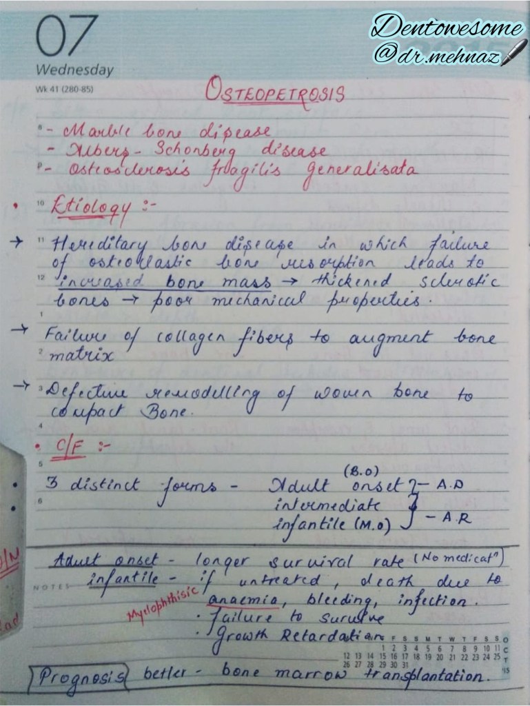
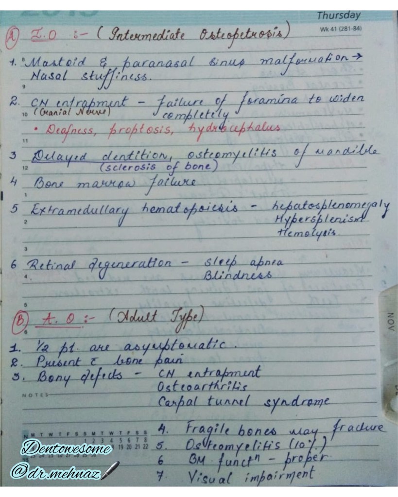
References: Shafer’sTextbook Of Oral Pathology
References: Shafer’sTextbook Of Oral Pathology


Dr. Mehnaz Memon🖊
References: Shafer’sTextbook Of Oral Pathology
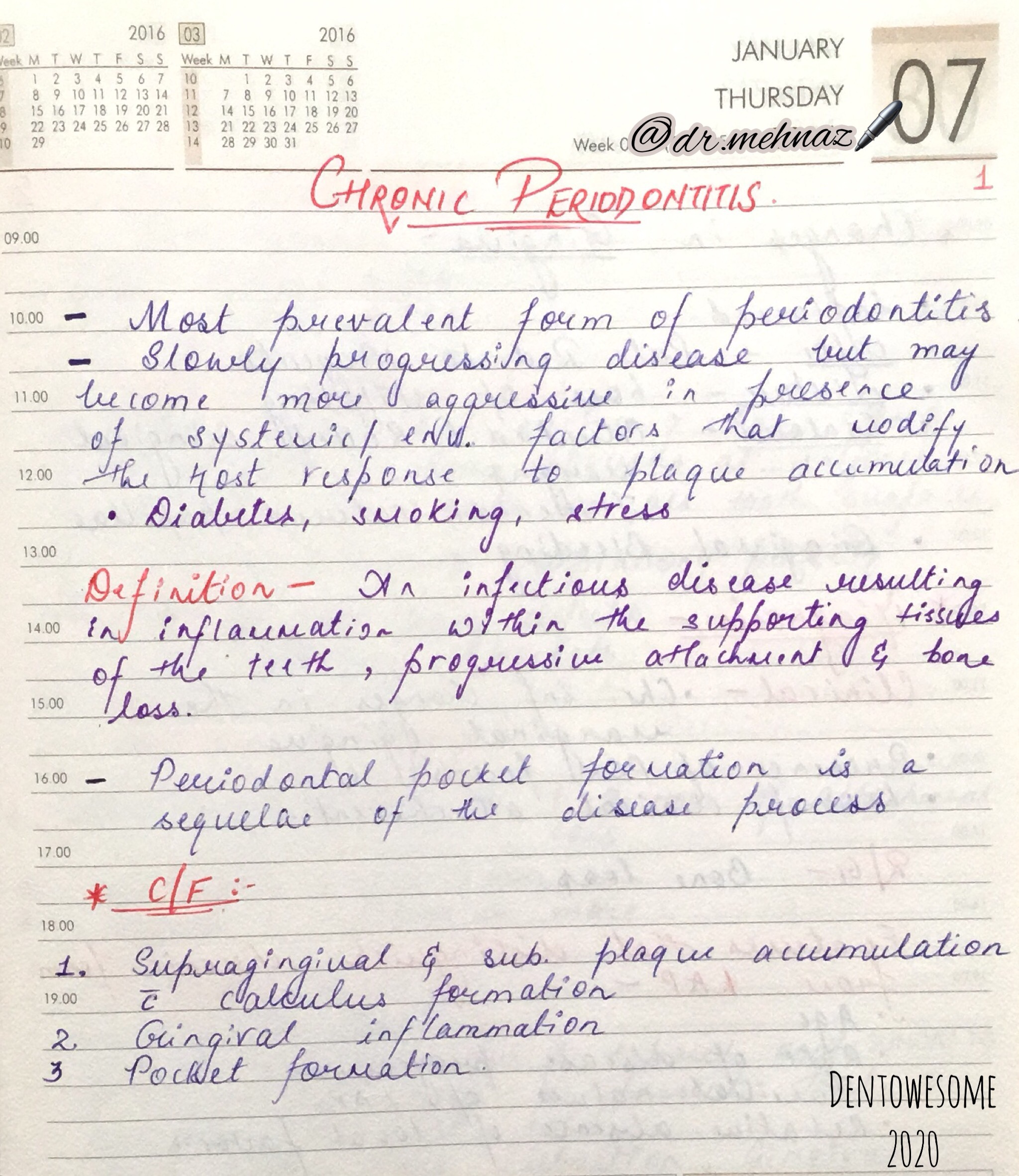
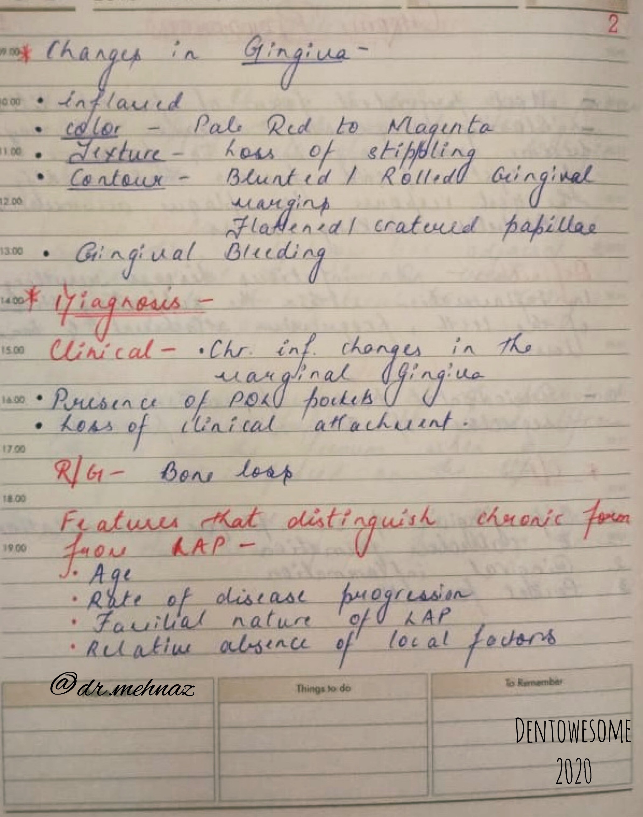

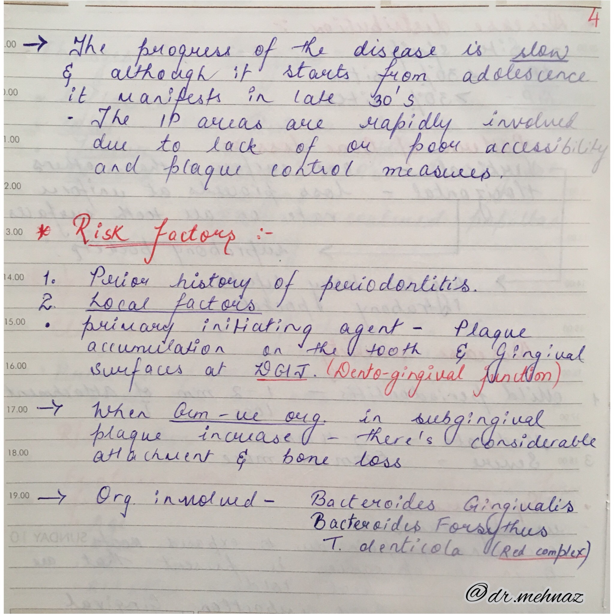

References: Essentials Of Periodontology by S Sahitya Reddy; Carranza’s clinical periodontology
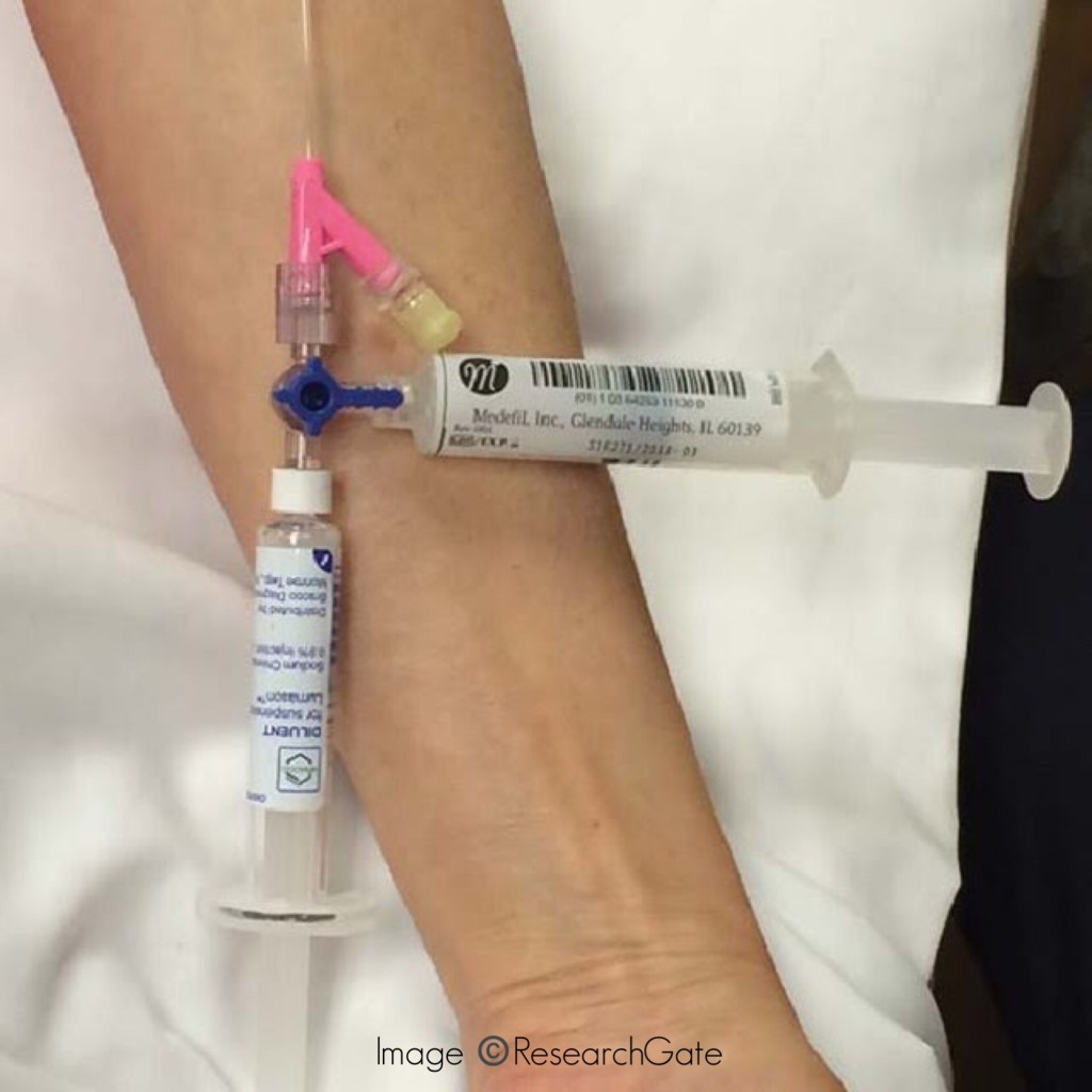
Size: 14G – 24G (Smaller the no., larger the bore of the needle)
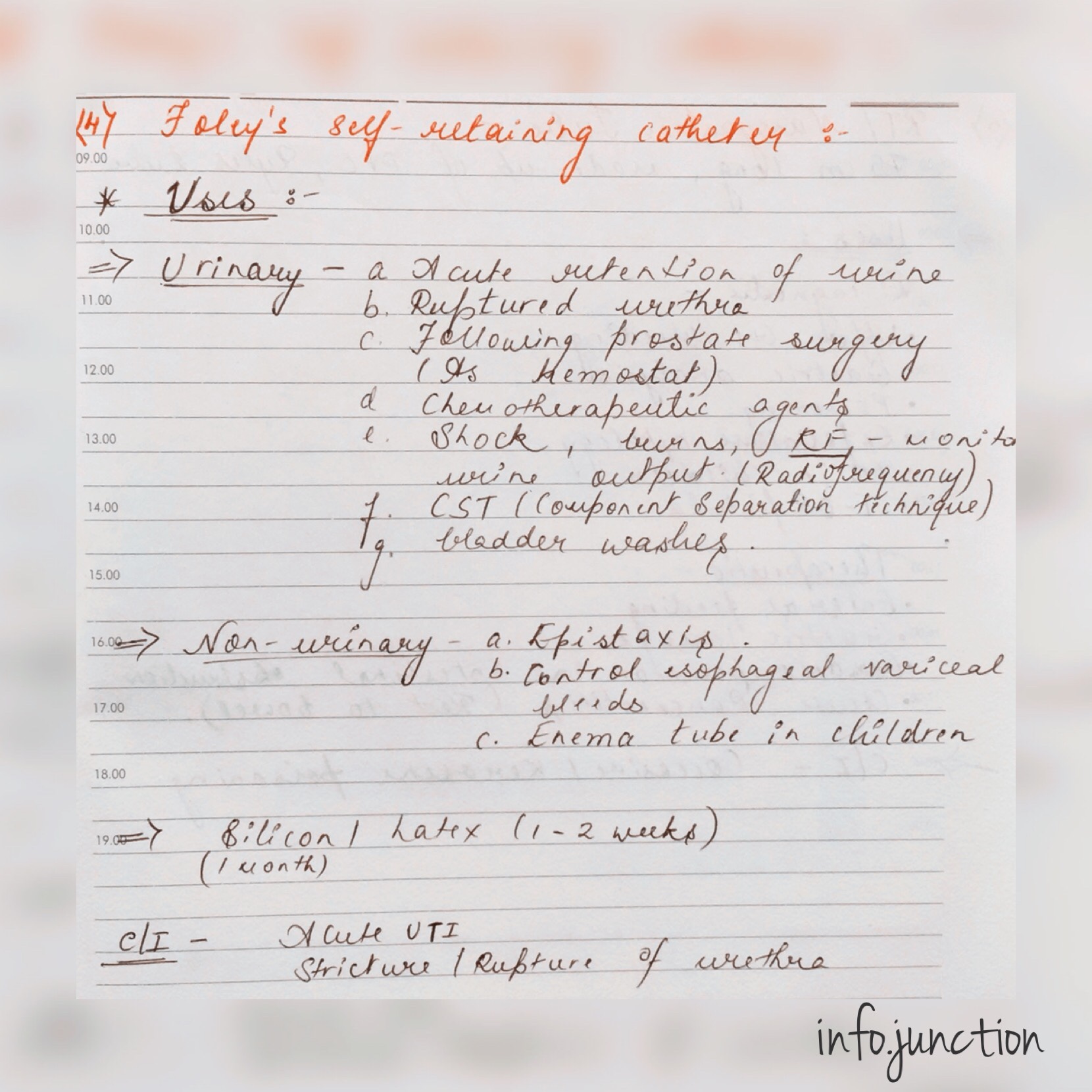
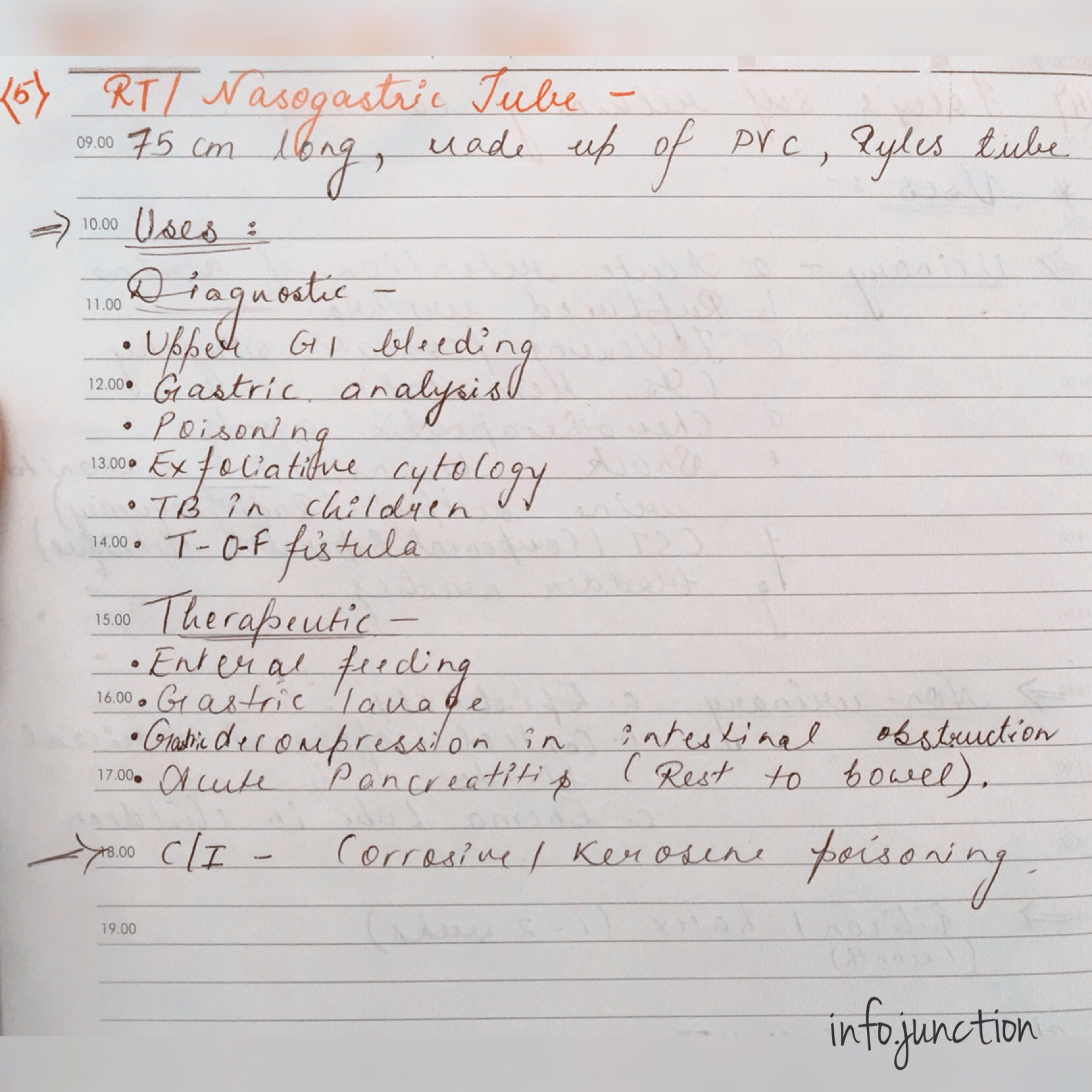
Use:
Source: Internet

References: Practical Medicine by P.J Mehta
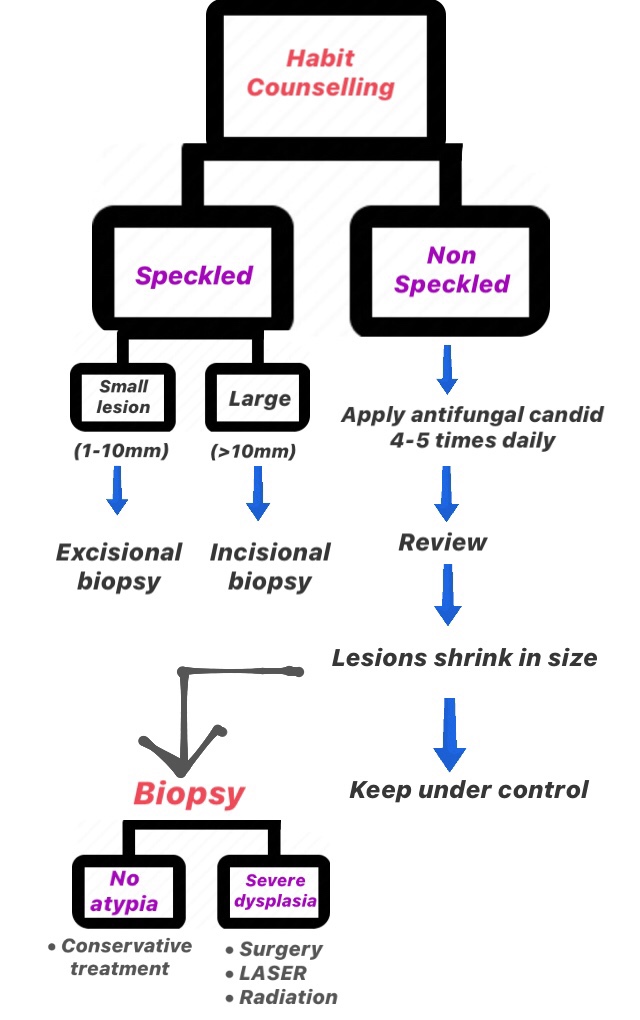
LEUKOPLAKIA

3) Surgical Approach
References: Ghoms, Textbook of Oral Medicine
Dr. Mehnaz Memon🖊
🔹Most common primary neoplasm of skeletal system.
1. Cells are closely packed in large sheets..👇🏻
2. Russell bodies: Russell bodies are multiple round cytoplasmic hyaline inclusions that are frequently seen in bone marrow aspirates in myeloma. They are composed of immunoglobulin molecules within vesicular structures derived from rough endoplasmic reticulum. Plasma cells containing them are sometimes referred to as Mott cells.
References: Shafer’sTextbook Of Oral Pathology
Dr. Mehnaz Memon🖊
1
2
3
References: Shafer’sTextbook Of Oral Pathology, Grossman’s Endodontic practice

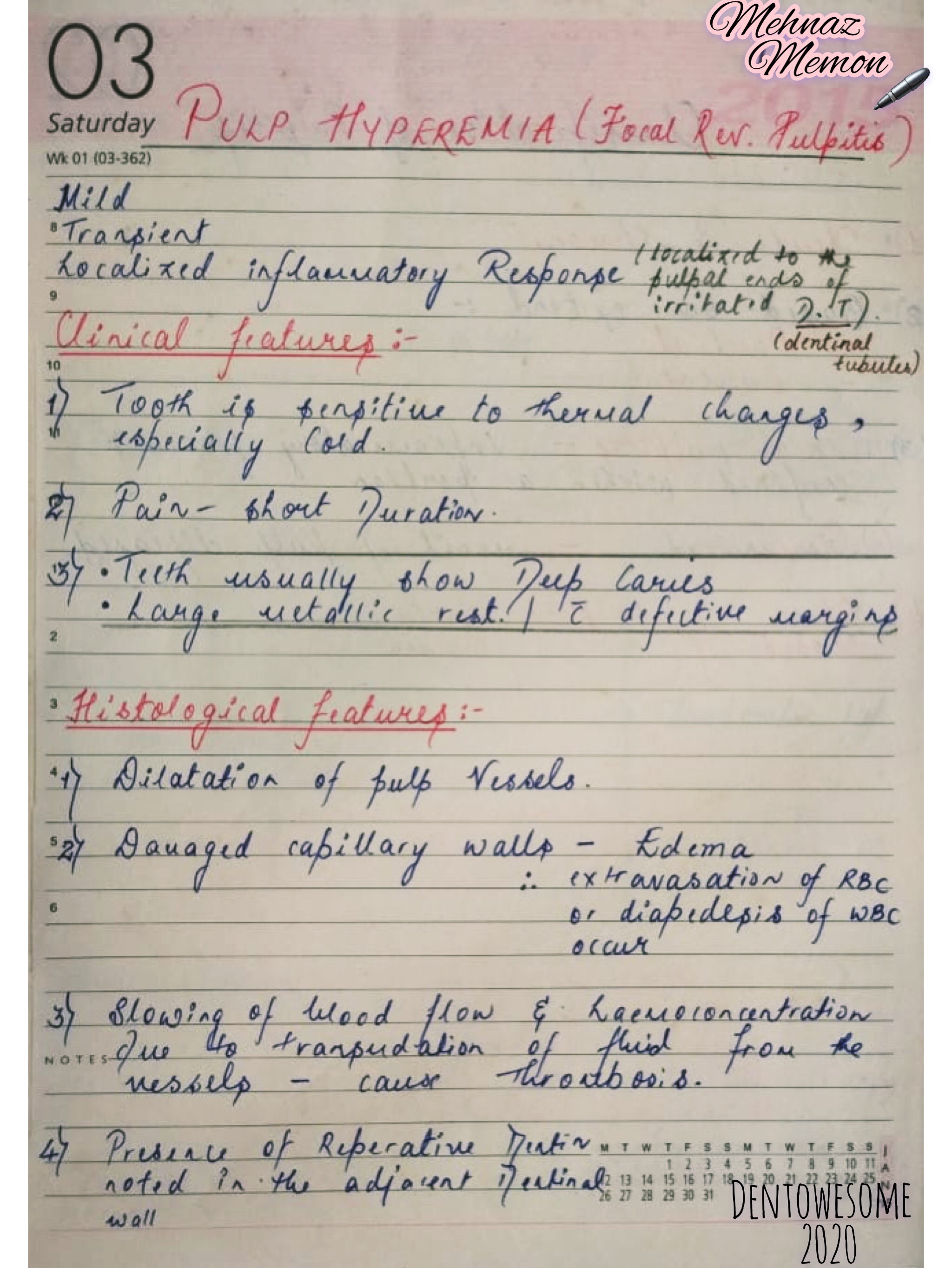
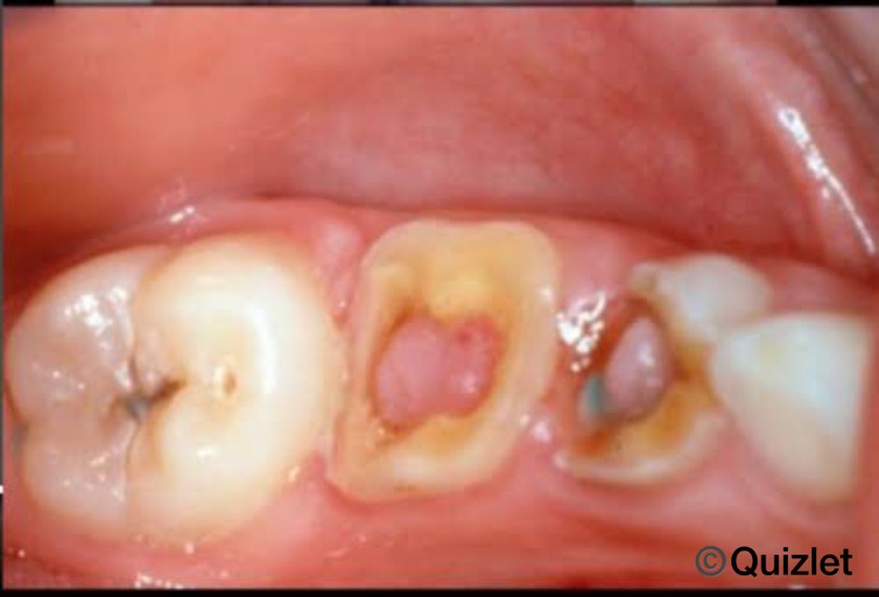
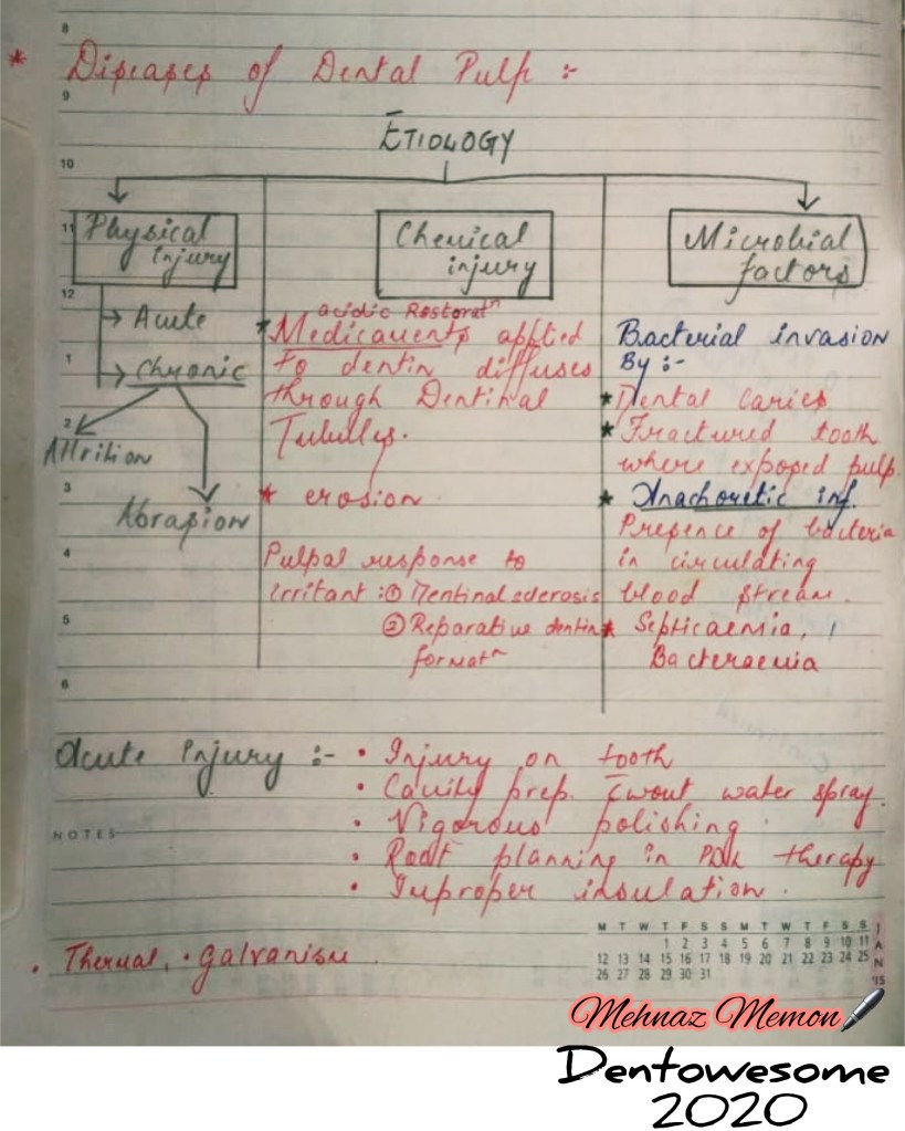
1) Pulp Hyperemia (Focal Reversible Pulpitis)
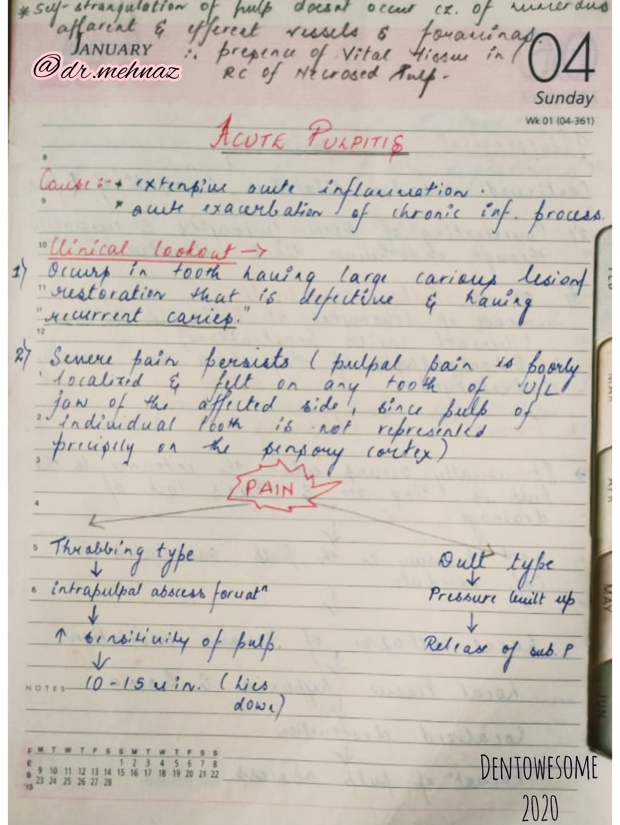
2) Acute Pulpitis


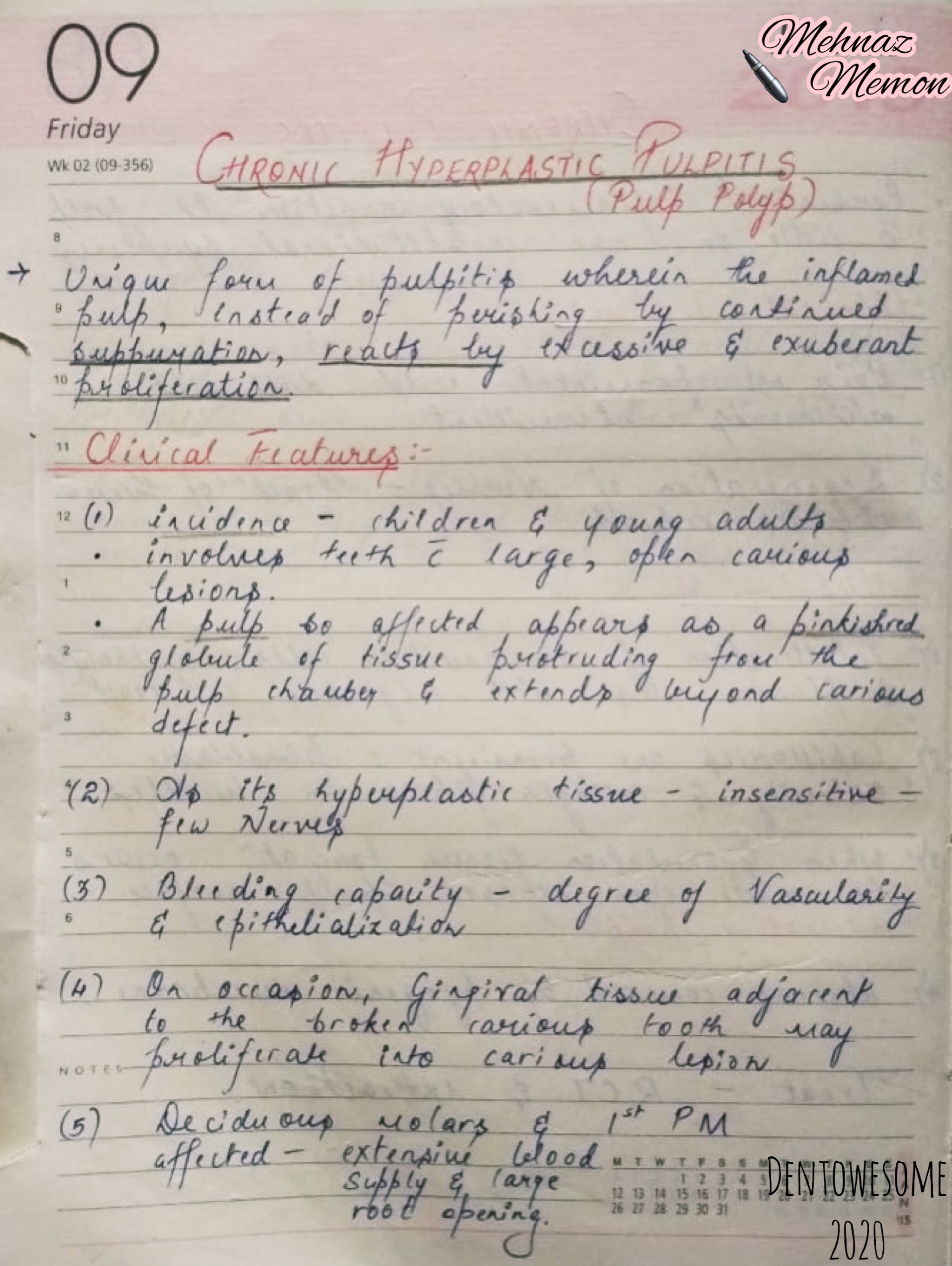
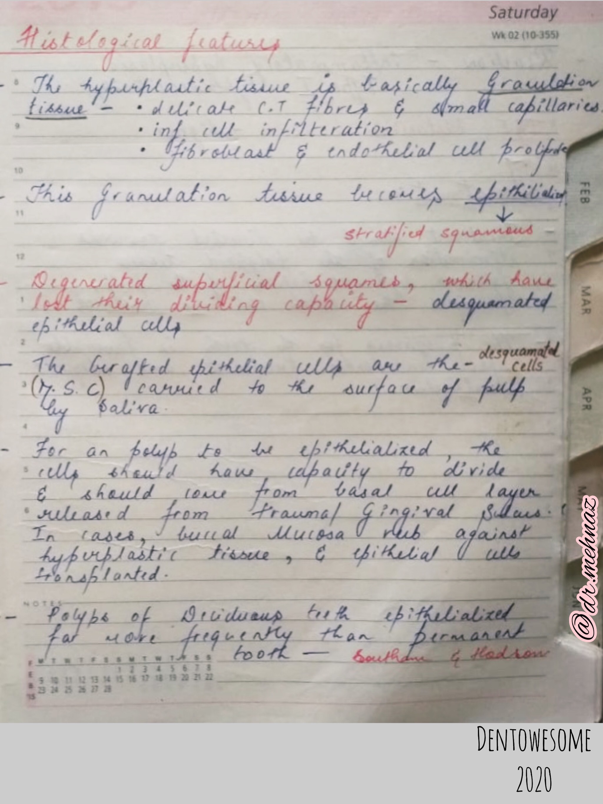
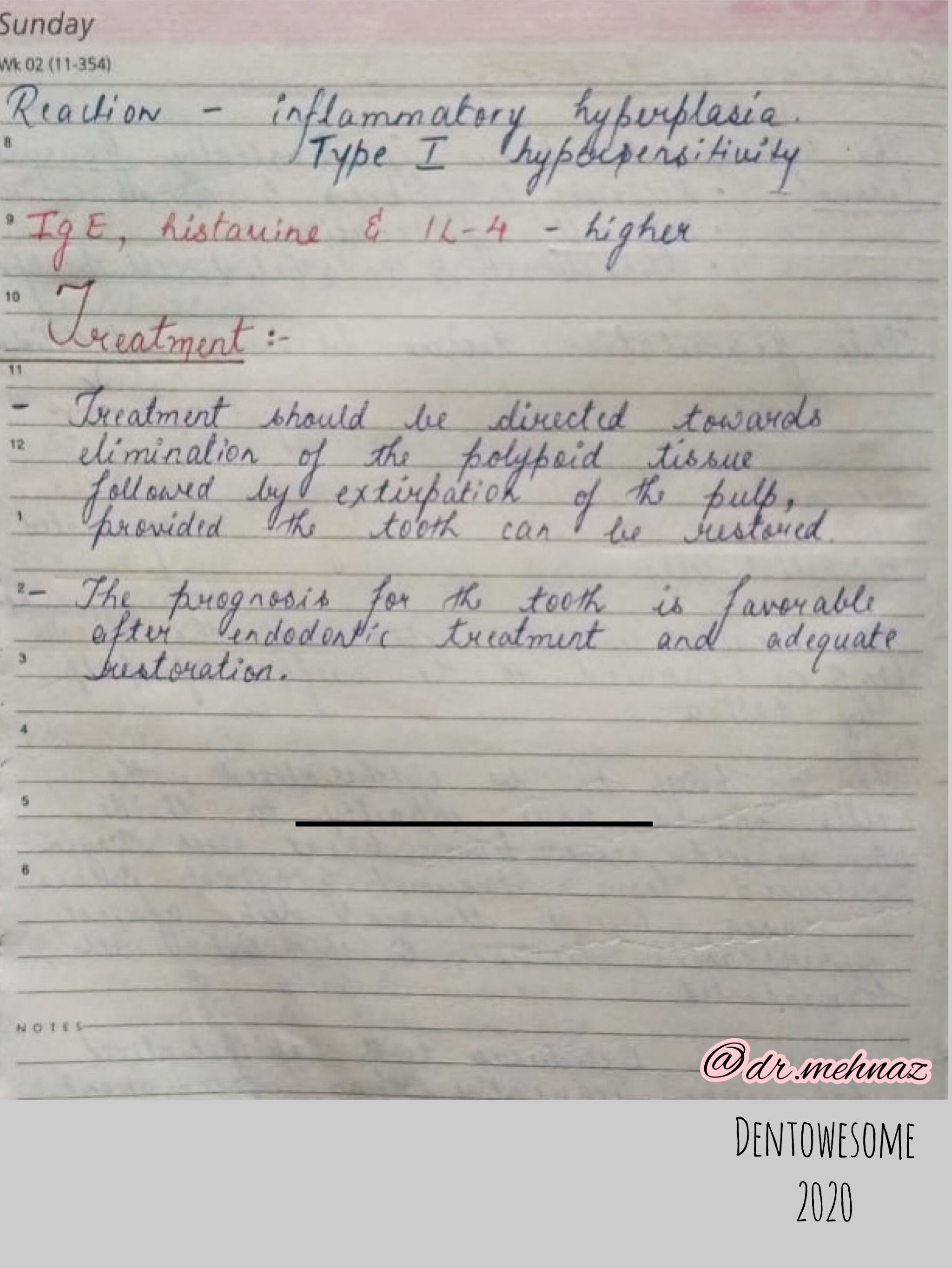
3) Chronic Pulpitis
References: Shafer’sTextbook Of Oral Pathology

References: Grossman’s Endodontic practice
