Highly elevated alkaline phosphatase is a good marker of Paget’s disease of bone in the appropriate clinical setting. Bone sequestration is often secondary to chronic osteomyelitis in the jaws. Bisphosphonates are used to treat Paget’s disease and also have been linked to jaw osteonecrosis.
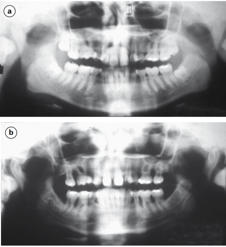
Study the two radiographs which are of the same patient at (a) 19 years and (b) 34 years of age.
1 . What is the most likely periodontal diagnosis at 34 years of age?
Generalised aggressive periodontitis. The severity of bone resorption and the radiographic absence of signs of resorption at 19 years preclude a diagnosis of chronic periodontitis.
2 . What does the initial phase of treatment involve?
Initially conventional cause-related treatment is instigated: instruction in toothbrushing and use of adjunctive aids for interproximal and subgingival cleaning, RSI, prophylaxis. Ultimately, treatments such as surgery and the adjunctive use of antimicrobials might be indicated, but conventional treatment is first in line.
3 . The patient has a sister who is aged 29 years. What advice might you offer?
Periodontal screening and radiographic examination. There is evidence that certain subjects are at high risk from developing aggressive periodontitis and this risk may be under genetic control. Siblings should be screened and affected individuals with children warned that early signs may develop from around puberty onwards
A 35-year-old man presents with gross loosening of both his lower left premolar teeth. The gingiva around them looks swollen and is purple–brown in colour. A radiograph shows irregular bone destruction to the apices. Incisional biopsy shows multinucleated osteoclast-like giant cells in a haemorrhagic fibrous stroma.
Which investigations should now be performed? The serum calcium level should be measured and radiographs reviewed to exclude hyperparathyroidism.
If these prove negative, what treatment should be undertaken? The lesion should be treated by local removal with curettage.
Which other lesions in the jaws contain multinucleate giant cells of this type? Osteoclast-like giant cells are found in
giant-cell granuloma
brown tumour of hyperparathyroidism
Pagetʼs disease of bone
aneurysmal bone cyst
some fibro-osseous lesions, particularly cherubism.
A 60-year-old man has been treated for a T2N0M0 squamous-cell carcinoma by radical radiotherapy. He has a history of chronic alcoholism and was a heavy smoker. Six years after treatment, he develops a painful ulcer in the alveolar mucosa in the treated area following minor trauma. His pain worsens and the bone became progressively exposed. He is treated by a partial mandibular resection with graft.
What diagnosis is most likely? The features suggest osteoradionecrosis. Recurrent carcinoma is possible but less likely.
How does radiotherapy damage tissues and what structural features might be seen in the bone? Radiotherapy damages tissues by producing free radicals. DNA damage may prevent cell division and repair. Endoarteritis obliterans results in reduced vascular supply to the tissues. Bone may become necrotic, showing osteocyte death, sequestration and breakdown of the matrix. Infection may result in osteomyelitis
What changes may arise in irradiated connective tissues 10 years after exposure? Mutations and other genetic damage may lead to neoplasia in irradiated tissues. Osteosarcoma can arise in this way.
A 58-year-old woman noticed that her front teeth had become spaced and seeks advice from her dentist. On entering the surgery, the dentist notices that she has difficulty in walking and does not respond to his questions. She has become increasingly deaf and her vision has also deteriorated. On examination, the maxilla and zygoma are enlarged and there is enlargement of the forehead.
1. What diagnosis would you suspect? Pagetʼs disease of bone results in enlargement of cranial bones and deformation of weight- bearing bones. The cranium is usually expanded in thickness and symptoms may arise from cranial nerve compression.
2. What information might be gained from oral radiographs and blood tests to support this diagnosis? Radiographs of the jaws may show hypercementosis, cemental masses, abnormal trabeculation and a cotton-wool appearance in the jaws. The alkaline phosphatase level is markedly raised.
3. What are the principal histological features of this disorder?
Disordered bone remodelling is seen; larger osteoclasts are present and the trabeculae show a scalloped outline. Numerous resting and reversal lines, resulting in a mosaic pattern, are seen and the vasculature may be increased. Globular cementum-like masses are seen in the jaws.