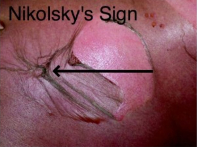
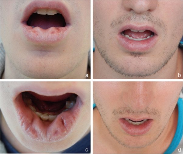
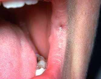
Nicolsky’s sign-application of pressure on normal skin produces a new lesion.Seen in pemphigus,TEN,Stevens Johnson syndrome ,Staphylococcla scaldedsyndrome
Auspitz sign- seen in psoriasis -peeling away scales result in pin point bleeding spots
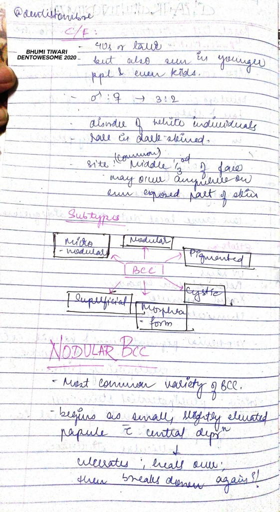
Albright sign -dimple at the metacarpelopharyngeal joint, due to short metacarpal.Seen in pseudohypoparathyroidism,Turner’s syndrome,basal cell carcinoma
Babinski’s sign- by stroking the lateral aspect of the dorsum of foot
Barber’s chair sign- seen in multiple sclerosis
Battle’s sign-in basal skull fracture(sphenoid bone)blood pigment stain behind the ear over the mastoid
Benda’s sign- in tuberculous meningitis.Spasm of the trapezius muscle that the shoulder on affected side is lifted and at times brought forward
Blue berry muffin sign-raised purple lesions seen in dermal metastases of neuroblastoma
Brim sign- seen in Paget’s disease
Brudzinski’s sign- seen in meningitis
Button hole sign- seen in neurofibromatosis
Carpet track sign- seen in discoid lupus erythematosis
Cerebriform tongue sign- seen in pemphigus veterans
Chandelier’s sign- seen in gonorrhea in women
Chvostek sign- elevation of corner of mouth
Coleman’s sign- in fracture of body of mandible ( hematoma seen in the floor of the mouth)
Corner’s sign- seen in scurvy
Crowe’s sign- seen in neurofibromatosis
Darier sign- seen in urticaria pigmentosa
Dubois sign- seen in congenital syphilis
Erb’s sign- seen in latent tetany
Paget’s sign- seen in yellow fever
Falling fragment sign- seen in solitary bone cyst
Fitzpatrick sign- seen in dermatofibroma
Flag sign- seen in kwashiorkor
Floating membrane sign-seen in hydatid cyst
Floating teeth sign- seen in eosinophilic granuloma
Forscheimer sign- seen in rubella
Head drop sign- seen in poliomyelitis
Heel pad sign- seen in acromegaly
Higoumanaki’s sign- seen in late syphilis
Hutchinson sign-seen in heroes zoster ophthalmicus
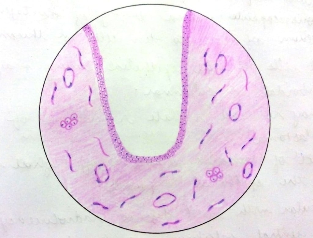
• The lining epithelium is derived from reduced enamel epithelium and hence appears as uniformly thin non-keratinized epithelium.
• The epithelium comprises of 2-3 layers of flattened cells and is characteristically devoid of rete ridges.
Connective tissue capsule is derived from dental follicle, consists of young fibroblasts widely separated by ground substance rich in mucopolysaccharide.
• Connective tissue capsule may show odontogenic epithelial remnants.
• Cystic lumen contains cystic fluid which is thin, watery or may be blood tinged.
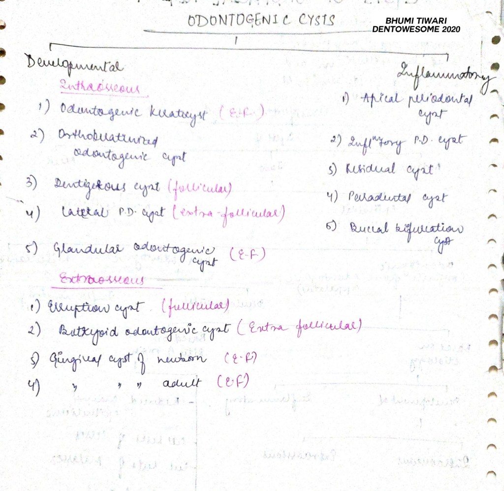
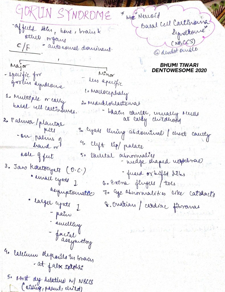
ODONTOGENIC KERATOCYST
• Typically shows a thin friable wall which is often difficult to enucleate from the bone in one piece.
• Cystic lumen may contain a clear liquid that is similar to a transidase of serum or it may be filled with a cheesy material, that on microscopic examination consists of keratinaceous debris.
• Microscopically, thin fibrous wall is essentially derated of any inflammatory infiltrate.
• Epithelium may show infoldings into the connective tissue capsule and may be separated from capsule in some areas.
CALCIFYING ODONTOGENIC CYST
• The lining epithelium is of variable thickness commonly around 5-10 layers.
• Spinous cells are loosely arranged.
• Presence of Ghost cells.
• Ghost cells are degenerating cells that appear as void, eosinophilic cells with nucleus showing different stages of degeneration.
• These cells are seen in thickened areas of lining epithelium.
• Connective tissue capsule is often scanty and sometimes contain discrete islands of odontogenic cells.
RADICULAR CYST
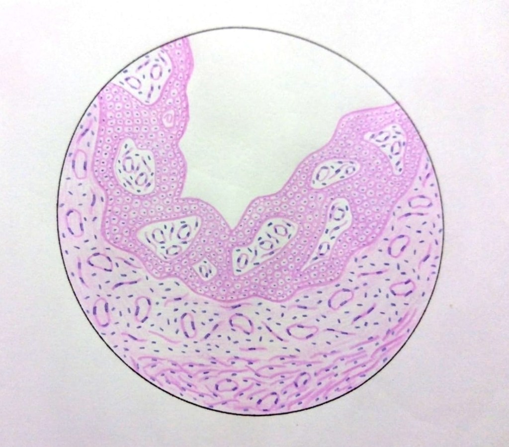
• The epithelial lining of the radicular cyst is stratified squamous in type of variable thickness.
• Epithelium shows spongiogenesis and inflammatory cell infiltration.
• Epithelium may show arc-shaped structures called Rushton bodies.
• The surrounding connective tissue shows granulomatous reaction due to the presence of Cholesterol clefts.