- SLE is a multisystem autoimmune inflammatory disorder of unknown etiology.
- Main feature is the formation of antibodies to DNA, which may initiate immune complex reactions, in particular a vasculitis.
- Female to male ratio of 9:1
- More common in persons of non-European descent.
- Etiology
- Geneticpredisposition—relativeofpatientshavehigher incidences of auto-antibodies, immune deficiency and connective tissue disease. This tendency is greatest among identical twins.
- Immunological abnormality possibly mediated by viral infection—immune complex consisting chiefly of nucleic acid and antibody account for majority of the tissue changes.
- Autoimmune disease—as these patients develop antibodies to many of their own cells.
- Endocrine—thereishighincidenceinfemalesinpreg- nancy. This finding suggestive of increased estrogen level.
- Biochemicalincreaseinexcretionofmetabolicproducts, particularly tyrosine and phenylalanine, in certain SLEpatient.
- CLINICAL MANIFESTATIONS
- Lupus is known as “the great mimic.”
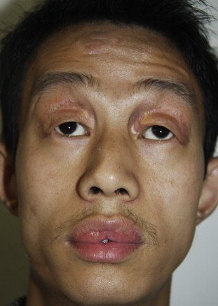
- Skin lesions of lupus can be classified
- lupus-specific (having diagnostic clinical or histopathologic features)
- nonspecific lesions.
- Three subtypes of lupus-specific
- Acute
- subacute
- chronic.
- Acute cutaneous lupus occurs in 30 to 50% of patients and is classically represented by the butterfly rash-mask-shaped erythematous eruption involving the malar areas and bridge of the nose
- Chronic cutaneous lupus occurs in 15 to 20% of cases and affects the skin of the face or scalp in about 80% of cases.
- The least common subtype, subacute cutaneous lupus, occurs in 10 to 15% of patients and includes papulosquamous (psoriasiform) and annular-polycystic eruptions, usually on the trunk and arms.
- Nonspecific but suggestive skin manifestations of lupus are common and include
- alopecia (both scarring following discoid lesions and non-scarring)
- Photosensitivity
- Raynaud’s phenomenon
- Urticaria
- Erythema
- Telangiectases
- cutaneous vasculitis.


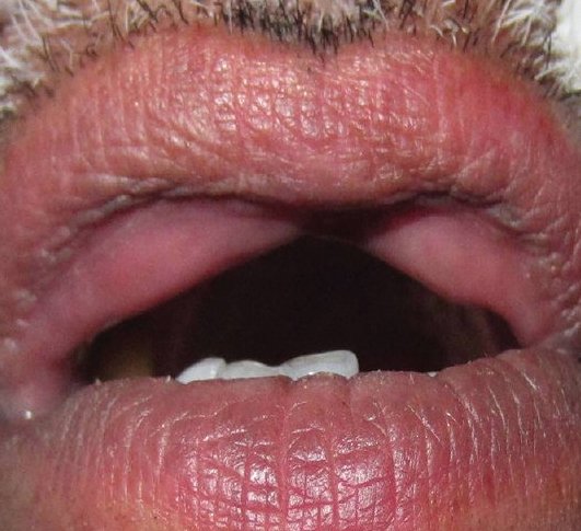
- ORAL MANIFESTATIONS
- Two predominant types of oral lesions are
- discoid lesions
- ulcerations.
- Oral ulcerations associated with SLE they occur with increased frequency on the palate and in the oropharynx and are characteristically painless.
- Histologically, they are characterized by lymphocytic infiltrate at the base of the ulcer and in the perivascular distribution, which is similar to that observed in discoid lesions.
- Discoid oral lesions, appear as whitish striae frequently radiating from the central erythematous area, giving a so-called “brush border.”
- Buccal mucosa, gingiva, and labial mucosa are the most commonly affected intraoral sites.
- Direct immunofluorescent staining for immunoglobulins and complement C3 factor is a useful aid to diagnosis. Granular deposition of IgM, IgG, and C3 along the basement membrane is characteristic

Diagnosis
• Clinical diagnosis—skin lesion with lesion present on oral mucosa which is atrophic and erythematous will suspect lupus erythematous. Oral and nasopharyngeal ulceration is major diagnostic criteria for SLE.
Laboratory diagnosis—L.E. cell inclusion phenomenon with surrounding pale nuclear mass apparently devoid of lymphocytes. Anemia, leukopenia and thrombocyto- penia, with sedimentation rate increased. Serum gamma globulin increased and Coomb’s test is positive.
Positive lupus band test—it shows deposition of IgG,IgM or complement component in skin.
- Differential Diagnosis
- Lichenplanus—homogenouspicture,nodarkerythema and no telangiectasia. Mucosal changes are usually extensive and symmetrical.
- Lichenoidreaction—historyofdrugisalwaysthere.
- Ectopic geographic tongue—systemic manifestation present is lupus erythematous, which is absent in ectopicgeographic tongue.
- Psoriasis—Auspitz’s’signispositive.
- Electrogalvanic lesion—dissimilar restorations are seenin oral cavity.
- Leukoplakiaanderythroplakia—lesionstendtomaintainsame appearance and there are no skin changes.
- Geographic stomatitis—no skin changes, mucosal lesionschange location rapidly.
- Benign mucous membrane pemphigoid—no systemiccomplain and serology test to be done.
- TREATMENT
- Corticosteriods are the cornerstone of therapy
- A pulse i.v cyclophosphamide regimen for remission induction followed by quarterly infusions
- Recently, mycophenolate mofetil and azathioprine
- NSAIDs for arthritis relief
- Antimalarial like hydroxychloroquinine – effective in cutaneous lupus
- DENTAL MANAGEMENT
- Recommended prophylactic antibiotics if ANC count falls below 500 – 1000 cells/mm3
- Adrenal supression –
- Adenocorticotropic hormone supression test is used to evalute
- Current guidelines – Replacement therapy with hydrocortisone is unnecessary
REFERENCE- ANIL GHOM TEXTBOOK OF ORAL MEDICINE; BURKIT TEXTBOOK OF ORAL MEDICINE AND GOOGLE[SLIDE SHARE]