
Source Borley Textbook
Source Borley Textbook

Source Borley Textbook
Reiter’s syndrome is associated with urethritis, balanitis, conjunctivitis, and mucocutaneous lesions.
It is a disease of unknown aetiology, although there is evidence of an infec- tious origin.
It is one of the most common complications of non-specific urethritis and it clinically mimicks gonorrhoea, although the urethral discharge is negative for Neisseria.

CLINICAL FEATURES
>Reiter’s syndrome is more prevalent in young adult men, usually between 20 and 30 years of age.
>The male-to-female ratio is 9:1.
>There is a typical tetrad of manifestations: non- gonococcal urethritis, arthritis, conjunctivitis, and mucocutaneous lesions.
>Urethritis may be the first sign. The urethral discharge is usually associated with itching and burning sensation.
>The arthritis is often bilaterally symmetrical and usually polyarticular.
>Conjunctivitis is often so mild as to be overlooked.
>The skin lesions are similar to those seen in keratoderma blennorrhagica and consist of red or yellow keratotic macules or papules which eventually desquamate.
Oral Manifestations
Sites—it is seen on the buccal mucosa, lips and gingiva.
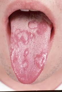
Oral lesions appear as painless, red, slightly elevated areas, some- times granular or even vesicular, with a white circinate border on the buccal mucosa, lips, and gingiva.
The palatal lesions appear as small, bright red purpuric spots, which darken and coalesce, while the lesions on the tongue closely resemble ‘geographic’ tongue.
Laboratory Findings
The patients usually have a mild leukocytosis, an elevated erythrocyte sedimentation rate, and pyuria.

REFERENCE- SHAFER’S TEXTBOOK OF ORAL PATHOLOGY [8TH ED} AND ANIL GHOM TEXTBOOK OF ORAL MEDICINE
Wegener’s granulomatosis is a disease of unknown aetiology, which basically involves the vascular, renal and respiratory systems. It involves the nose, paranasal air sinuses, lower respiratory tract, gut, joints, nervous system, and kidneys. Involvement of the kidney is the common cause of death.
This disease is caused by an abnormal immune reaction secondary to a nonspecific infection or a hypersensitivity reaction to an unknown antigen.

Clinical Features
>Wegener’s granulomatosis may occur at any age, although the majority of cases are in the fourth and fifth decadesof life.
>There is a slight predilection for occurrence in males.
>It is best described as a multisystem disease, which is usually first characterized clinically by the development of rhinitis, sinusitis, and otitis or ocular symptoms.
>The patient soon develops a cough and hemoptysis as well as fever and joint pain.
>Hemorrhagic or vesicular skin lesions are also commonly present.
>Granulomatous lesions of the lungs are found on the chest radiograph, while the glomerulonephritis, which develops ultimately, leads to uraemia and terminal renal failure.
>In nervous system, sensory neuropathy may be an occasional finding.
>Prognosis—the disease is usually fatal, with mean survival time of 5 months. Death occurs due to involvement of kidney.
Oral Manifestations
Involvement of the gingiva has been the most common and characteristic manifestation, and is termed as strawberry gingivitis.Gingival lesions may manifest as ulcerations, friable granular lesions, or simply enlargements of the gingiva.
The inflammatory process starts in the interdental papilla and spreads rapidly to the periodontal structure and leads to bone loss and tooth mobility.
Palate—orallesionstypicallyincludeulcerationofthe palate by extension of nose lesions and destruction of nasal septum. This will lead to perforation of palate.
• Teeth—theremaybelooseningofteethwithinsomecases spontaneous exfoliation. After extraction of teeth patient is usually noticed poor healing.

Laboratory Findings
Laboratory findings include anaemia, leukocytosis, elevated erythrocyte sedimentation rate, and hyperglobulinaemia. Because of kidney involvement, haematuria is common, as well as the finding of albumin, casts, and leukocytes in the urine. Circulating immune complexes have been demonstrated in some patients, but this is not a consistent finding.
Histologic Findings
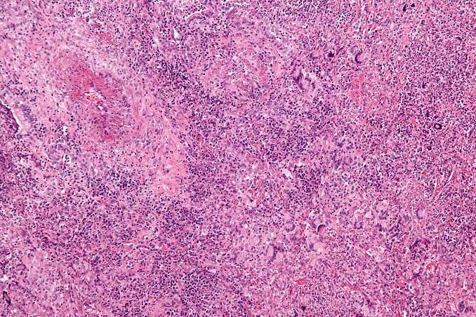
Wegener’s granulomatosis presents a pattern of mixed inflammation centred around the blood vessels.
The lesions in the upper respiratory tract and lungs consist of giant cell necrotizing granulomatous lesions showing vasculitis.
Oral biopsy specimens show pseudoepitheliomatous hyperplasia and subepithelial abscesses. The gingival and other lesions show a nonspecific granulomatous process with scattered giant cells.
Diagnosis
REFERENCE- SHAFER’S TEXTBOOK OF ORAL PATHOLOGY {8TH ED} AND ANIL GHOM TEXTBOOK OF ORAL MEDICINE
1. Recurring oral ulcers,
2.Recurring genital ulcers, and
3.Eye involvement.
ETIOLOGY
CLINICAL MANIFESTATIONS
ORAL MANIFESTATIONS
Histologic Features
The microscopic findings are not diagnostic. They consist of parakeratosis, acanthosis, and polymorphonuclear leukocyte infiltration of epithelium, sometimes with mi- croabscess formation similar to psoriasis. The connective tissue shows a lymphocyte and plasma cell infiltrate.
Laboratory Findings
The patients usually have a mild leukocytosis, an elevated erythrocyte sedimentation rate, and pyuria.
DIFFERENTIAL DIAGNOSIS
TREATMENT
The management of Behçet’s syndrome depends on the severity and the sites of involvement.
REFERENCE- BURKET TEXTBOOK OF ORAL MEDICINE AND SHAFER’S TEXTBOOK OF ORAL PATHOLOGY {8TH EDITION}






Source – Bhalajhi and Google images
Mouth rehabilitation is defined as restoration of form and function of the masticatory apparatus to as nearly normal as possible.
# Adjunctive care :Lesions like ulcer, abscess should be cure and be given time for healing. During their healing period adjunctive therapies like tissue massage, use of mouth washes etc. are used. For normal patients, 48 hour rest with tissue massage is sufficient.
#Removal of retained dentition : An OPG gives a clear idea whether to remove the retained dentition or not.
Removal of unerupted teeth::
The unerupted teeth lying close to ridge should be removed but the one which deeply submerged and non symptomatic can be left untouched.
Removal of retained roots::
All the retained roots should be removed but if it’s removal can cause large bony defect and is asymptomatic , then it can be left untouched.
# Correction of Hypermobile Ridge tissue :
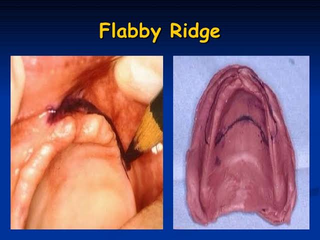
Hypermobile tissues are caused by excessive residual ridge resorption. Small Hypermobile tissues which do not affect functioning of denture can be left untouched but the large pendulous Hypermobile tissues should be removed.
#Removal of soft tissue interfaces : Firm soft tissue interference, which do not affect the stability of the denture,can be left intact. In case of maxillary tuberosity with excessive fibrous tissue, there is need for surgical removal.
#Removal of Hypertrophic maxillary labial frenum:

In case of high frenum attachment ,where the frenum is not so close to crest of the ridge , surgery is not mandatory, only labial notch in the denture is sufficient. But when this notch breaks the peripheral seal , in that case surgical procedure is done.
( ref : Textbook of prosthodontics: Deepak nallaswamy , images : Google images)
#Removal of a Hypertrophic Lingual frenum:
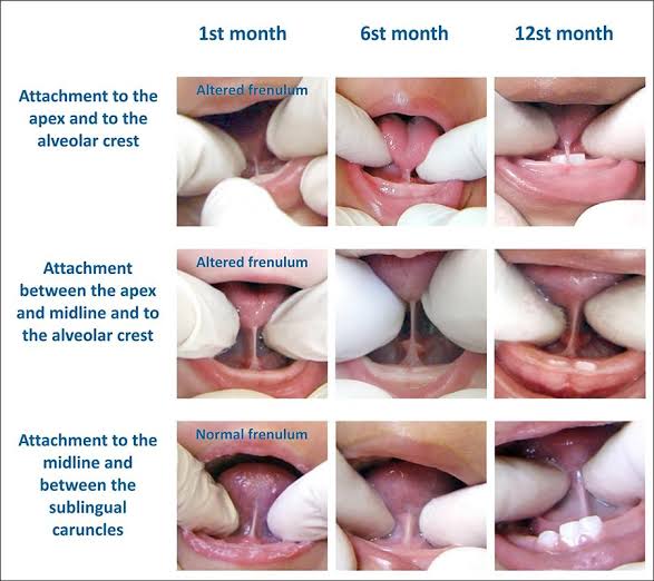
Frenectomy is indicated in this case. Tongue tie test is performed.Patient is asked to touch his upper lip with tongue, if the denture is displaced by doing so then frenectomy is to be done.
#Correction of prominent buccal frenum: Removal is generally not necessary as they are not present as so large. Buccal frenum is soft and it can be easily displaced by denture without any injury.
#Removal of Papillary hyperplasia:

These hyperplastic lesions occur due to irritation. It can be large or small . Small lesions are treated by curettage and large by split thickness supraperiosteal excision.
#Treatment of Epulis fissuratum:

It is an soft tissue reaction in sulcular regions due to overextension of denture flanges. No specific treatment except shortening and smoothening of denture borders.Give it time to heal.
# Removal of ridge undercuts: Small and moderate unilateral undercuts can be left untouched , while severe undercuts or bilateral moderate requires surgical correction ( alveoloplasty).
( ref: textbook of prosthodontics , Deepak nallaswamy , images: Google images.)
#Management of prominent Mylohyoid and internal oblique ridges: They are seen in lingual surfaces of mandible. Sometimes they become prominent due to ridge resorption. If there is repeated ulceration, loss of peripheral seal , they should be surgically reduced.
#Reduction of Maxillary tuberosity: Reduction of wide tuberosity is easier compared to vertically large tuberosity. Before it’s reduction, radiological evaluation is mandatory,as maxillary sinus tends to expand in tuberosity.
#Treatment of sharp spiny ridges: These type of ridges usually occur in lower anterior region due to resorption of labial and lingual cortical plates. For this ,ridge augmentation can be done.
Meyer’s classification for knife edge ridges:
⛔Saw-tooth
⛔Razor-like
⛔Ridge with discrete spiny projections
#Excision of Tori :

Tori are small bony projections of unknown etiology. Indications for maxillary Tori removal:
Interference of speech
Loss of posterior palatal seal
Poor denture stability
All the mandibular Tori should be excised .
#Vestibuloplasty:

It is the surgical procedure to increase the vestibular depth. Techniques for vestibuloplasty are::
Mucosal Advancement
Secondary Epithelialisation
Epithelial Graft Vestibuloplasty
( ref : textbook of prosthodontics,Deepak nallaswamy, image: Google images)
Source Nikhil Marva textbook


