RADIOLOGICAL APPEARANCES & FEATURES(PART II)

Many lesions that occur on the jaw present with similar radiographic appearances making it difficult to differentiate among them . Despite development of various cross sectional imaging modalities ,the radiographs are still remain the first and most important investigations.

So some of the common features that we as dentist might come across are listed below.I believe this would come handy for your radiodiagnosis😊.

  • Ghost teeth appearance-regional odontodysplasia
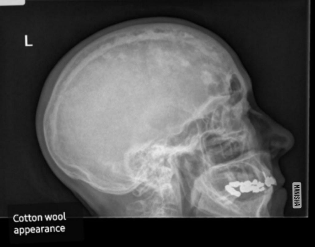
  • Ground glass appearance-monostotic fibrous dysplasia, hyperparathyroidism, middle stage of paget’s disease & ossifying fibroma,sickle cell anemia
  • Garrington sign-osteosarcoma (localised symmetrical widening of PDL
  • Hanging drop appearance-orbital blow out fracture
  • Heart shaped radiolucency-nasopalatine /incisive canal cyst
  • Honey comb appearance-anerysmal bone cyst,odontogenic myeloma,central giant cell granuloma,central hemangioma
  • Lincoln sign-paget’s disease (increased uptake in the mandible mimicking Lincoln’s beard.
  • Mass of color/ cauliflower like appearance-calcified lymph nodes
  • Moth eaten appearance-chronic osteomyelitis, early stage of ostesarcoma,radiolucent stage of fibrous dysplasia,squamous cell carcinoma,leukemia,malignant lymphoma
  • Mottled appearance-ossifying fibroma,fibrous dysplasia
  • Onion peel /skin appearance-Garre’s osteomyelitis, Ewing’s sarcoma,eosinophilic granuloma,caffey’s disease.
  • Orange peel appearance-mixed stage of fibrous dysplasia (peud-de-orange)
  • Pear shaped appearance-globulomaxillary cyst
  • Pepper pot appearance-hyperparathyroidism
  • Peripheral cuffing of bone-peripheral giant cell granuloma
  • Pressure type appearance-squamous cell carcinoma of gingiva
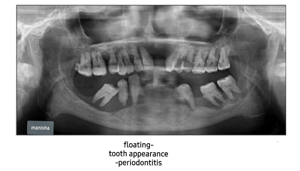
  • Punched out appearance-multiple myeloma
  • Rootless teeth appearance-dentinal dysplasia
  • Salt and pepper appearance-hyperparathyroidism, thalassaemia
  • Sausage like appearance-sialographic appearance of sialodochitis
  • Sialectasis-sialographic appearance of sialadenitis
  • Sharpened pencil appearance-osteoarthritis /rheumatoid arthritis of TMJ
  • Shell teeth appearance-type III dentinogenesis imperfecta
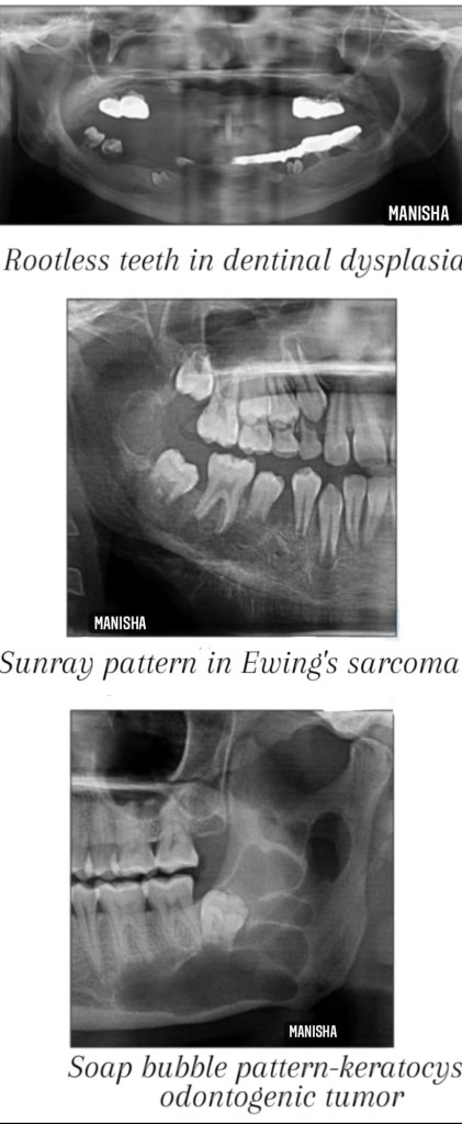
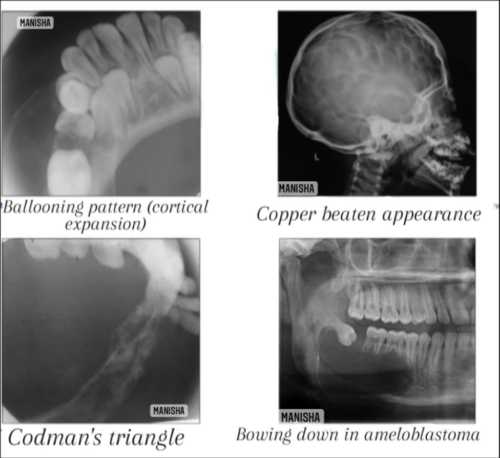
  • Soap bubble appearance-ameloblastoma,aneurysmal bone cyst,central hemangioma,central giant cell granuloma, odontogrnic keratocyst
  • Step ladder appearance-sickle cell anemia
  • Sunburst/sunray appearance-central hemangioma,osteosarcoma,Ewing’s sarcoma
  • Target sign-(rounded radiopacity with a central radiolucency)-impacted tooth in bucco lingual direction(lower 3rd molar)
  • Tennis racket appearance-odontogenic myxoma
  • Thistle tube appearance-type II dentinal dysplasia
  • Thumb print /fingerprint appearance-mixed stage of fibrous dysplasia
  • Tram track sign/calcifications-Sturge Weber syndrome
  • Trap door appearance-orbital blow out fracture
  • Tree in winter appearance-normal sialographic appearance of parotid gland

Some of the radiographic features are pathogonomic to a specific disease, thus can be helpful in narrowing down of differential diagnosis.

🖋Manisha M.A

Sources:White and Pharoah’s -Oral radiology textbook,Shafer’s textbook of oral pathology (8th edition) , Websites-www.ijournalhs.org, Article-Phore S,Panchal RS,Bhagla P, Nabi N.Dental radiographic signs.Indian j health sci 2015;8:85-90.

RADIOLOGICAL APPEARANCES & FEATURES(PART I)

The real importance of learning radiographic signs associated with specific disease is of relevance to clinical examination of the head & neck ; at the same time aiding in differentiating – what is normal from abnormal & hence appropriate treatment can be instituted for such conditions /abnormalities.

Here are the list of few radiographic appearances .

Cotton wool appearance seen on the lateral skull in Paget’s disease
  • Antral halo appearance-acute sinusitis
  • Ball in hand appearance-sialographic appearance of intrinsic benign tumor
  • Balloon like appearance-follicular cyst
  • Beaten silver/Copper beaten appearance-Crouzen syndrome,hypophosphatasia,craniofacial dysostosis ,obstructive hydrocephalus
  • Bush in winter appearance-normal sialographic appearance of submandibular gland
  • Candlestick appearance-Progressive systemic sclerosis,pycnodysostosis
  • Cherry blossom/branchless fruit laden tree/snowstorm/punctate sialectasis-sialographic appearance of Sjogren’s syndrome
  • Codman’s triangle-osteogenic sarcoma,Ewing sarcoma,carcinoma of alveolar ridge
  • Cotton wool appearance-paget’s disease,fibrous dysplasia(thick,amorphous radiopaque stage),cemmento-osseous dysplasia,chronic diffuse sclerosis osteomyelitis
  • Crew cut/Hair-on-end appearance-sickle cell anemia,thalassemia
  • Driven snow appearance-calcifying epithelial odontogenic tumor /cyst(Pindborg tumor)
  • Downward bowing appearance-cemento-osseous fibroma,ameloblastoma
  • Ely’s cyst-osteoarthritis
  • Filling defect appearance-salivary gland tumor
  • Floating tooth appearance-squamous cell carcinoma,malignant lymphoma,periodontitis ,eosinophilic granuloma,osteomyelitis

🖋Manisha M.A

Sources:White and Pharoah’s -Oral radiology textbook,Shafer’s textbook of oral pathology (8th edition).Websites-www.dreamstime.com, www.ijournalhs.org,Article-Phore S,Panchal RS,Bhagla P, Nabi N.Dental radiographic signs.Indian j health sci 2015;8:85-90.

SUPERNUMERARY TEETH

A supernumerary tooth is an additional tooth to the normal set of teeth. It may closely resemble the teeth of the group to which it belongs, i.e, molars, premolars or anterior teeth, or it may bear little resemblance in size or shape to the teeth with which it is associated.

It is also called as ‘hyperdontia’. It is defined as any tooth or tooth substance in the excess of the usual configuration of twenty deciduous or thirty two permanent teeth.

Pathogenesis and etiology

  • The various factors which are responsible for formation of supernumerary teeth as follows:
  • Phylogenetic reversion theory—this theory is nowadaysdiscarded.
  • Dichotomy theory—this theory states that tooth bud issplit to form two different tooth,
  • Hyperactivity of dental lamina—a supernumerary toothdevelops from 3rd tooth bud arising from dental lamina near the permanent tooth bud.
  • Hereditary—it is inherited as an autosomal dominant trait, when associated with syndromes. It is inherited as an autosomal recessive trait when associated with only supernumerary teeth. Supernumerary teeth are most commonly found in relative of affected children than in general population.

Four different morphological types of supernumerary teeth have been described:

• Conical
• Tuberculate
• Supplemental
• Odontome

Conical. This small peg-shaped conical supernumerary tooth is most commonly found in the permanent dentition. It develops with root formation ahead of or at an equivalent stage to that of permanent incisors and usually presents as a mesiodens. The conical supernumerary can result in rotation or displacement of the permanent incisor, but rarely delays eruption.

Tuberculate. The tuberculate type of supernumerary possesses have more than one cusp or tubercle. It is frequently described as barrel-shaped and may be invaginated. Root formation is delayed compared to that of the permanent incisors. Tuberculate supernu- meraries are often paired and are commonly located on the palatal aspect of the central incisors. They rarely erupt and are frequently associated with delayed eruption of the incisors.

Supplemental. The supplemental supernumerary refers to a duplication of teeth in the normal series and is found at the end of a tooth series. The most common supplemental tooth is the permanent maxillary lateral incisor, but supplemental premolars and molars also occur. The majority of supernumeraries found in the primary dentition are of the supplemental type and seldom remain impacted.

Odontome. The term ‘odontoma’ refers to any tumour of odontogenic origin. Odontoma represents a hamartomatous malformation rather than a neoplasm. The lesion is composed of more than one type of tissue and consequently has been called a composite odontoma. Two separate types have been described, the diffuse mass of dental tissue which is totally disorganized is known as a complex composite odontoma; whereas, the malformation which bears some superficial anatomical similarity to a normal tooth is referred to as a compound composite odontoma.

According to location
• Mesiodens—it is located at or near the midline in the incisal region of maxilla between central incisors . It is a small tooth with cone shaped crown and short root. It may cause retarded eruption, displacement or resorption of adjacent root. It frequently causes improper alignment.

• Distomolar—it is found in molar region frequently located distal to 3rd molar. Generally, these teeth are smaller than normal 2nd and 3rd molar, crown morphology is highly abnormal.

• Paramolar—it is supernumerary molar, usually small and rudimentary and is situated buccally or lingually to one of the maxillary molars or interproximally between 1st, 2nd and 3rd maxillary molars.

• Peridens—supernumerary teeth that erupt ectopically, either buccally or lingually to the normal arch are referred as peridens.

Radiographic features

When it is needed—if abnormal clinical signs are present you can go for OPG examination, IOPA, occlusal radiographic examination.

Significance—radiograph will aid in determining the location and number of unerupted teeth. It can also used to see if there is any cyst formation.

Appearance—theirradiographicpictureischaracteristic of teeth.

Management

• Surgical extraction—it depends on potential effect on normal dentition, their position, number and complications that may result from surgical removal. If required, they should be extracted.

REFERENCE- SHAFER’S TEXTBOOK OF ORAL PATHOLOGY 8TH ED AND ANIL GHOM TEXTBOOK OF ORAL MEDICINE TEXTBOOK OF ORAL MEDICINE

Macrognathia

General factors which would influence and tend to favour mandibular prognathism are as follows:

• Increased height of the ramus

• Increased mandibular body length

• Increased gonial angle

• Anterior positioning of the glenoid fossa

•Decreased maxillary length

• Posterior positioning of the maxilla in relation to the cranium

• Prominent chin button

•Varying soft-tissue contours

Source- textbook of oral pathology Shafers and Google images