Differential Diagnosis of Sinus, Fistula & Abscess

Sinus

Latin: Hollow (or) a bay

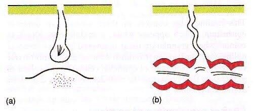
A sinus is a blind tract leading from the surface down to the tissue. There may be a cavity in the tissue which is connected to the surface through a sinus. The sinus is lined by granulation tissue which may be epithelized.

A sinus has one open draining end and the channel ends in a blind ending. An example would be a dental sinus draining from a dental abscess to either the inside of the mouth or the skin.

Fistula

Latin: flute(or) a pipe (or) a tube

It is a communicating tract between 2 epithelial surfaces commonly between hollow viscous & skin or between 2 hollow viscera. The tract is lined by granulation tissue which is subsequently epithelized. A fistula may be an abnormal communication between vessels.

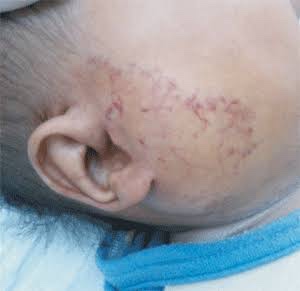
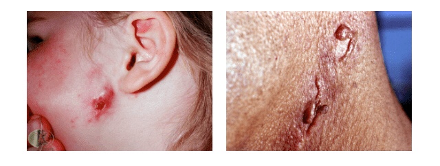
An example would be from the mouth (oral cavity) to the skin surface, usually of the face or neck, and this specific type is called an orocutaneous fistula.

These defects can cause aesthetic and functional problems due to the continuous leakage of saliva.
It is an abnormal epithelized communcation between maxillary sinus and oral cavity through perforation in the sinus wall

Fistulas and sinuses of the neck and face: classification

Fistulas and sinuses of the neck and face may be classified by cause.

Developmental

Fistulas and sinuses due to developmental causes are usually present at birth.

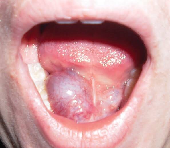
  • Thyroglossal duct cyst – the most common developmental cyst in the neck. The cyst characteristically moves upwards when the tongue is poked out or with swallowing. It may burst to form a sinus which usually opens just below the hyoid bone in the midline of the neck. It drains mucus.
  • Branchial cleft cyst – the most common developmental cyst of the side of the neck. A sinus may drain mucus or pus following rupture of an abscess. It usually opens on the side of the neck.
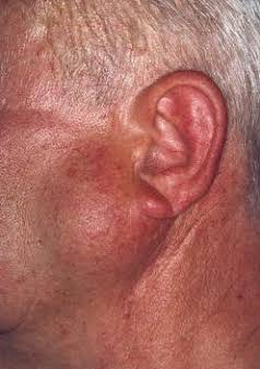
  • Preauricular sinus

Cysts

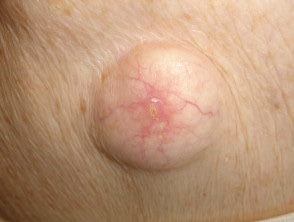
Cysts are lumps in the skin containing fluctuant contents. They may have an opening to the skin surface.

  • Dermoid cyst
  • Epidermal cyst

Traumatic

  • Accidental
  • Radiotherapy
  • Surgical

Infective

  • Actinomycosis
  • Bone infection
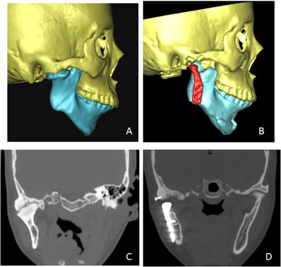
    Chronic osteomyelitis – most commonly associated with poorly controlled diabetes mellitus or following radiotherapy to the jaw for cancer or Paget disease of the bone. It may also complicate a chronic dental infection.
  • Dental infection
    • Chronic dentoalveolar abscess
    • Dental implant
    • Failed endodontic procedure
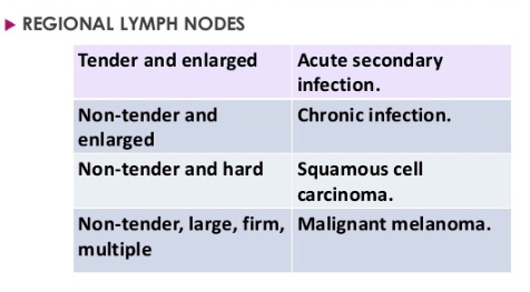
Lymph node

  • Cat scratch disease
  • Dental infection
  • Tuberculosis (scrofuloderma)

Neoplastic

  • Oral squamous cell carcinoma is the most common
  • Benign tumours of the mouth rarely form a fistula

Causes for persistence of sinus/fistula

  • Presence of a foreign body e.g. suture material
  • Presence of a necrotic tissue underneath e.g. sequestrum
  • Insufficient or non-dependent drainage. e.g. TB sinus
  • Distal obstruction e.g. biliary fistula
  • Persistant drainage like urine/faeces/CSF
  • Lack of rest
  • Epithelialisation of the tract e.g. AVF
  • Malignancy
  • Dense fibrosis
  • Irradiation
  • Malnutrition
  • Specific causes e.g. TB, actinomycosis
  • Ischemia
  • Drugs e.g. steroids
  • Interference by the patient

How is a fistula or sinus diagnosed?

In addition to careful history and examination, one or more of the following tests will usually be required to confirm the diagnosis and determine the cause:

  1. passing a probe into the channel
  2. radiology – may include plain x-rays, x-rays using contrast medium, CT or MRI scans
  3. microbiological assessment of swabs or biopsy material
  4. biopsy and pathology
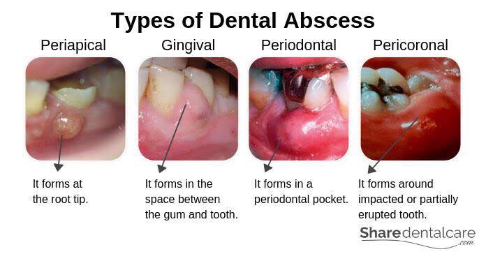
Abscess

Collection of pus

Clinical Features & Diagnosis:

Features of acute inflammation; The four cardinal signs of inflammation are:

  • redness (Latin rubor) – below localization of abscess
  • heat (calor) – inflammed area is hot
  • swelling (tumor) – pus present inside abscess cavity
  • and pain (dolor) – throbbing type

Diagnosis of Dental Abscess:

  • The location of abscess will cause tenderness with palpation test
  • The abscessed tooth will be very sensitive to percussion
  • IOPA findings will suggest slight thickening of PDL space with radiolucency at apex

Dentowesome|@drmehnaz🖊


References:

  1. dermnetnz.org
  2. SlideShare
  3. Google.com
  4. Study Notes✍🏻

EXAMINATION OF A SWELLING & ULCER

A swelling is a value term that denotes only enlargement or protuberance in body due to any cause.

According to the cause a swelling may be congenital, traumatic, inflammatory, Neoplastic or miscellaneous.

Examination of a swelling should be accompanied by a complete history of the patient. Following points should be noted:

Duration: The clinician may ask ‘when was the swelling first
noticed’? Swellings that are painful and of shorter duration are mostly inflammatory (acute), whereas those with longer duration and without pain are chronic, e.g. a chronic periapical abscess.

Mode of onset: The clinician may ask ‘how did the swelling start’? The history of any injury or trauma or any inflammation may contribute to the diagnosis and nature of the swelling.

Progression: The clinician should ask ‘has the lump changed in size since it was first noticed? Benign growths such as bony swellings grow in size very slowly and may remain static for a long period of time. If the swelling decreases in size, this suggests of an inflammatory lesion.

Site of swelling: The original site where it started must be assessed.

Other symptoms: Pain, fever, difficulty in swallowing, difficulty in respiration, disfigurement, bleeding or pus discharge are the common symptoms associated with swellings in the orofacial region.

Recurrence of the swelling: many swellings do recur after removal of the tissue, indicating the presence of precipitating factor, e.g. ranula.

LOCAL EXAMINATION🔍

(A) Inspection

  • Situation: few swellings are peculiar in their position
  • Color: Black – Naevus/Melanoma; Red/purple: Hematoma; Bluish: Ranula
  • Shape: Ovoid, pear shaped, kidney shaped, spherical or irregular
  • Size: Mention in cms. – the vertical horizontal dimension
  • Surface: Cauliflower as in Squamous Cell Carcinoma; Filliform – Papilloma
  • Edge: Sessile/pedunculated/indistinct
  • Number: Multiple/diaphyseal
  • Pulsation: The swelling which is superficial to artery, in close relation with it will be pulsatile. Pulsatile nature of swelling is assessed with 2 fingers on mass.
Mnemonic: SETTLE
  • Skin: Red & edematous. Pigmentation of skin is seen in moles or after repeated exposure to X-rays. Skin over a growth looks like the peel of an orange.

(B) Palpation

  • Temperature: Local temperature is raised due to extensive vascularity of the swelling; best felt with back of fingers.
  • Tenderness: Patient complains of pain due to pressure exerted by swelling.
  • Size, shape & extent: Mention in cms. – the vertical horizontal dimension
  • Fluctuation: If swelling contains liquid or gas it fluctuates.
Fluctuation test is positive if the two digits are pushed away in both directions.
  • Translucency: contains clear fluid
  • Compressibility: When pressure is applied to a swelling it decreases in size and when pressure is released swelling regains its size itself. Characteristic sign of VASCULAR HEMANGIOMA
  • Reducibility: Swelling reduces and ultimately disappears when pressed upon.

Surface: Can be👇🏻

• Smooth (cystic swellings)
• Lobular with smooth lumps (lipoma)
• Nodular (multinodular goitre)
• Matted (lymph nodes)
• Irregular (carcinoma)

Margins: Well defined/indistinct👇🏻

• Malignant growth - irregular margin
• Acute inflammatory swelling - ill defined margin
• Benign tumor - swelling slips & is indistinct

Edge👇🏻

• Well defined & regular-Benign Neoplasms
• Well defined & irregular-Malignant Neoplasms
• Ill defined & diffuse - Inflammatory swellings

Consistency: 👇🏻

• Soft - lipoma
• Cystic - Cyst or chronic abscesses
• Firm - Fibroma
• Hard - Chondroma
• Bony hard - Osteoma
• Stony hard - Carcinoma
• Variable consistency - Malignancy

EXAMINATION OF ULCER:

An ulcer is break in continuity of epithelium, skin or mucous membrane. A proper
history must be taken in case of an ulcer:

Mode of onset: The clinician may ask ‘how has the ulcer developed’? The patient may provide significant information about the nature and etiology of the ulcer such as any trauma or spontaneously.

Duration: The clinician may ask ‘how long is the ulcer present here’? It determines the chronicity of the ulcer. For example, traumatic ulcers in oral cavity are acute (occurring for a short period), but if the agent persists; it may become a chronic ulcer.

Pain: The clinician may ask ‘is the ulcer painful’? Most of the ulcers, being inflammatory in nature, produce pain. Painless ulcers usually suggest nerve diseases (such as peripheral neuritis, syphilis, etc).

Discharge: Any blood, pus or serum discharge must be noted.

Associated disease: Any associated generalized systemic problem may be associated with the ulcers of oral cavity (such as
tuberculosis, squamous cell carcinoma, etc).

LOCAL EXAMINATION🔍

(A) Inspection

Size & Shape:

• Tuberculous ulcer - oval with irregular border
• Varicose ulcer - vertical & oval in shape
• Carcinomatous ulcer - irregular

Number: Tuberculous, inflammatory ulcer may be more than one in number

Position:

• Arterial ulcer: Tip of the toes, dorsum of the foot
• Varicose ulcer: lower limb
• Perforating ulcers: over the sole at pressure points
• Non-healing ulcers: over the shin
• Rodent ulcer: upper part of face

Edge: An area between margin & floor. In spreading ulcer, edge is inflamed. Undermined edges destroy subcutaneous tissue faster than skin.

Beaded: As seen in rodent ulcer

Floor: This is the part of the ulcer which is exposed or seen.

• Red granulation tissue - Healing ulcer
• Necrotic tissue, slough - spreading ulcer
• Pale, scanty granulation tissue - tuberculous ulcer
• Wash-leather slough - Gummatous ulcer

Discharge:

• Serous discharge - Healing ulcer
• Purulent discharge - Spreading ulcer
• Bloody discharge - Malignant ulcer
• Discharge with bony spicules - Osteomyelitis
• Greenish diacharge - Pseudomonas infection

(B) Palpation

Tenderness: Characteristic of infected ulcers and arterial ulcers.

Induration: The edge, base and the surrounding area should be examined for induration

• Maximum induration - Squamous cell carcinoma
• Minimal induration - Malignant melanoma
• Brawny induration - Abscess
• Cyanotic induration - Chronic venous congestion as in varicose ulcer

Mobility: Malignant ulcers are usually fixed, benign ulcers are not.

Bleeding: Malignant ulcer is friable like a cauliflower. On gentle palpation, it bleeds. Granulation tissue as in a healing ulcer also causes bleeding.

Surrounding Area:

  • Thickening and induration is found in squamous cell carcinoma.
  • Tenderness and pitting on pressure indicates spreading inflammation surrounding the ulcer.

Relevant Clinical Examination:

Dentowesome|@drmehnaz🖊


References:

  1. A Practical Manual of Public Health Dentistry by CM Marya
  2. Slideshare.net
  3. https://www.medcampus.io/mnotes/examination-of-a-swelling-transmitted-vs-expansile-pulsations-
  4. medinaz.com, http://www.rxpg.com
  5. Study Notes✍🏻

Swellings in the angle of Mandible, Floor of Mouth & Palate

Swellings at the angle of Mandible include: ✍🏻👇🏻

🔅Congenital disease

• Branchial Cleft Cyst

🔅Neoplasm

(i) Benign

  • Hemangioma
  • Lymphangioma, Cystic hygroma
  • Pleomorphic adenoma (mixed tumor)
  • Warthin tumor
  • Neurofibroma
  • Angiolipoma
  • Adenoma
  • Hamartoma
  • Lipoma
  • Oncocytoma

(ii) Malignant

  • Mucoepidermoid carcinoma
  • Squamous cell carcinoma
  • Adenoid cystic carcinoma
  • Acinic cell carcinoma
  • Adenocarcinoma
  • Rhabdomyosarcoma
  • Lymphoma, leukemia
  • Metastatic adenopathy

🔅Inflammation/Infection

  1. Parotitis
  2. Parotid Abscess
  3. Tuberculosis
  4. Sarcoidosis
  5. Sjögren disease
  6. HIV

Detailed View🔍

1) Branchial Cleft Cyst:

  • Failure of involution of clefts and pouches lead to cysts, fistulas or sinus tracts.
  • Its a painless fluctuant swelling
  • First branchial cleft cysts are rare usually located at parotid gland or periparotid region.
  • Second branchial cleft cyst – Type II are the most common
  • Typically, second branchial cleft cysts present as a rounded swelling just below the angle of mandible, anterior to the sternocleidomastoid

2) Hemangiomas:

They are the most common benign salivary gland mass. Capillary hemangiomas involve parotids

3) Lymphangiomas

They are congenital malformations of the lymphatic system that may involve the parotid gland (Soft asymptomatic neck mass associated with facial asymmetry)

4) Pleomorphic Adenoma:

Hard painless slow growing mass

5) Warthin Tumor:

Incorporation of heterotopic salivary gland ductal epithelium within intraparotid & periparotid nodes

6) Parotitis & Parotid Abscess:

  • Most common in children
  • Mumps is the most common viral cause of parotitis
  • The condition manifests tender swelling at the angle of Mandible
  • Sialadenitis is most commonly due to bacterial infections caused by Staphylococcus aureus.
  • Premature neonates and immunosuppressed individuals are affected.

Swellings in the floor of Mouth: 👇🏻✍🏻

Ranula presents as a translucent blue, dome-shaped fluctuant swelling & contains viscid, glairy jelly like fluid
  1. Ranula – a type of mucocele found on the floor of the mouth. Present as a swelling of connective tissue consisting of collected Mucin from a ruptured salivary gland by local trauma.
  2. Swellings in the floor of the mouth are more likely to arise from structures above the Mylohyoid muscle. The commonest swellings in the floor of the mouth are denture induced hyperplasia & salivary calculus.
  3. Swellings in the floor of the mouth may inhibit swallowing & speech.
  4. Mandibular tori produce bony hard swelling lingual to the lower premolars.

Differential diagnosis of swellings of the floor of the mouth or neck (Jham et al., 2007): https://www.researchgate.net/figure/Differential-diagnosis-of-swellings-of-the-floor-of-the-mouth-or-neck-Jham-et-al-2007_tbl1_287206404


Swellings on the Palate: 👇🏻✍🏻

  1. Torus palatinus is an intrinsic bone lesion whereas a dental abscess pointing on the palate (usually from the palatal roots of the 1st & 2nd maxillary molars or from upper lateral incisors) is extrinsic.
  2. Salivary neoplasms
  3. Invasive carcinoma from the maxillary sinus may produce a palatal swelling.
  4. Kaposi’s sarcoma, typical of HIV/AIDS may also present as lump on palate.
  5. Paget’s disease.

Differential diagnosis of palatal swellings: https://www.researchgate.net/figure/Differential-diagnosis-of-palatal-swellings_tbl1_221967546

Dentowesome|@drmehnaz🖊


Image source: Google.com

PARATHYROID MEDICINE

Muhad Noorman p, Final year, Team Dentowesome

Hyperparathyroidism

Disorder of parathyroid gland characterised by excess secretion of PTH hormones resulting in clinical and biochemical hypercalcemia.

The most common cause of excess hormone production (hyperparathyroidism) is the development of a benign tumor in one of the parathyroid glands. This enlargement of one parathyroid gland is called a parathyroid adenoma which accounts for about 70 percent of all patients with primary hyperparathyroidism. The other causes comprises of parathyroid hyperplasia and PARATHYROID carcinoma (rarely).

Types

1) Primary hyperparathyroidism occurs when there is a disorder of the parathyroid glands themselves.

2) Secondary hyperparathyroidism results when a condition elsewhere in the body affects the parathyroid glands, causing them to produce too much hormone

3) Teritiary hyperparathyroidism causes when long standing secondary hyperparathyroidism become autonomous gland

CLINICAL FEATURES

Commonly found in middle aged women’s.

Hyperparathyroidism may or may not cause symptoms. When symptoms do appear, they are often mild, such as
weakness,
fatigue,
depression,( psychic moans) or
body aches and pains.
In primary hyperparathyroidism, the elevated levels of PTH cause elevated levels of blood calcium (hypercalcemia). Increased calcium and phosphorus excretion in the urine may cause kidney stones.(Nephrocalcinosis and nephrolithiasis).

Diagnosis of hyperparathyroidism relies on blood tests to measure hormone and calcium levels. Elevated Serum Calcium and phosphorus level .

Surgery is the main treatment for hyperparathyroidism. Surgical resection of adenoma and transplantation in hand muscles is most followed protocol .

Reference; Internet, SRB Textbook of surgery

CLEFT LIP AND PALATE- PART 2 – CLASSIFICATION, CLINIC FEATURES AND MANAGEMENT

Muhad Noorman P, Final year Team dentowesome.

CLASSIFICATIONS OF CLEFT LIP AND PALATE

Davis & Ritchie classification (1922)

Veau’s (1931)

Harkins and associates(1962)

Kernahan’s Classification (1971)

Spina (1974)

Tessiers’s Classification.

VEAU’S CLASSIFICATION

Group 1- Cleft of soft palate only
Group 2 – Cleft of hard and soft palate extending
no further extending than involving incisive foramen, (secondary palate only)
Group 3 – Complete unilateral cleft, extending from uvula
to incisive foramen in the midline, then deviating the one side and usually extending through the alevolus at the position of the future lateral incisor
Group 4- Complete bilateral cleft, extending forward through incisive foramen to alevolus. Premaxilla, suspended from the nasal septum .
Asian population have highest frequency often 

CLINICAL FEATURES:-

Incidence- 1in 500,With african population the lowest at 1in 250. Cleft lip alone more common in males. Isolated Cleft palate more common in female. 50% are syndromic, and are born with other congenital abnormalities.Cleft lip appear as unilateral / bi lateral. Line of cleft start on lateral part of upper lip and continues through philtrum of alveolus between lateral incisor and canine. When cleft lip continues from incisive foramen through palatal suture middle in palate,cleft lip with palate (unilateral/bilateral) present . . Cleft palate appears with involving soft palate only, involving uvula (bifid uvula),isolated cleft palate also.
.Patient have significant physical and physiological effects like, difficulty in eating and drinking with regurgitation of food to nose.
. Speech problem
. Ear infection: Malposition of Eustachian tubes result in middle ear infection
.Cosmetic deformities.

MANAGEMENT 

Management of Cleft lip and palate require, multidisciplinary coordinated approach by specialist including maxillofacial surgeon, pediatric surgeon, anesthetist, Prosthodontist, Orthodontist, Speech pathologist, otolaryngology ,audiologist  etc..

Management is aimed at closure and correction of lip and palate, secondary correction of palatal fistulae, orthodontic management of malocclusion, Orthognathic surgery, Rhinoplasty,and providing prosthesis for patients.

Pre operative criteria selected by physicians for surgery is Millards Rule of 10’s
1) 10lb weight
2) 10mg/l of haemoglobin
3) 10 weeks of age

  SURGICAL MANAGEMENT
1) Primary : Closure of lip & palate
2) Secondary : Closure of palatal fistula, Pharygoplasty  ,Bone grafting, orthodontic management Rhinoplasty and Scar revision.

Reference: Oral and Maxillofacial surgery, Balaaji. Textbook of general surgery for dental students, SRB

Dysplasia :-

It is histological cannotation to premalignance marked by abbarent and uncoordinated cellular proliferation depicted at cellular level as atypia which is reflected as dysplasia.

An oral precancerous lesion, also called dysplasia, is a growth that contains abnormal cells confined to the lining of the oral cavity, or mouth. This lining is called the mucosa. It covers the inside of the cheeks, the inside of the lips, the gums, the tongue, and the roof and floor of the mouth.

Dysplasia can be mild moderate and severe.

Clinical features :-

  1. Increased mitosis
  2. Loss of basal polarity
  3. Hyperchromatism of cell
  4. Increased nuclear cytoplasmic ratio
  5. Nuclear atypia
  6. Large prominent nucei
  7. Epithelial pearl
  8. Individual cell keratinisation

Treatment :-

  • NSAIDs for pain and inflammation medical treatment.
  • Excision of the part which is affected along with some part of healthy tissue. This is done under anesthesia.

Reference :-

Image :- Google
Writing :- notes made from mastering bds and Sanjay Kumar Purkait books

Luxation of TMJ :-

TMJ has hinge like junction.

Luxation occurs when :-

  1. Overextension of TMJ
  2. Isolated injury or associated with maxillofacial injuries

Luxation is unilateral but bilateral Luxation also occurs.

Clinical features :-

  • Inability to close mouth
  • Head trauma
  • No deviation of mandible
  • Lower jaw deviated away from luxated side
  • Pain may occur in some cases
  • Inflammation may be seen

Treatment :-

  • NSAIDS should be the medical treatment
  • Closed reduction
  • Fulcrum should be placed
  • Open reduction or mandibular condylectomy are indicated

Reference :-

Image :- Google
Writing :- notes made from mastering bds and Sanjay Kumar Purkait books

Ankylosis of TMJ :-

It is one of most incapacitating of all diseases involving this structure.

Types :-

  • In Complete Ankylosis i.e there is limitation of motion.
  • In Unilateral Ankylosis i.e chin is displaced.
  • In Intra-articular Ankylosis, joint undergoes progressive destruction.
  • In Extra-articular Ankylosis, splitting of TMJ.

Clinical features :-

  • Occurs before 10 years of age
  • Equal sex distribution
  • Restriction in opening of mouth
  • Injury at infancy or childhood, there is associated facial deformity.
  • Pain may occur in some cases
  • Inflammation may be seen

Treatment :-

  • NSAIDS can be use as medical treatment.
  • Surgical methods like condylectomy intraoral coronoidectomy, ramus osteotomy, high condylectomy, forceful opening of the jaw under general anesthesia, lysis of adhesions of the pterygoid space.

Reference :-

Image :- Google
Writing :- notes made from mastering bds and Sanjay Kumar Purkait books

CANCRUM ORIS

Don’t let Cancrum Oris take your pretty cheeks away! Stay nourished! 😂

STUDY NOTES ⚕️

Cancrum oris is a special type of gangrene.

Causes:

🔍 Malnourishment.

🔍 Major diseases like diptheria, Whooping cough, measles, kala azar, typhoid, etc.

🔍 These factors lead to invasion of opportunistic organisms like the Vincent’s organisms – Borrelia vincentii and fusiforms causing ulcerations, erosions and eventually fibrosis.

Clinical Presentation:

👩‍⚕️ It is an extensive ulcerative disease of the cheek mucosa usually occuring in malnourished children.

👨‍⚕️ As the disease progresses, there may be complete reduction of the cheek thickness.

Treatment:

💊 Ryle’s tube feeding.

💊 Improve the nutrition.

💊 Appropriate antibiotics: Metronidazole 400mg thrice daily for 7-10 days.

💊 Surgical reconstruction of the cheek.

Complications:

⚠️ Fibrosis leading to restricted movement of the jaw.

⚠️ Sepsis, toxaemia and death.

SOURCE: Manipal Manual of Surgery (3rd edition)


~Sunantha✍️

GLASSGOW COMA SCALE

Assess and then decide! 😂

STUDY NOTES ⚕️

Glassgow Coma Score is used to assess the level of consciousness properly instead of using vague terms like semi-conscious, obtundant, etc.

Hence it is widely used thereby avoiding observer errors in the observation patients.

NEUROLOGICAL ASSESSMENT USING GLASSGOW COMA SCALE:

1️⃣ Eyes Open:

Spontaneously – 4

To speech – 3

To pain – 2

None – 1

2️⃣ Best Verbal Response:

Oriented – 5

Confused – 4

Inappropriate words – 3

Incomprehensible sounds – 2

None – 1

3️⃣ Best Motor Response:

Obeys commands – 6

Localises pain – 5

Withdrawal to pain – 4

Flexion to pain – 3

Extension to pain – 2 (due to raised intracranial pressure)

None – 1

~*~

⭐ Maximum score is 15.

⭐ Minimum score is 3.

⚠️ If a patient has a score of 7 or less than 7, then he/she is said to be in coma.

SOURCE: Manipal Manual of Surgery (3rd edition)


~Sunantha✍️