EXAMINATION OF A SWELLING & ULCER

A swelling is a value term that denotes only enlargement or protuberance in body due to any cause.

According to the cause a swelling may be congenital, traumatic, inflammatory, Neoplastic or miscellaneous.

Examination of a swelling should be accompanied by a complete history of the patient. Following points should be noted:

Duration: The clinician may ask ‘when was the swelling first
noticed’? Swellings that are painful and of shorter duration are mostly inflammatory (acute), whereas those with longer duration and without pain are chronic, e.g. a chronic periapical abscess.

Mode of onset: The clinician may ask ‘how did the swelling start’? The history of any injury or trauma or any inflammation may contribute to the diagnosis and nature of the swelling.

Progression: The clinician should ask ‘has the lump changed in size since it was first noticed? Benign growths such as bony swellings grow in size very slowly and may remain static for a long period of time. If the swelling decreases in size, this suggests of an inflammatory lesion.

Site of swelling: The original site where it started must be assessed.

Other symptoms: Pain, fever, difficulty in swallowing, difficulty in respiration, disfigurement, bleeding or pus discharge are the common symptoms associated with swellings in the orofacial region.

Recurrence of the swelling: many swellings do recur after removal of the tissue, indicating the presence of precipitating factor, e.g. ranula.

LOCAL EXAMINATION🔍

(A) Inspection

  • Situation: few swellings are peculiar in their position
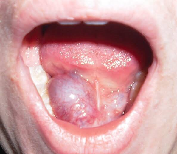
  • Color: Black – Naevus/Melanoma; Red/purple: Hematoma; Bluish: Ranula
  • Shape: Ovoid, pear shaped, kidney shaped, spherical or irregular
  • Size: Mention in cms. – the vertical horizontal dimension
  • Surface: Cauliflower as in Squamous Cell Carcinoma; Filliform – Papilloma
  • Edge: Sessile/pedunculated/indistinct
  • Number: Multiple/diaphyseal
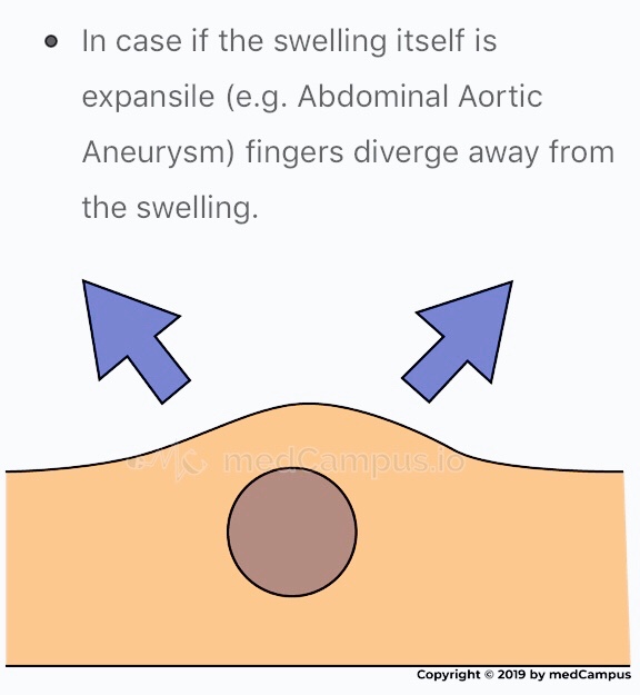
  • Pulsation: The swelling which is superficial to artery, in close relation with it will be pulsatile. Pulsatile nature of swelling is assessed with 2 fingers on mass.
Mnemonic: SETTLE
  • Skin: Red & edematous. Pigmentation of skin is seen in moles or after repeated exposure to X-rays. Skin over a growth looks like the peel of an orange.

(B) Palpation

  • Temperature: Local temperature is raised due to extensive vascularity of the swelling; best felt with back of fingers.
  • Tenderness: Patient complains of pain due to pressure exerted by swelling.
  • Size, shape & extent: Mention in cms. – the vertical horizontal dimension
  • Fluctuation: If swelling contains liquid or gas it fluctuates.
Fluctuation test is positive if the two digits are pushed away in both directions.
  • Translucency: contains clear fluid
  • Compressibility: When pressure is applied to a swelling it decreases in size and when pressure is released swelling regains its size itself. Characteristic sign of VASCULAR HEMANGIOMA
  • Reducibility: Swelling reduces and ultimately disappears when pressed upon.

Surface: Can be👇🏻

• Smooth (cystic swellings)
• Lobular with smooth lumps (lipoma)
• Nodular (multinodular goitre)
• Matted (lymph nodes)
• Irregular (carcinoma)

Margins: Well defined/indistinct👇🏻

• Malignant growth - irregular margin
• Acute inflammatory swelling - ill defined margin
• Benign tumor - swelling slips & is indistinct

Edge👇🏻

• Well defined & regular-Benign Neoplasms
• Well defined & irregular-Malignant Neoplasms
• Ill defined & diffuse - Inflammatory swellings

Consistency: 👇🏻

• Soft - lipoma
• Cystic - Cyst or chronic abscesses
• Firm - Fibroma
• Hard - Chondroma
• Bony hard - Osteoma
• Stony hard - Carcinoma
• Variable consistency - Malignancy

EXAMINATION OF ULCER:

An ulcer is break in continuity of epithelium, skin or mucous membrane. A proper
history must be taken in case of an ulcer:

Mode of onset: The clinician may ask ‘how has the ulcer developed’? The patient may provide significant information about the nature and etiology of the ulcer such as any trauma or spontaneously.

Duration: The clinician may ask ‘how long is the ulcer present here’? It determines the chronicity of the ulcer. For example, traumatic ulcers in oral cavity are acute (occurring for a short period), but if the agent persists; it may become a chronic ulcer.

Pain: The clinician may ask ‘is the ulcer painful’? Most of the ulcers, being inflammatory in nature, produce pain. Painless ulcers usually suggest nerve diseases (such as peripheral neuritis, syphilis, etc).

Discharge: Any blood, pus or serum discharge must be noted.

Associated disease: Any associated generalized systemic problem may be associated with the ulcers of oral cavity (such as
tuberculosis, squamous cell carcinoma, etc).

LOCAL EXAMINATION🔍

(A) Inspection

Size & Shape:

• Tuberculous ulcer - oval with irregular border
• Varicose ulcer - vertical & oval in shape
• Carcinomatous ulcer - irregular

Number: Tuberculous, inflammatory ulcer may be more than one in number

Position:

• Arterial ulcer: Tip of the toes, dorsum of the foot
• Varicose ulcer: lower limb
• Perforating ulcers: over the sole at pressure points
• Non-healing ulcers: over the shin
• Rodent ulcer: upper part of face

Edge: An area between margin & floor. In spreading ulcer, edge is inflamed. Undermined edges destroy subcutaneous tissue faster than skin.

Beaded: As seen in rodent ulcer

Floor: This is the part of the ulcer which is exposed or seen.

• Red granulation tissue - Healing ulcer
• Necrotic tissue, slough - spreading ulcer
• Pale, scanty granulation tissue - tuberculous ulcer
• Wash-leather slough - Gummatous ulcer

Discharge:

• Serous discharge - Healing ulcer
• Purulent discharge - Spreading ulcer
• Bloody discharge - Malignant ulcer
• Discharge with bony spicules - Osteomyelitis
• Greenish diacharge - Pseudomonas infection

(B) Palpation

Tenderness: Characteristic of infected ulcers and arterial ulcers.

Induration: The edge, base and the surrounding area should be examined for induration

• Maximum induration - Squamous cell carcinoma
• Minimal induration - Malignant melanoma
• Brawny induration - Abscess
• Cyanotic induration - Chronic venous congestion as in varicose ulcer

Mobility: Malignant ulcers are usually fixed, benign ulcers are not.

Bleeding: Malignant ulcer is friable like a cauliflower. On gentle palpation, it bleeds. Granulation tissue as in a healing ulcer also causes bleeding.

Surrounding Area:

  • Thickening and induration is found in squamous cell carcinoma.
  • Tenderness and pitting on pressure indicates spreading inflammation surrounding the ulcer.

Relevant Clinical Examination:

Dentowesome|@drmehnaz🖊


References:

  1. A Practical Manual of Public Health Dentistry by CM Marya
  2. Slideshare.net
  3. https://www.medcampus.io/mnotes/examination-of-a-swelling-transmitted-vs-expansile-pulsations-
  4. medinaz.com, http://www.rxpg.com
  5. Study Notes✍🏻

HEMATOLOGICAL INVESTIGATIONS

Also known as: CBC; Hemogram

Sample Required?

  • A blood sample drawn from a vein in your arm or a fingerstick or heelstick (newborns)

Test Preparation Needed?

  • None

Why get tested?

  • To determine your general health status; to screen for, diagnose or monitor any one of a variety of diseases and conditions that affect blood cells, such as anemia, infection, inflammation, bleeding disorder or cancer.

Also known as: Hgb; Hb; H and H (Hemoglobin and Hematocrit)

Sample Required?

  • A blood sample drawn from a vein in your arm or a fingerstick or heelstick (newborns)

Test Preparation Needed?

  • None

Why get tested?

  • To evaluate the hemoglobin content of your blood as part of a general health check-up; to screen for and help diagnose conditions that affect red blood cells (RBCs); If you have anemia (low hemoglobin) or polycythemia (high hemoglobin), to assess the severity of these conditions and to monitor response to treatment

• When to get tested?

  • With a hematocrit or as part of a complete blood count (CBC), which may be ordered as a component of a general health screen; when you have signs and symptoms of anemia (weakness, fatique) or polycythemia (dizziness, headache); at regular intervals to monitor these conditions or response to treatment

Also known as: Thrombocyte count; PLT; Platelet distribution width; PDW; Mean Platelet volume; MPV.

Sample Required?

  • A blood sample drawn from a vein in your arm or a fingerstick or heelstick (newborns)

Test Preparation Needed?

  • None

Why get tested?

  • To determine the number of platelets in a sample of your blood as part of a health exam; to screen for, diagnose, or monitor conditions that affect the number of platelets, such as a bleeding disorder, a bone marrow disease, or other underlying condition.

• When to get tested?

  • As part of a routine complete blood count (CBC); when you have episodes of unexplained or prolonged bleeding or other symptoms that may be due to a platelet disorder

What is being tested?

  • Platelets, also called thrombocytes, are tiny fragments of cells that are essential for normal blood clotting. They are formed from very large cells called megakaryocytes in the bone marrow and are released into the blood to circulate. The platelet count is a test that determines the number of platelets in a person’s sample of blood. When there is an injury to a blood vessel or tissue and bleeding begins, platelets help stop bleeding.

Also known as: Leukocyte differential count; Peripheral differential; WBC count differential; Diff; blood differential; Differential Blood Count

Formal name: White blood cell differential

• Why get tested?

  • To help determine the cause of abnormal results on a WBC count; to help diagnose or monitor an illness affecting your immune system, such as an infection or inflammatory condition, or cancers that affect your white blood cells, such as leukemia.

• When to get tested?

  • As part of a CBC; when you have a routine health examination; when results of a CBC fall outside the reference range; when you have any number of signs and symptoms that may be related to a condition affecting white blood cells, such as infection, inflammation, or cancer, when you are receiving treatment that is known to affect WBCs, such as chemotherapy.

• What is being tested?

  • WBCs, also called leukocytes, are cells that circulate in the blood and the lymphatic system that help protect the body against infections. They are an important part of the body’s immune system and also have a role in inflammation, allergic responses, and protection against cancer. A WBC differential totals the number of each of the different types of WBCs in a person’s sample of blood.
  • There are five types of white blood cells, each with different functions.

  • Also known as: TLC; WBC count
  • Total WBC count is used as one of the index of presence of systemic infection and to rule out the possibility of leukemia & malignant neutropenia
  • Calculated with haemocytometer/ automated cell counts
  • RBCs are lysed by diluting the blood sample with dilute acetic acid leaving the WBCs intact.

  • Also known as: Red Blood Cell Count, RBC count
  • Red blood cells, also known as erythrocytes, make up the cellular part of blood, giving it its red color and also the ability to bind and carry oxygen to all parts of the body. Under a microscope, they appear to be circular and biconcave in shape.
  • Gives us the number of erythrocytes per cubic mm in circulating blood & Hb in blood.
  • Procedure done by office or chairside method and also automated procedure.
  • Hematological diseases of RBCs are anemia & polycythemia.

  • Categorized by mean corpuscular volume, anemia can be differentiated into microcytic, macrocytic and normocytic anemias. Normocytic anemia can be further divided into intrinsic and extrinsic RBC defect and blood loss.
  • MCV – Mean corpuscular volume is the average volume of red blood cells and is reflective of RBC size. When RBCs increase or decrease in size, the mean corpuscular volume changes; this helps physicians determine the type of anemia and its causes. Normal MCV is 80–96 µm³.
  • MCH stands for “mean corpuscular hemoglobin.” An MCH value refers to the average quantity of hemoglobin present in a single red blood cell.
  • MCHC is short for mean corpuscular hemoglobin concentration. MCHC refers to the average amount of hemoglobin inside a single red blood cell.
  • Hematocrit is the measure of the total volume % of red blood cells in the blood. The normal value for hematocrit is 45% for men and 40% for women. It is an important component of a patient’s complete blood profile.

Indications:

  • To prepare smears from paper points removed from root canals for evaluation of microcytic status of canal prior to filling.
  • A scraping or swab of an oral lesion is needed to confirm diagnosis of thrush
  • A scraping of gingival region or mucosal ulcer is sometimes used to confirm diagnosis of Acute Necrotising ulcerative stomatitis.
  • Identification of giant cells that accompany vesicular infections
  • Identification of Acantholysis

Dentowesome|@drmehnaz🖊


References: Google.com, lecturio.com, Study Notes✍🏻

Swellings in the angle of Mandible, Floor of Mouth & Palate

Swellings at the angle of Mandible include: ✍🏻👇🏻

🔅Congenital disease

• Branchial Cleft Cyst

🔅Neoplasm

(i) Benign

  • Hemangioma
  • Lymphangioma, Cystic hygroma
  • Pleomorphic adenoma (mixed tumor)
  • Warthin tumor
  • Neurofibroma
  • Angiolipoma
  • Adenoma
  • Hamartoma
  • Lipoma
  • Oncocytoma

(ii) Malignant

  • Mucoepidermoid carcinoma
  • Squamous cell carcinoma
  • Adenoid cystic carcinoma
  • Acinic cell carcinoma
  • Adenocarcinoma
  • Rhabdomyosarcoma
  • Lymphoma, leukemia
  • Metastatic adenopathy

🔅Inflammation/Infection

  1. Parotitis
  2. Parotid Abscess
  3. Tuberculosis
  4. Sarcoidosis
  5. Sjögren disease
  6. HIV

Detailed View🔍

1) Branchial Cleft Cyst:

  • Failure of involution of clefts and pouches lead to cysts, fistulas or sinus tracts.
  • Its a painless fluctuant swelling
  • First branchial cleft cysts are rare usually located at parotid gland or periparotid region.
  • Second branchial cleft cyst – Type II are the most common
  • Typically, second branchial cleft cysts present as a rounded swelling just below the angle of mandible, anterior to the sternocleidomastoid

2) Hemangiomas:

They are the most common benign salivary gland mass. Capillary hemangiomas involve parotids

3) Lymphangiomas

They are congenital malformations of the lymphatic system that may involve the parotid gland (Soft asymptomatic neck mass associated with facial asymmetry)

4) Pleomorphic Adenoma:

Hard painless slow growing mass

5) Warthin Tumor:

Incorporation of heterotopic salivary gland ductal epithelium within intraparotid & periparotid nodes

6) Parotitis & Parotid Abscess:

  • Most common in children
  • Mumps is the most common viral cause of parotitis
  • The condition manifests tender swelling at the angle of Mandible
  • Sialadenitis is most commonly due to bacterial infections caused by Staphylococcus aureus.
  • Premature neonates and immunosuppressed individuals are affected.

Swellings in the floor of Mouth: 👇🏻✍🏻

Ranula presents as a translucent blue, dome-shaped fluctuant swelling & contains viscid, glairy jelly like fluid
  1. Ranula – a type of mucocele found on the floor of the mouth. Present as a swelling of connective tissue consisting of collected Mucin from a ruptured salivary gland by local trauma.
  2. Swellings in the floor of the mouth are more likely to arise from structures above the Mylohyoid muscle. The commonest swellings in the floor of the mouth are denture induced hyperplasia & salivary calculus.
  3. Swellings in the floor of the mouth may inhibit swallowing & speech.
  4. Mandibular tori produce bony hard swelling lingual to the lower premolars.

Differential diagnosis of swellings of the floor of the mouth or neck (Jham et al., 2007): https://www.researchgate.net/figure/Differential-diagnosis-of-swellings-of-the-floor-of-the-mouth-or-neck-Jham-et-al-2007_tbl1_287206404


Swellings on the Palate: 👇🏻✍🏻

  1. Torus palatinus is an intrinsic bone lesion whereas a dental abscess pointing on the palate (usually from the palatal roots of the 1st & 2nd maxillary molars or from upper lateral incisors) is extrinsic.
  2. Salivary neoplasms
  3. Invasive carcinoma from the maxillary sinus may produce a palatal swelling.
  4. Kaposi’s sarcoma, typical of HIV/AIDS may also present as lump on palate.
  5. Paget’s disease.

Differential diagnosis of palatal swellings: https://www.researchgate.net/figure/Differential-diagnosis-of-palatal-swellings_tbl1_221967546

Dentowesome|@drmehnaz🖊


Image source: Google.com

Diagnosis of Dental Caries

Introduction:

Early detection and diagnosis of dental caries reduces irreversible loss of tooth structure, the treatment costs and the time needed for restoration of the teeth. Dental caries often initiates at the fissures in the occlusal surface of the tooth. Conventional examination for caries detection is primarily done using visual inspection, tactile sensation and radiographs. While these methods give satisfactory results in detection of cavitated lesions, they are usually inadequate for the detection of initial lesions. Because of these deficiencies, new detection methods have been developed to aid better diagnosis.

Conventional Methods Used in Diagnosis of Dental Caries

It is one of the most common diagnosis methods implemented by dentists. In order to make an accurate assessment, the teeth should be clean, dry and examined under a light source. In visual examination, changes in tooth structure such as; enamel dissolution, white spot lesions, discoloration, surface roughness and presence of cavitation are assessed. When illuminated, the carious tissues scatter the light and make enamel look whiter and opaque. This is due to increased porosity caused by demineralization. Similarly, when dentin undergoes demineralization, a shadow is observed under the intact enamel. When caries progress, the surface breaks down and a cavitation is formed.

The explorer and the dental floss are used for tactile examination but the use of an explorer is not preferred because:

  1. Sharp tip of the explorer can produce traumatic defects on the enamel surface,
  2. The cariogenic bacteria may be transferred from one tooth surface to another,
  3. Probing may cause cavitation and fracture in the incipient lesions,
  4. Explorers have low sensitivity resulting in undetected lesions.

If the explorer catches or resists removal when moderate pressure is applied, and when this is accompanied by one of the following;

  • Softness at the base of the lesion,
  • Opacity adjacent to the pit or fissure,
  • The enamel is softened adjacent to the pit and fissure, we can conclude that the area is carious.

Pickard, proposed the use of dental floss for the detection of caries. When there is food packing between the teeth and the floss is frayed when passed through the contact area, this might be the indication of caries.

Clinically “sound” and apparently intact occlusal surfaces, however, may develop lesions which penetrate into the dentin, which can be observed only through radiographic examination

Radiographic examination has great value in detecting caries lesions especially when they are not clinically visible. In low caries population, as a result of fluoride use, the surface of enamel does not break down, making the caries detection harder. In recent years, the incidence of such lesions has increased dramatically. According to studies, bitewing radiography has been proven to be an effective method in the detection of proximal caries and hidden caries.
Besides its advantages, radiographs also have some limitations too. For this reason, it is advisable to use clinical evaluation along with radiographic imaging. The disadvantages of radiography are as follows:
• Proximal contacts are overlapped,
• The lesion depth may appear to be increased due to angulation and this may lead to false diagnosis,
• Occlusal lesions may not be detected because of the superposition of the buccal and lingual cusps,
• The real cause of the radiolucency can’t be determined whether it is due to caries, resorption or wear,
• The superficial demineralization of the buccal and lingual surfaces may seem like proximal caries,
• Active and arrested caries can’t be distinguished in the radiographs.
• Radiographs may give false positive results due to a phenomenon called “Mach band effect”. In this perceptual phenomenon, the contrast between the dark and lighter areas has increased, resulting in a dark demarcation band. This effect causes formation of a radiolucent area in dentin enamel junction.


Cervical burn out is another optical phenomenon where a wedge shaped radiolucent area is seen between the bone and the cemento-enamel junction. This effect is due to tissue density and the low penetration of X-rays at the cervical region.

Despite the disadvantages, radiographs are the most commonly used diagnosis tool and with the development of new techniques many of the problems are solved.

There are two layers of decalcification in carious dentin. The first one is the soft and infected layer which doesn’t have the capacity of remineralization. The second one is hard, intermediately decalcified and has the ability of remineralization. Many studies were carried out to differentiate these layers. Although there are opinions stating the benefit of caries detection dyes, there are also opinions that dyes can lead to over-reduction in the dentin.

Most clinical investigations have concluded that, caries detection dyes don’t stain bacteria but stain the less mineralized organic matrix. In a study of Demarco et al. they suggested that dye remnants that remained on the walls of the cavity may cause a decrease in the shear bond strength between the composite restorations and the enamel.

In the upcoming blog post we will learn about the Novel Methods for Caries Detection..

Happy Reading😊

References: Meandros Med Dent J 2018;19:1-8, Image source: Google

CORTICOSTEROIDS IN DENTISTRY

🔷 Introduction:

Steroids are a group of hormones produced naturally in the body.

The adrenal cortex consists of 3 zones:

  1. Zona Glomerulosa synthesizes Aldosterone, the most potent Mineralocorticoid in humans.
  2. Zona Fasciculata – produces hydrocortisone (a glucocorticoid)
  3. Zona Reticularis produces Adrenal Androgens

• Glucocorticoid secretion is regulated by ACTH produced in the anterior pituitary. Cortisol has a -ve feedback on ACTH production.

• In Addison diseases, Glucocorticoid secretion impaired & ACTH is ⬆️

🔷 Corticosteroid Action & its regulation

  • Glucocorticoid – Hepatic Glycogen deposition
  • Mineralocorticoid – Sodium, electrolyte-fluid balance.
  • Glucocorticoid play critical role in body’s response to stress.

Stress

⬇️

Release of cytokines (IL-1)

⬇️

⬆️ cortisol levels

🔷 Classification:

Adrenocortical Hormones & Related Drugs

🔷 Steroid synthesis:

The substrate for steroid production is cholesterol. It is mobilized from the outer to the inner mitochondrial membrane by the steroidogenic acute regulatory (StAR) protein.

🔷 Glucocorticoids:

1) Hydrocortisone

Action: Anti-allergy, anti-inflammatory

Therapeutic Uses:

  • intralesional injection in dose of 20-50 mg/mL
  • Topical application
  • Tablet in dose of 100mg/day can be given systemically to relieve burning sensation.
  • Tab. Cortisol(25mg) combined with dexamethasone(90mg) can be given at biweekly interval.*

Action: Fibrinolytic,Anti-allergic, anti-inflammatory*

It causes ⬇️ fibroblastic production & deposition of collagen*

  • Topical application with orabase
  • Intralesional injection of hydrocortisone acetate (25mg/ml)
  • Topical application (0.25-0.5%) to the affected area
  • Intra-articular injection (25mg/ml)
  • 20-30mg/day in divided doses

Trade Name: CORTEF, ORABASE-HCA

• Preparation:

  1. Oral: 5 – 20 mg tab.
  2. Topical – 1% eye drop solution; 0.025 nasal drops, 0.25-2.5% skin creams

• Hydrocortisone acetate 25-50 mg/mL for soft tissue suspensions

• Hydrocortisone sodium phosphate: 50mg/mL IV, IM, SC

2) Cortisone:

• Uses:

  1. OSMF
  2. Addison disease
  3. Hodgkin lymphoma

3) Prednisolone:

Action: Anti-allergic, anti-inflammatory, immunosuppressive

• Uses:

  1. Rheumatoid arthritis: 10mg/day in divided doses
  2. Collagen disease: 1mg/kg
  3. SLE: IM/IV; Topical application 2-3 times daily
  4. Leukemia: ALL as maintenance dose
  5. Erythema multiforme, pemphigus, bullous pemphigoid, behchet’s syndrome
  6. Bronchial asthma: 40-60 mg
  7. Post-herpetic neuralgia
  8. Amyloidosis, cyclic neutropenia, purpura

Trade name: DELTA-CORTEF, PRELONE

4) Triamcinolone:

• Uses:

  1. Lichen planus: Topical application 3-4 times daily
  2. Erythema multiforme: 40-100 mg/day or inhalation doses
  3. Recurrent aphthae: Oral (2-4 mg/day)
  4. Desquamative gingivitis, OSMF, contact chelitis – intralesional (2-3 mL/day)

Trade name: Kenolog cream

5) Dexamethasone:

Uses:

👉🏻Mainly used for adrenal cortical suppression

  • Allergic diseases, serum sickness, urticaria, hay fever, angioneurotic edema: IV; topically 2-4 times a day
  • Benign migratory glossitis: Topical application
  • Shock, cerebral edema, occular diseases.

Trade name: DECADRON

6) Betamethasone:

Action: Anti-allergy, anti-inflammatory

Uses: Lichen planus, pemphigus, Aphthae ulcer

Dr. Mehnaz Memon🖊


References: Self Notes, Image chart source: Classification of Drugs with DOC by Vikas Seth (Third Edition)

Types Of Caries

• Clinical Classification of Caries:

1️⃣ According to Anatomical Site –

  • Pit & fissure caries
  • Smooth Surface Caries
  • Cervical
  • Root caries

2️⃣ According to rate of caries progression –

  • Acute dental caries
  • Chronic dental caries

3️⃣ According to nature of attack-

  • Primary
  • Secondary

4️⃣ Based on chronology –

  • Infancy caries
  • Adolescent caries

A. Pit & Fissure Caries:

https://dentowesome.wordpress.com/2020/05/11/pit-fissure-caries/

B. Smooth surface caries:

  • On proximal surface of teeth or gingival 3rd of buccal & lingual preceded by formation of plaque.
  • Early while chalky spot – decalcification of enamel.

C. Linear Enamel Caries:

  • Atypical form
  • Found in primary dentition
  • Gross destruction of labial surface of incisor teeth

https://dentowesome.wordpress.com/2020/05/07/dental-caries/

D. Root caries:

  • Soft progressive lesion that is found everywhere on root surface that has least connective tissue attachment & is exposed to oral enviornment.
  • Older age group & gingival recession

E. Acute Dentinal Caries:

  • Rapid clinical course
  • Early pulp involvement
  • Initial lesion is small, while rapid spread of process at DEJ & diffuse involvement of dentin produce large internal excavation.

F. Rampant Caries:

Sudden, rapid & almost uncontrolled destruction of teeth affecting surface that are relatively caries free.

G. Nursing bottle caries (Baby bottle syndrome)

Affect deciduous teeth due to prolonged use of nursing bottle containing milk, sugar or honey.

💬 What is 👶 bottle decay? What causes it and how to prevent it? 👇🏻

H. Chronic dental caries: (Slower progress)

I. Recurrent caries: (Presence of leaky margins)

J. Arrested caries:

  • No tendency of future progression, caries become static.
  • Brown pigmentation in the hard tissue.

Dentowesome|@drmehnaz🖊


Image Source: Google.com

CHOOSING APPROPRIATE ANTIBIOTIC

Antibiotics can be divided into 2 classes based on their mechanism of action:-

  • Bactericidal
  • Bacteriostatic

➡️ Bactericidal antibiotics kill bacteria by inhibiting cell wall synthesis. Example:

  • Beta-lactams (Penicillins, Cephalosporins, Carbapenems, Monobactams)
  • Glycopeptides viz. Vancomycin
  • Aminoglycosides
  • Fluoroquinollines
  • Others: Bacitracin, Cycloserine, Metronidazole

➡️ Bacteriostatic antibiotics limit the growth of bacteria by interfering with bacterial protein production, DNA replication or other aspects of bacterial cellular metabolism.

They must work together with immune system to remove the micro-organisms from the body. Example:

  • Tetracyclins
  • Sulphonamides
  • Macrolides
  • Lincosamides
  • Chloramphenicol
  • trimethoprim

Most antimicrobial agents in clinical use are bactericidal,

Note that while it is rational to favor bactericidal agents over bacteriostatic agents, neither has ever been shown to be superior (probably because true recovery from infection cannot occur until the body is able to mount an appropriate immune response, thus “buying time” may be just as good as active killing)

Minimum inhibitory concentration (MIC) versus minimum bactericidal concentration (MBC).

➡️ The MBC is the minimum concentration of drug which can kill the micro-organisms.

➡️ The MIC is the minimum concentration of drug which can inhibit the growth of micro-organisms.

🔷 CHOOSING APPROPRIATE ANTIBIOTIC (A Clinician’s guide to the CARAT criteria)

Council for Appropriate and Rational Antibiotic Therapy (CARAT) criteria for accurate use of antibiotic therapy
● Evidence-based results ●Therapeutic benefits
● Safety
● Cost-effectiveness
● Optimal drug dose and duration —Shorter-course, more aggressive therapy

(i) Evidence-based results:

In choosing an antibiotic, clinicians should consider the clinical evidence demonstrating that the drug is clinically and microbiologically appropriate, the efficacy of that drug in well-designed clinical trials, and the antibiotic resistance patterns of the local region. Clinicians should then use their professional judgment to choose the optimal antibiotic.

(ii) Therapeutic benefits:

If possible, the clinician should identify the causative pathogen and use surveillance data on regional antibiotic resistance patterns in selecting the optimal therapeutic agent.

(iii) Safety:

In treating patients with a particular drug, safety must be weighed against efficacy. Clinically applicable treatment strategies should be chosen to maximize efficacy while minimizing side effects.

(iv) Optimal drug for optimal duration:

Optimal drug selection requires finding the antimicrobial class and the specific member of that class that is best suited to treat a particular infection. Because empiric therapy is necessary in most cases, multiple factors have to be considered. Among these are whether the etiologic agent is likely to be gram-positive or gram-negative, whether a narrow or broad-spectrum agent should be chosen, the resistance patterns of the likely pathogen to this drug, both nationally and regionally, and the individual patient’s medical history, including recent antibiotic exposure.

Optimal duration means prescribing the selected drug for the shortest amount of time required for clinical and micro- biologic efficacy. There are many reasons for reducing an- timicrobial therapy to the shortest appropriate duration. They include the potential for reduced occurrence of adverse effects, increased patient adherence, decreased promotion of resistance, and decreased costs.

(v) Cost-effectiveness:

Choosing inappropriate therapy is associated with increased costs, including the cost of the antibiotic and increases in overall costs of medical care because of treatment failures and adverse events.

Dentowesome | @drmehnaz🖊


References:

  1. https://www.amjmed.com/article/S0002-9343(05)00381-5/pdf
  2. https://microbeonline.com/minimum-inhibitory-concentration-and-minimum-bactericidal-concentration-mbc/

Selection of NSAID

Mild/Moderate Pain• Paracetemol
• Low dose Ibuprofen
Post op. or short lasting pain• Ketorolac, diclofenac
Musculoskeletal pain• Paracetemol
• Ibuprofen, naproxen, ketoprofen
RA,AS,Acute gout, Acute Rh. fever• Naproxen, piroxicam
• Indomethacin, high dose aspirin
GI irritation• Paracetemol
• Cox-2 inhibitors
H/O HS reaction to NSAIDs• Paracetemol/
• Cox-2 inhibitors
Paediatric pt.• Paracetemol, Ibuprofen & naproxen
Pregnancy• Paracetemol
Selection of NSAID

Dentowesome 2020|@drmehnaz 🖊

ANALGESICS AND ANTI-INFLAMMATORY DRUGS(NSAID’s) IN DENTISTRY

💊Analgesic is a drug that selectively relieves pain by acting in the CNS or on the peripheral pain mechanisms without significantly altering consciousness.

💊They are divided into 2 groups:

  1. Opiod/Narcotic/Morphine like
  2. Non-opiod/Non-Narcotic/antipyretic (Aspirin like drugs)

The antipyretic analgesics & NSAID’s are more commonly employed for dental pain because tissue injury and inflammation due to abscess, caries, tooth extraction etc. is major cause of acute dental pain.

🔆 CLASSIFICATION:

Image: 1
Image: 2

🔆 ANALGESIC/NSAID’s IN DENTISTRY:

  • The antipyretic analgesics used mainly for dental pain is Paracetemol.
  • Paracetemol (Acetaminophen) is most frequently used to relieve toothache in Pregnancy. It has week anti-inflammtory effect & causes no teratogenesis in the developing fetus.
  • Pain during invasive dental procedures is alloyed by a local anesthesic before & after is treated with NSAID’s.

💊 NSAID’s use during Pregnancy: (The devastating effects to the infant) 👇🏻

Image: 3

🔆Use of OPIOIDS in Dental Pain:

  • Less used than analgesics
  • Mostly codeine is used for dental patients because other opioids cause dullness & short lasting pain.
  • Other alternative Opioid Analgesics used are – Tramadol, Pentazocine.

💊MORPHINE – Depressant actions‼️

🔻Adverse effects:
  1. Sedation, mental clouding, lethargy, nausea, vomitting, diarrhoea.
  2. Respiratory depression, blurring of vision.
  3. Allergic conditions, rashes, urticaria, itching, swelling.
  4. May develop tolerance & dependance.
🔻Contraindications:
  1. Urinary retention – infants & elderly
  2. Asthma patients
  3. Hypotensive states
  4. Hypovolaemic states

💊NSAID’s :

  1. Analgesic, antipyretic, anti-inflammatory effect.
  2. Effectively relieves inflammatory tissue, injury related pain, signs of inflammation like pain, tenderness, swelling are suspected.
  3. Cellular metabolism is increased & due to increased Glucose utilization there is decrease in blood sugar.
  4. Has teratolytic & mild-antiseptic properties
  5. Irritates gastric mucosa
  6. Interferes with platelet aggregation & bleeding time prolonged.
🔻Adverse effects:
  • Nausea, vomiting, diarrhoea, blood loss in stools.
  • Haemolysis in G6PD deficient patients.
  • Nephrotoxicity & hepatotoxicity in long term use.
  • Allergic reactions – rashes, urticaria, photosensitivity.
  • Pregnancy & Infancy – Refer Image 3
🔻Contraindications:
  • Nursing and pregnancy
  • Serious bleeding
  • Allergy/Asthma/Angioedema
  • Impaired renal function
  • Drug (anticoagulant)

Dr. Mehnaz Memon🖊


References:

  1. Flowcharts: Classification of Drugs with DOC by Vikas Seth (Third Edition)
  2. KD TripathiEssentials of Medical Pharmacology 7th Edition; Internet

POSTOPERATIVE CARE AFTER DENTOALVEOLAR SURGERY

Good aftercare to prevent complications and unnecessary suffering, with loss of valuable time, is as important as a good operation.

The main purpose of aftercare is to expedite healing and prevent or relieve pain and swelling.

Rest is necessary for the prompt healing of wounds. Ambulatory patients should be directed to go home & remain quiet for several hours, preferably sitting in a comfortable chair or, if lying down, keeping the head elevated on several pillows.

  • Only liquids and soft solids should be eaten the first day. They may be warm or cold but not extremely hot.
  • Food intake should not begin until several hours after surgery to avoid disturbing the blood clot.
  • If the extractions were limited to one side, chewing can be done on the unoperated side, but when local anesthesia has been used, chewing should be avoided until sensation has returned.
  • Fluids should be taken in greater amounts than usual to prevent dehydration from limited food intake.
  • A normal diet should be resumed as soon as possible, since this facilitates healing.
  • The teeth should be brushed as usual, and on the day after surgery rinsing of the mouth should begin.
  • A saline solution (1/2 teaspoon of salt in a glass of warm water) is best for this purpose.
  • Commercial mouthwashes if used should be diluted with water due to the high alcohol content that can irritate the wound.
  • Hydrogen peroxide rinses should not be used initially as this agent can remove the blood clot.❌
  • Some degree of postoperative pain accompanies many exodontic procedures and begins after the effects of anesthetic have left. This is considered a normal response to the unavoidable trauma of surgery.
  • In most cases, such pain lasts no more than 12 to 24 hours, although a traumatic periostitis may persist for several days.
  • Ordinarily this type of pain can be controlled by the use of cold packs (30 minutes per hour) during the first 24 hours & the proper administration of analgesic drugs.
  • For mild pain, as after a routine extraction, one of the antipyretic analgesics is usually adequate.
  • For moderate pain, such as after removal of an impacted tooth, a drug such as codeine or meperidine (Domerol) should be used.
  • Narcotics are needed only in rare instances.
  • The combination of a sedative drug with an analgesic agent can also be used but barbiturate alone should never be used to relieve pain as it can result in mental disorientation in a patient suffering from extreme pain.
  • The degree of swelling that occurs is generally in direct proportion to the degree of surgical trauma.
  • The application of cold to the operated site helps diminish postoperative swelling. If a rubber ice bag is not available, the ice can be placed in a plastic bag.
  • Cold can be applied intraorally by holding an ice cube in the mouth.
  • Pressure dressings can also be beneficial in limiting postoperative swelling.
  • Once swelling has reached the maximum (usually after 24 to 48 hours), cold is no longer effective, and heat, in the form of moist compresses, should be applied. It too should be used only 30 minutes per hour. The area should be lubricated with petroleum jelly to avoid burning the skin.
  • Intraoral heat is achieved by the use of hot isotonic saline rinses.
  • Cigarette smoking should be avoided after tooth extraction because it has been shown to increase the incidence of alveolar osteitis

Dr. Mehnaz Memon🖊


References: Textbook of Oral Surgery – Daniel M Laskin