Written by : Dr. Urusa I Inamdar
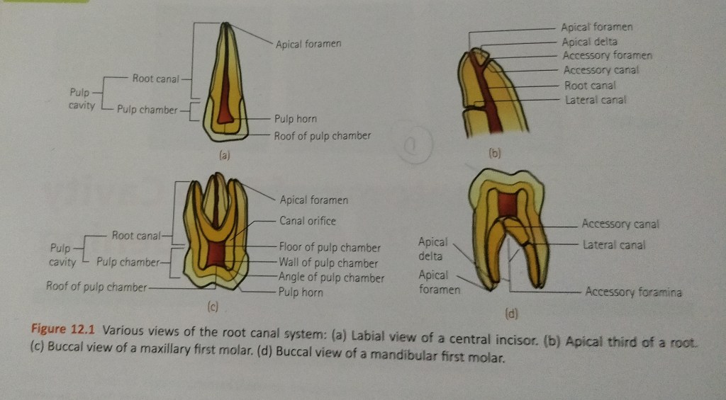
It is the portion of the pulp cavity from the canal orifice to the apical foramen.
It is divided into 3 sections :
- Coronal
- Middle
- Apical
- Accessory canals or lateral canals : lateral branching of the main root canal generally occurring in the apical third or furcation area of a root.
- Lateral canal : accessory canal that branches to the lateral surface of the root and may be visible on a radiograph.
- Apical foramen : aperture at or near the apex of a root through which the blood vessels and nerves of the pulp enter or leave the pulp cavity.
- Accessory foramina : openings of the accessory and lateral canals in the root surface.
A straight root canal extending the entire length of the root is uncommon. Either a constriction is present before the apex is reached or , as is often the case , a curvature is present.
The curvature may be :
- A straight canal extending with minimal apical curvature.
- A gradual curvature of the canal with a straight apical ending.
- A gradual curvature of the entire canal.
- A sharp curvature of the canal near the apex.
A curvature of about 20° in a narrow root canal may be difficult or even impossible to negotiate with endodontic instruments, whereas a curvature of even 30° may be negotiated if the root canal is wide.
Success in negotiating a narrow , curved canal depends on following :
- Degree of curvature.
- Size and constriction of the root canal.
- Size and flexibility of the endodontic instrument blade.
- Skill of the operator.
The various classification proposed are as follows :
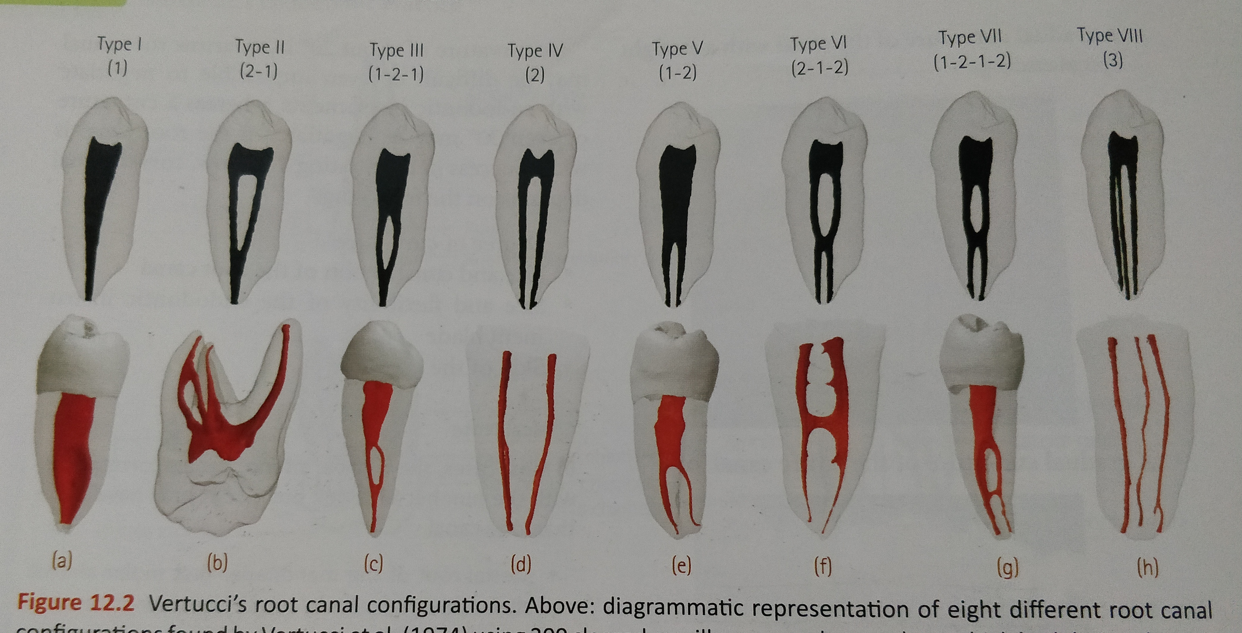
- Vertucci’s Classification:
- Type I : Single canal extends from the pulp chamber to the apex (1)
- Type II : Two separate canals leave the pulp chamber and join short of the apex to form one canal (2-1)
- Type III : One canal leaves the pulp chamber and divides into two in the root , the two then merge to exit as one canal (1-2-1)
- Type IV : Two separate distinct canals extend from the pulp chamber to the apex (2)
- Type V : One canal leaves the pulp chamber and divides short of the apex into two separate distinct canals with separate apical foramina (1-2)
- Type VI : Two separate canals leaves the pulp chamber , merge in the body of the root , and redivide short of the apex to exit as two distinct canals (2-1-2)
- Type VII : One canal leaves the pulp chamber, divides and then rejoins in the body of the root , and finally redivides into two distinct canals short of the apex (1-2-1-2)
- Type VIII : Three separate distinct canals extend from the pulp chamber to the apex (3)
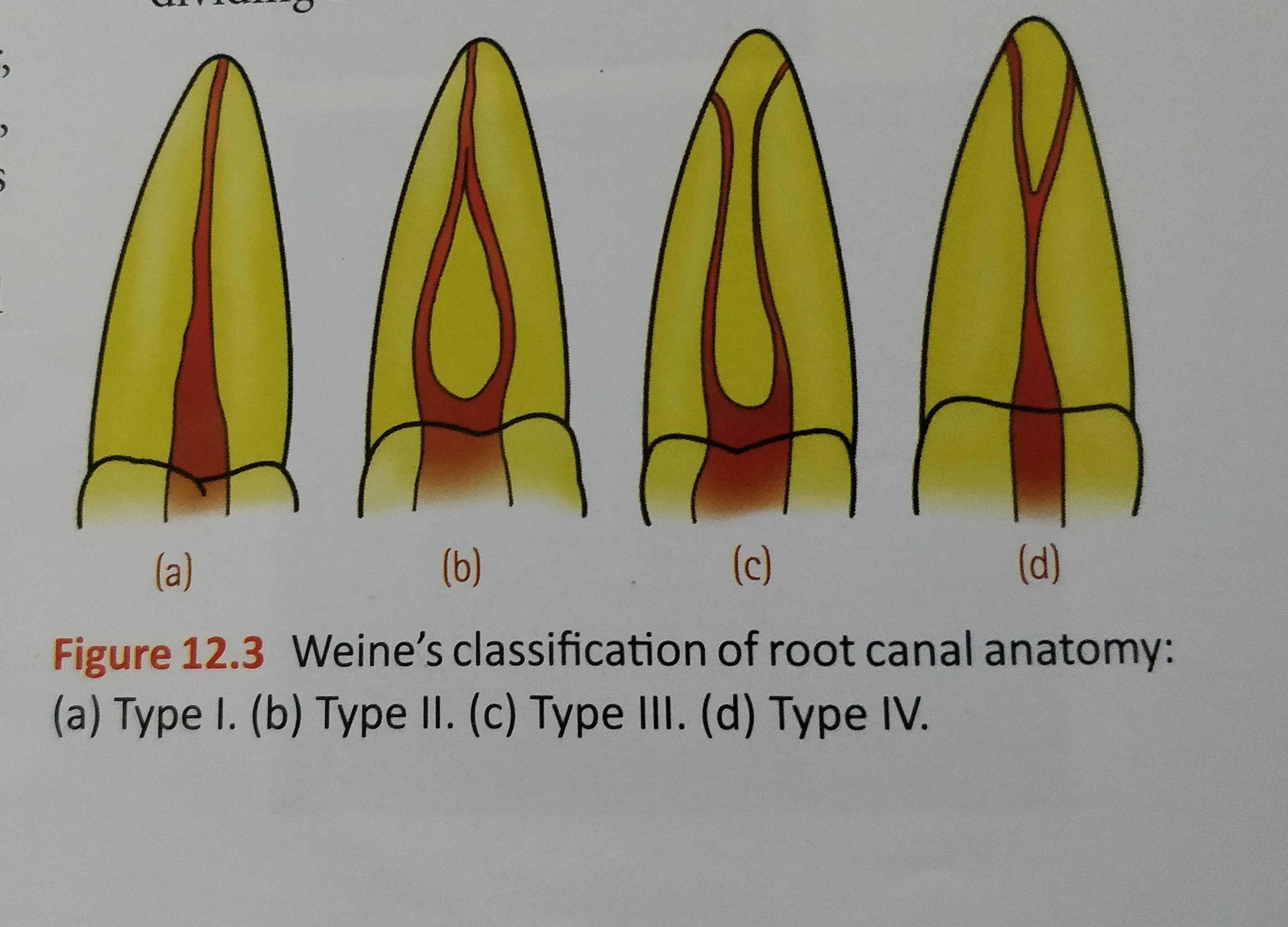
- Weine’s classification :
- Type I : Single canal from pulp chamber to apex
- Type II : Two canals leaving from the chamber and merging to form a single canal short of the apex
- Type III : Two separate and distinct canals from chamber to apex
- Type IV : One canal leaving the chamber and dividing into two separate and distinct canals
- Classification based on canal cross – section:
- Round (circular)
- Oval
- Long oval
- Flattened (flat/ribbon)
- Irregular
References:
- Dental notes
- Grossman’s Endodontic Practice (13th edition)

