🔹 Introduction:
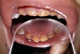
👉🏻 The affected teeth (both dentitions) are grey to yellow brown with broad crowns and constriction at cervical area resulting in tulip shape.
👉🏻 The enamel is easily broken, exposure of dentin ➡️ accelerated attrition.
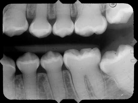
👉🏻 Normal non-mineralized pulp chambers and canals
👉🏻 This condition is inherited in an autosomal dominant pattern, as a result of mutations on chromosome 4q21, in the dentine sialophosphoprotein gene (DSPP).
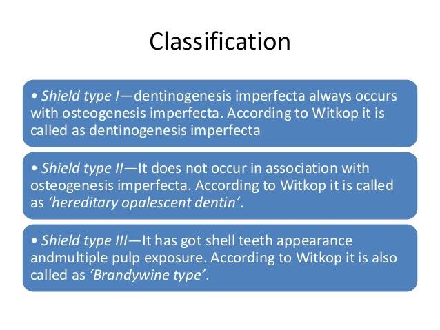
🔹Classification:
🔹Radiographic Features:
🔹Histopathological Features:
👉🏻 Mesodermal disturbance.
👉🏻 Dentin composed of irregular tubules with large areas of unclassified matrix. The tubules are larger in diameter & less numerous.
👉🏻 Odontoblasts – dentinal matrix not layed properly & they are entrapped within this matrix.
CHEMICAL FEATURES:
• Increased water content (60 times the normal)
• Decreased inorganic content
🔹Treatment: Cast Metal crowns & Jacket crowns
Dr. Mehnaz Memon🖊
References:
- Shafer’sTextbook Of Oral Pathology (7th Ed)
- Image Source: International Journal of Medicine Research; Dr G’s Toothpix; Google

