Also termed:
- Vincent’s infection
- Trench mouth
- Fusospirochetal gingivitis
- Phagedenic gingivitis
Etiology:
1. Role of bacteria:
- B. Vincentii
- F. Nucleatum
- P. Intermidia
- Treponema species
2. Predisposing factors:
| LOCAL | SYSTEMIC |
| Smoking | Poor nutritional status |
| Psychological stress | Leukemia, AIDS |
| Poor oral hygiene | Syphilis |
| Marginal gingivitis | Aplastic Anemia |
| Faulty restoration | Vitamin C, B2 deficiency |
| Deep periodontal pockets | Inadequate sleep |
| Local trauma | Immunosuppressant |
Clinical Features:
1. Age: Young & Middle-aged
2. No sex predilection
3. Site:
a. Interdental papillae
b. Free gingival margin
c. Crest of gingiva
d. Soft palate & tonsillar areas-Vincent’s Angina
4. Diagnostic Triad –
| Pain |
| Interdental ulceration |
| Gingival bleeding |
5. Signs & Symptoms:
a. Onset is sudden with pain, tenderness, profuse salivation & peculiar metallic taste.
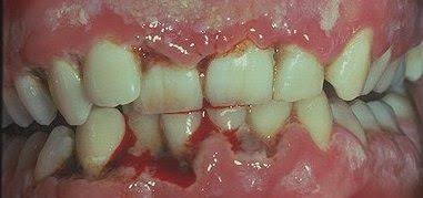
b. Spontaneous bleeding
c. Fetid Odor
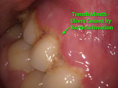
d. The interdental papillae are blunted, inflamed, edematous and hemorrhagic. Show Punched out crater like necrotic areas covered by grayish pseudo membrane
e. Lymphadenopathy, fever & malaise
f. Involvement of PDL leads to NUP
g. Cancrum oris (Noma)
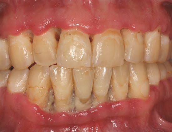
h. Gingiva is stained and teeth seem slightly to be extruded & moveable
i. Patient is unable to eat properly
• Histopathology:
– Interdental papillae show surface ulceration covered by a fibrinopurulent membrane.
– Underlying connective tissue shows acute/mixed inflammatory infiltrate along with extensive hyperemia.
• Treatment:
ANUG results from impaired host response to a potentially pathogenic microflora. Compared to other PDL diseases, ANUG resolves quickly after removal of bacterial infection.
1. Alleviation of acute inflammation by reducing the microbial load and removal of necrotic tissue.
2. Treatment of underlying chronic disease.
3. Alleviation of generalized symptoms.
4. Correction of systemic conditions or factors that contribute to the initiation and progression of gingival changes.
Sequence of treatment:
First Visit:
– Evaluation
– Comprehensive medical history
– H/O acute disease its onset and duration
– Examination of the patient’s oral cavity
Treatment of acutely involved areas:
– Isolate with cotton rolls. Apply topical anesthetic and after 2-3 min. the area are gently swabbed with moistened cotton pellet to remove pseudo membrane and surface debris.
– Cleanse the area with warm water and remove superficial calculus with ultrasonics.
– Contraindication:
• Subgingival scaling & curretage.
• Extractions & surgery
– 4 weeks: Waiting Period
– Patient with severe cases, lymphadenopathy/other systemic conditions – Antibiotic regimen (Amoxycillin, erythromycin & Metronidazole) & NSAID’s.
Patient Instructions:
1. Avoid tobacco, alcohol
2. Rinse with a glassful of an equal mixture of 3% H2O2 and warm water every 2 hours or twice daily with 0.12% chlorhexidine solution.
3. Get adequate rest. Avoid exertion
4. Brushing with ultrasoft brush with a bland dentifrice for removal of surface debris only.
5. Follow-up after 1-2 days.
• Second visit:
– Evaluate the condition
– Pain usually subsides but the gingival margins are still erythematous.
– Scaling can be done
– Follow-up after 5 days
• Third visit:
– Plan for the management of patient’s periodontal conditions.
– Plaque control procedures
– Nutrition
– Cessation of habit
– Chlorhexidine rinses for 2-3 weeks
– Recall after a month.
References: Essentials Of Periodontology by S Sahitya Reddy; Carranza’s clinical periodontology; Internet images


[…] https://dentowesome.wordpress.com/2020/04/07/271/ […]
LikeLike