ODONTOTOMY

The extraction of a tooth requires the separation of its attachment to the alveolar bone via the crestal and principal fibers of the periodontal ligament. Also involved is a process of expanding the alveolar socket.

After the tooth has been removed, the form of alveolar process is restored by finger pressure. Bleeding is arrested by means of a pressure pack placed between the jaws, and the wound is allowed to fill with a blood clot.

EXODONTIC PROCEDURES:

The following techniques may be used for tooth removal:

  1. the forceps technique
  2. the elevator technique (https://dentowesome.wordpress.com/2020/06/18/dental-elevators/)
  3. the open view technique

Odontotomy can be used to facilitate any of these three procedures.

ODONTOTOMY

  • In some instances the extraction may be simplified by cutting a tooth apart. This is especially desirable in the case of multi-rooted deciduous or permanent teeth with severely divergent roots.
  • It is also useful in cases where the crown is so decayed that only a shell remains. (As shown in the figure👇🏻)
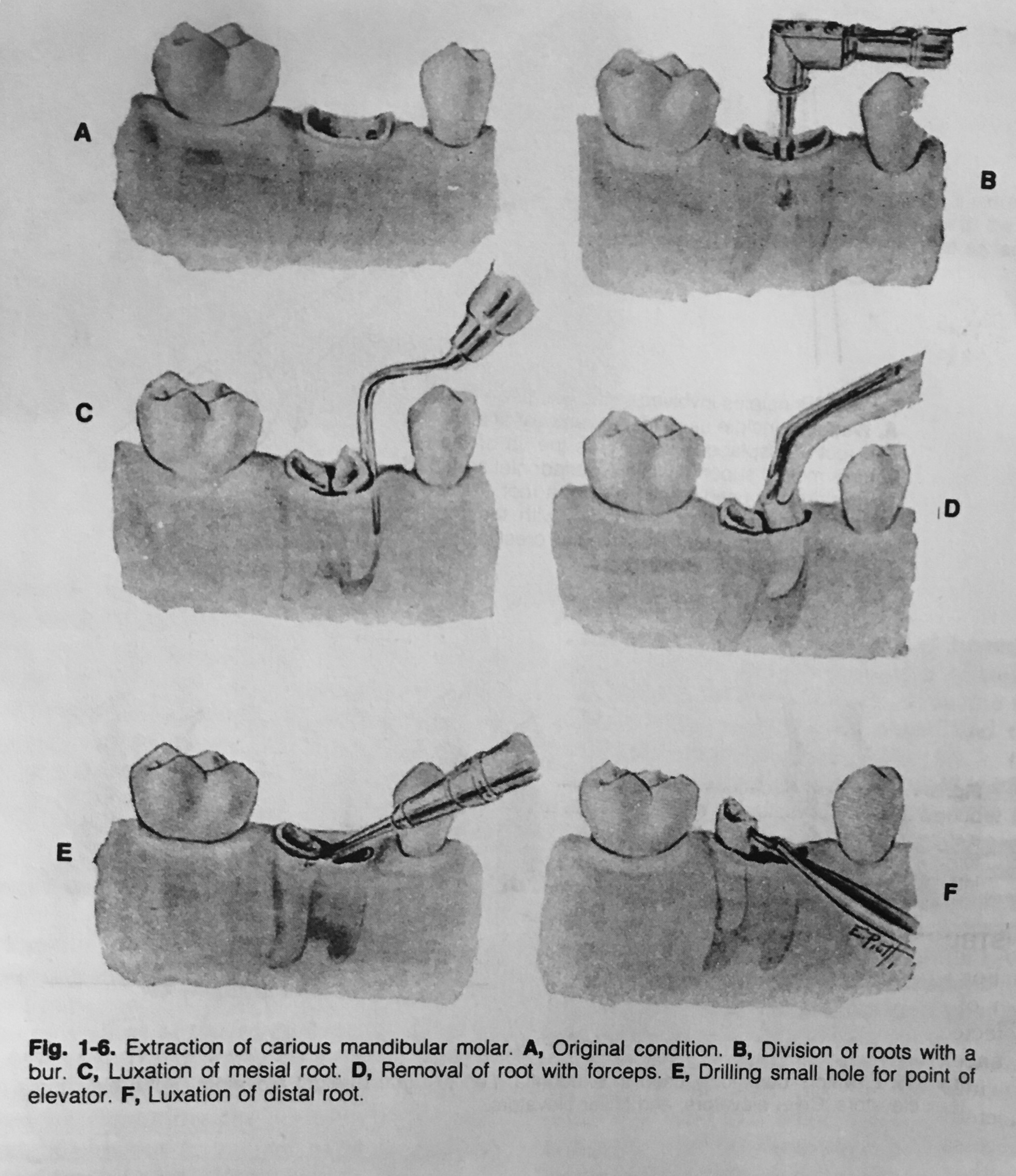
  • If the roots are divergent, the bur cut, instead of being vertical, may be made parallel with one of the roots, and this part of the tooth then is extracted first.
  • The other roots may be removed either with forceps or an elevator.

Dentowesome|@drmehnaz🖊


References: Textbook of Oral Surgery – Daniel M Laskin

POSTOPERATIVE CARE AFTER DENTOALVEOLAR SURGERY

Good aftercare to prevent complications and unnecessary suffering, with loss of valuable time, is as important as a good operation.

The main purpose of aftercare is to expedite healing and prevent or relieve pain and swelling.

Rest is necessary for the prompt healing of wounds. Ambulatory patients should be directed to go home & remain quiet for several hours, preferably sitting in a comfortable chair or, if lying down, keeping the head elevated on several pillows.

  • Only liquids and soft solids should be eaten the first day. They may be warm or cold but not extremely hot.
  • Food intake should not begin until several hours after surgery to avoid disturbing the blood clot.
  • If the extractions were limited to one side, chewing can be done on the unoperated side, but when local anesthesia has been used, chewing should be avoided until sensation has returned.
  • Fluids should be taken in greater amounts than usual to prevent dehydration from limited food intake.
  • A normal diet should be resumed as soon as possible, since this facilitates healing.
  • The teeth should be brushed as usual, and on the day after surgery rinsing of the mouth should begin.
  • A saline solution (1/2 teaspoon of salt in a glass of warm water) is best for this purpose.
  • Commercial mouthwashes if used should be diluted with water due to the high alcohol content that can irritate the wound.
  • Hydrogen peroxide rinses should not be used initially as this agent can remove the blood clot.❌
  • Some degree of postoperative pain accompanies many exodontic procedures and begins after the effects of anesthetic have left. This is considered a normal response to the unavoidable trauma of surgery.
  • In most cases, such pain lasts no more than 12 to 24 hours, although a traumatic periostitis may persist for several days.
  • Ordinarily this type of pain can be controlled by the use of cold packs (30 minutes per hour) during the first 24 hours & the proper administration of analgesic drugs.
  • For mild pain, as after a routine extraction, one of the antipyretic analgesics is usually adequate.
  • For moderate pain, such as after removal of an impacted tooth, a drug such as codeine or meperidine (Domerol) should be used.
  • Narcotics are needed only in rare instances.
  • The combination of a sedative drug with an analgesic agent can also be used but barbiturate alone should never be used to relieve pain as it can result in mental disorientation in a patient suffering from extreme pain.
  • The degree of swelling that occurs is generally in direct proportion to the degree of surgical trauma.
  • The application of cold to the operated site helps diminish postoperative swelling. If a rubber ice bag is not available, the ice can be placed in a plastic bag.
  • Cold can be applied intraorally by holding an ice cube in the mouth.
  • Pressure dressings can also be beneficial in limiting postoperative swelling.
  • Once swelling has reached the maximum (usually after 24 to 48 hours), cold is no longer effective, and heat, in the form of moist compresses, should be applied. It too should be used only 30 minutes per hour. The area should be lubricated with petroleum jelly to avoid burning the skin.
  • Intraoral heat is achieved by the use of hot isotonic saline rinses.
  • Cigarette smoking should be avoided after tooth extraction because it has been shown to increase the incidence of alveolar osteitis

Dr. Mehnaz Memon🖊


References: Textbook of Oral Surgery – Daniel M Laskin

How to prevent excessive bleeding during Dentoalveolar Surgery❓

Types of bleeding are:

  1. Primary (during or immediately after surgery)
  2. Reactionary (Upto 48 hours due to a defective suture or as clot in the vessels has got disturbed)
  3. Secondary (8-14 days due to wound getting infected and capillaries have eroded surfaces)

To prevent excessive blood loss during surgery we need to understand the source of bleeding i.e. possible reason for bleeding.


Dr. Mehnaz Memon🖊

Complications of Local Anesthesia

🔷 Local Complications:

  • Needle breakage
  • Persistant anesthesia or paresthesia
  • Facial Nerve Paralysis
  • Trismus
  • Soft-tissue injury
  • Hematoma
  • Pain on injection
  • Burning on injection
  • Infection
  • Edema
  • Sloughing of tissues
  • Post-anesthetic intraoral lesions

🔷 Systemic Complications:

  • Vasodepressor syncope
  • Over dosage (toxic reaction)
  • Allergy
  • Psychogenic reactions

Clinical Tip💡

👉🏻Several preventive measures can be followed:📝

1) Select a local anesthetic with a duration of action that is appropriate for the length of the planned procedure.

2) Advise the patient and accompanying adult about the possibility of injury if the patient bites, sucks or chews on the lips, tongue and cheek. They should delay eating and avoid hot drinks until the effects of the anesthesia are totally dissipated.

3) Reinforce the warning with patient stickers and by placing a cotton roll or rolled up gauze (“Bite on the ghost”) in the mucobuccal fold if anesthesia symptoms persist.

4) The management of soft tissue trauma involves reassuring the patient and parent (it’s okay if the tissue turns white), allowing up to a week for the injury to heal, and lubricating the area with petroleum jelly or antibiotic ointment to prevent drying, cracking and pain.

References: Internet; Handbook of LA, 6e by Stanley F Malamed


Dr. Mehnaz Memon🖊

Inferior Alveolar Nerve Block (IANB)


➡️ The inferior alveolar nerve along with its 2 terminal branches – the mental and incisive nerves innervates the mandibular teeth as well as the buccal soft tissue anterior to 1st molars. This also provides anesthesia to the lip and chin on the side of injection.

➡️ Buccal Nerve – Buccal mucosa and cheek from 3rd molars to 1st molar.

➡️ Structures/Landmarks:

  1. Coronoid Notch
  2. Internal Oblique Line
  3. Lingula
  4. Pterygomandibular raphe
  5. Retromolar triangle
  6. Retromolar fossa
  7. Mandibular fossa
  8. Posterior border of ramus
  9. Occlusal plane

➡️ A plane passing through the deepest portion of coronoid notch parallel to occlusal plane will pass just over the lingualar notch. This determines the height of injection.

➡️ The internal oblique line serves as the attachment for deep tendon of temporalis muscle is important as it determines the AP point of injection.

➡️ Also the distance from internal oblique line to lingula is 9-11 mm in most adults provides the clinician a reference of depth of needle penetration.

➡️ The finger is placed to the depth of coronoid notch to determine vertical height of injection.

➡️ The barrel of the syringe is positioned from opposite the premolar region parallel to occlusal plane with the needle at the predetermined height.

➡️ Posterior to Internal Oblique line and anterior to lingula – contact of the needle.


▪️Clinical Tip👇🏻

References: Internet; Handbook of LA, 6e by Stanley F Malamed; MONHEIMS LOCAL ANESTHESIA AND PAIN CONTROL IN DENTAL PRACTICE 7ED