@dr.mehnaz🖊
References: Shafer’sTextbook Of Oral Pathology
References: Shafer’sTextbook Of Oral Pathology


References: Shafer’sTextbook Of Oral Pathology


Dr. Mehnaz Memon🖊
References: Shafer’sTextbook Of Oral Pathology
By KRITI NAJA JAIN:-
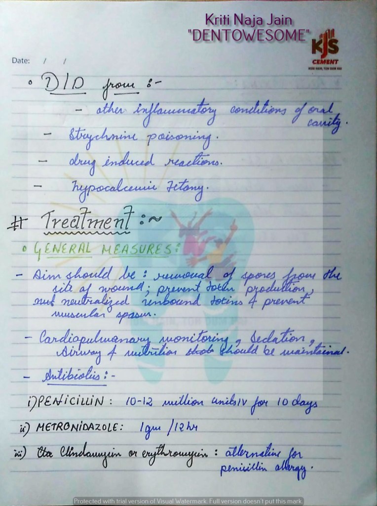
REFERENCES:-
BY Dr. KRITI NAJA JAIN :-
1. FIBROUS DYSPLASIA :-
Def:- Fibrous dysplasia is an uncommon nonhereditary, developmental anomaly of the bone due to a defect in osteoblastic differentiation and maturation.
HISTOPATHOLOGY:


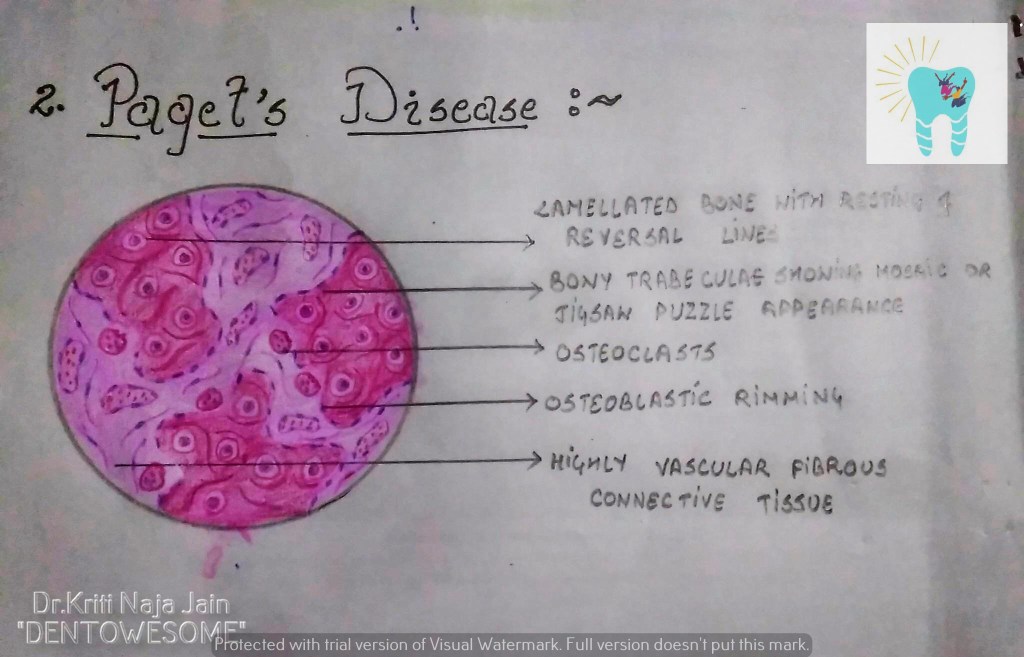
2. PAGET’S DISEASE (OSTEITIS DEFORMANS):-
Def:- Paget’s disease of bone is a condition characterized by abnormal and anarchic resorption and deposition of bone, resulting in distortion and weakening of the
affected bones.
HISTOPATHOLOGY:-

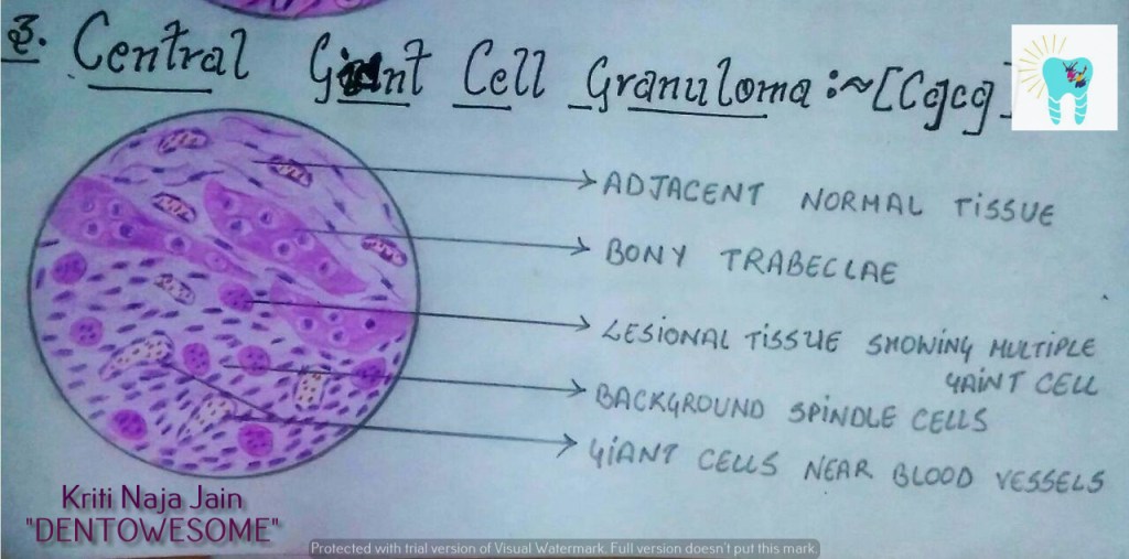
3. CENTRAL GAINT CELL GRANULOMA(GIANT CELL LESION; GIANT CELL TUMOR):-
Def :- Central giant cell granuloma (CGCG) is an uncommon, benign and proliferative lesion whose aetiology is not defined. Central giant cell granuloma is a relatively common benign intraosseous destructive giant cell lesion, which often affects the anterior part of the jawbone. By seeing clinical and radiographically , CGCG is divided into two types:-
1. Nonaggressive lesions make up most cases, exhibit few or no symptoms, demonstrate slow growth, and do not show cortical perforation or root resorption of teeth involved in the lesion.
2. Aggressive lesions are characterized by pain, rapid growth, cortical perforation, and root resorption. They show a marked tendency to recur after treatment, compared with the nonaggressive types.
HISTOPATHOLOGY:-

REFERENCE:-
1.Maji Jose 2nd edition
Source: Shafer’s Textbook of Oral Pathology, 8th Edition
BY: Dr.Kriti Naja Jain :-
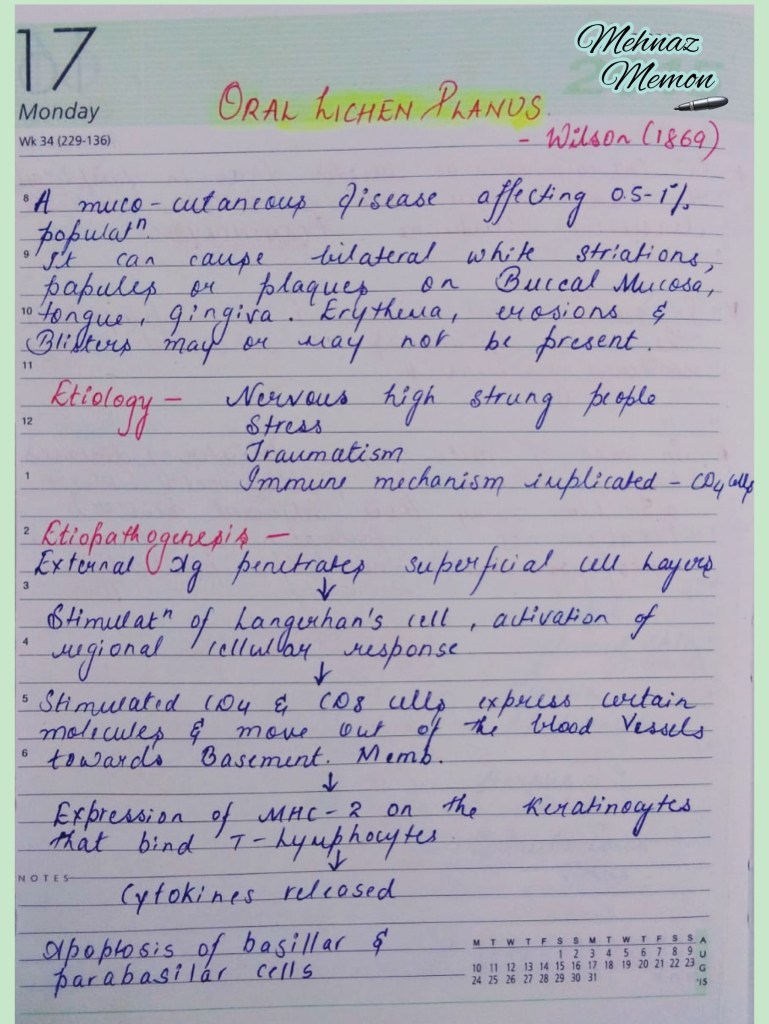
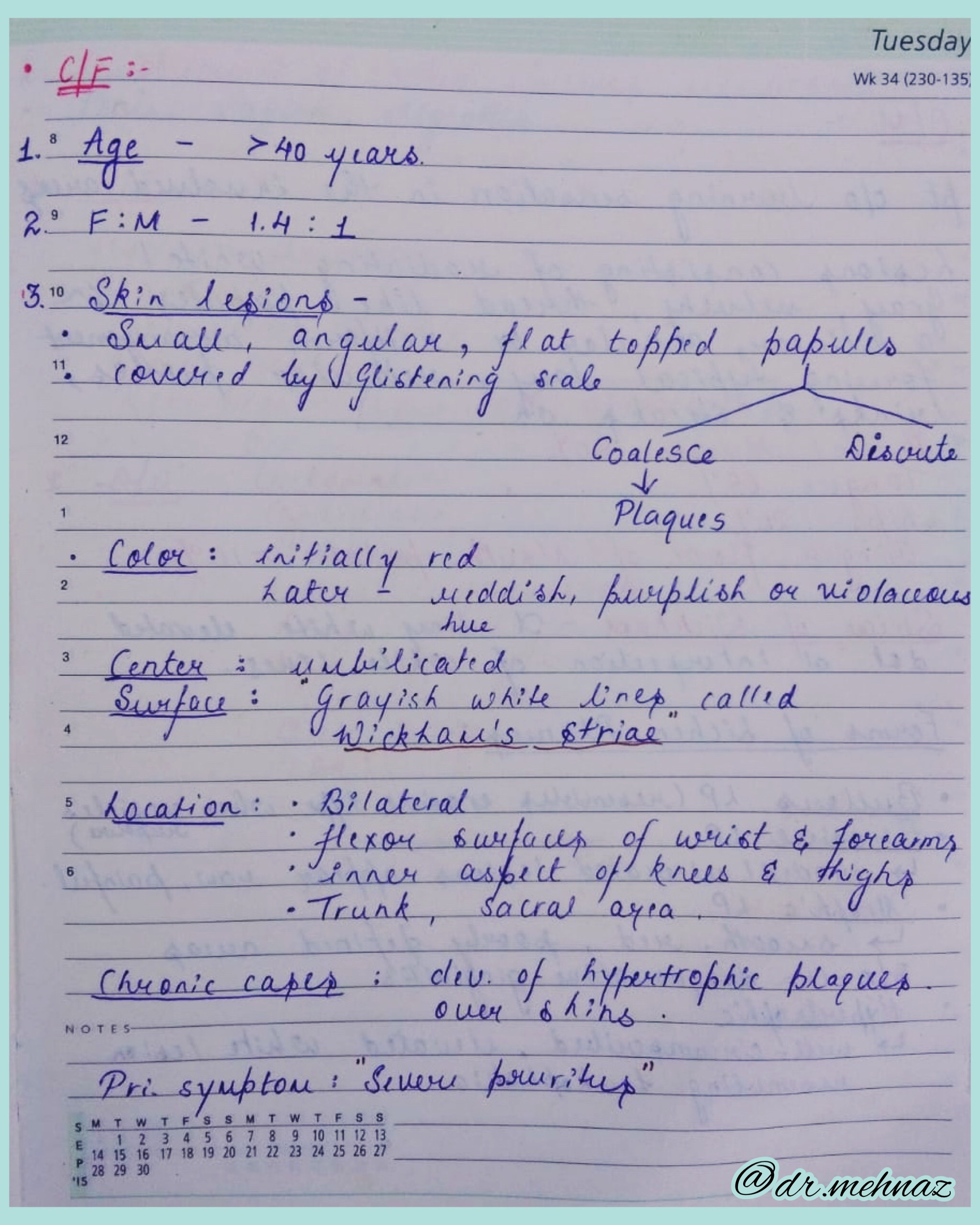
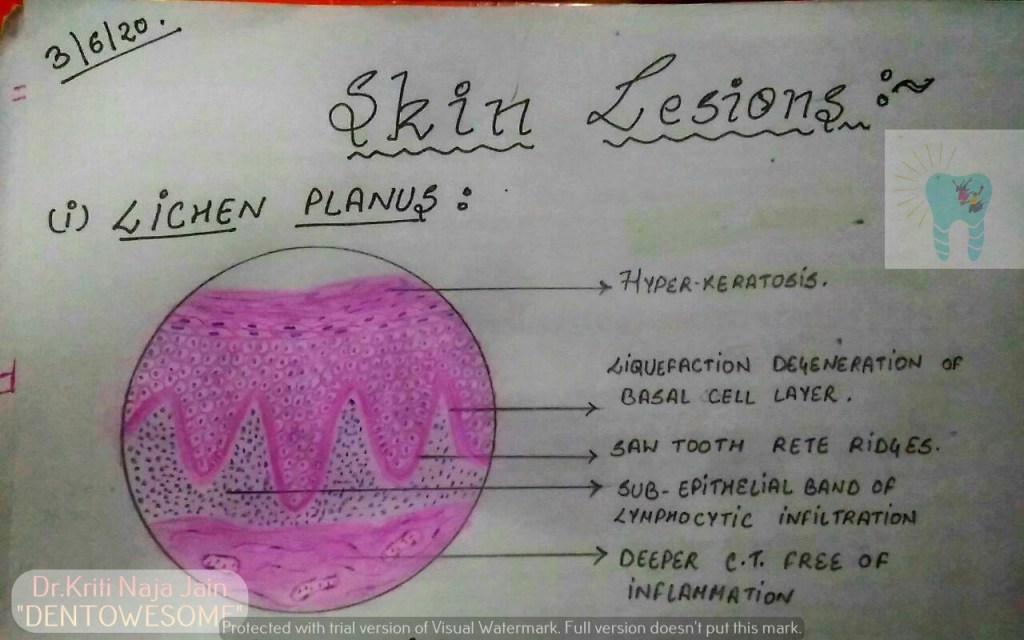
1.LICHEN PLANUS:-
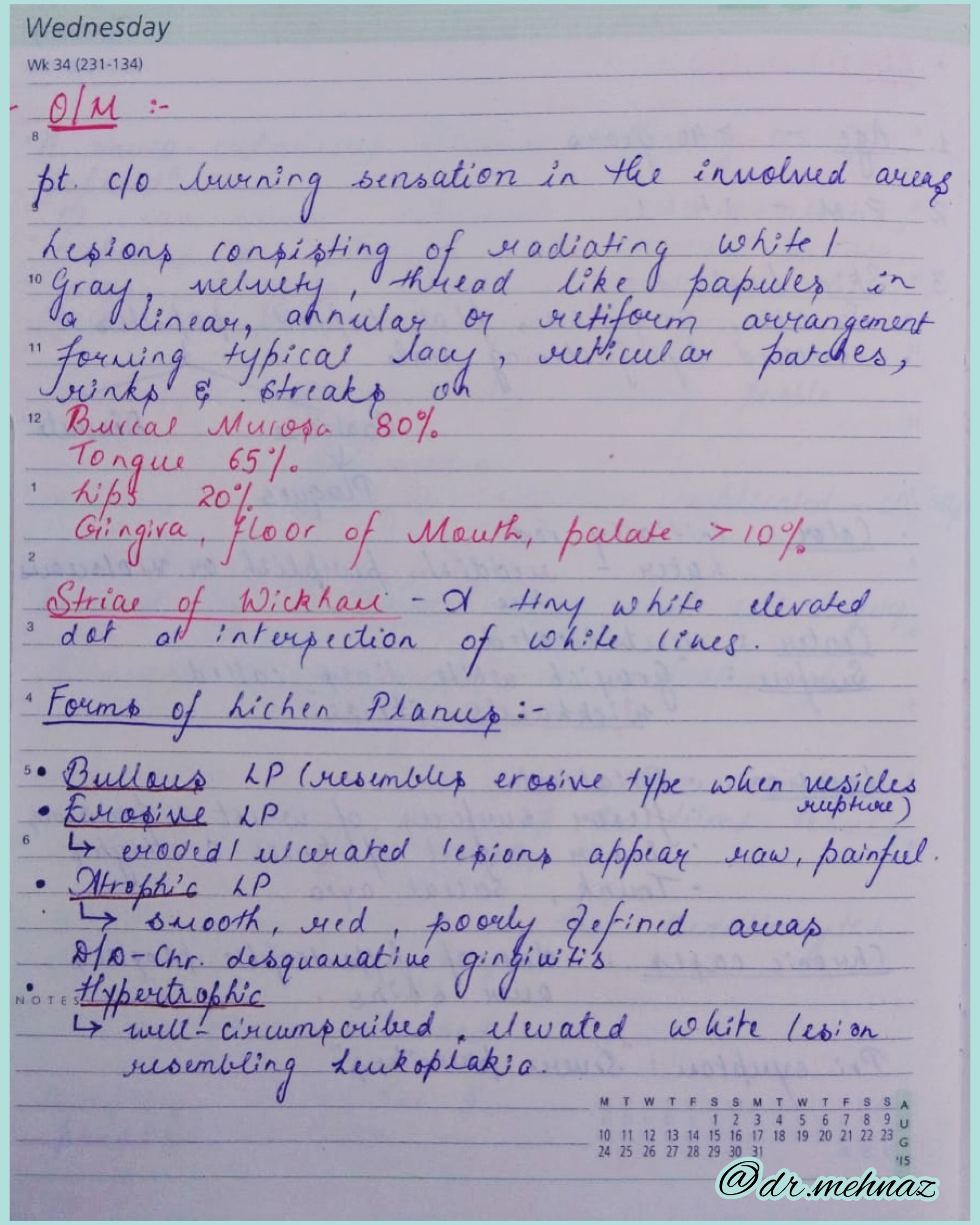
*Lichen planus is a chronic mucocutaneous disorder manifested in a various forms in the oral cavity.
*The most characteristic pattern is” RETICULAR TYPE” with the interlacing white stripe called “WICKHAM’S STRIAE”.
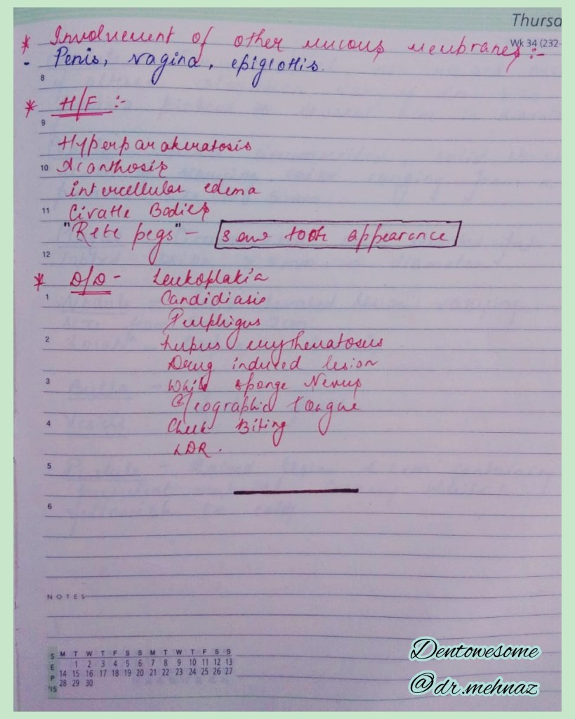
*HISTOPATHOLOGY:-

2.PEMPHIGUS :-
Pmphigus is a tissue specific autoimmune disease affecting the skin and mucosa. Clinical manifestations is in the from of “vesiculobullous lesions” that rupture to form ulcer and erosions .
*Vesiculobullous lesions develop due to immune mediated acantholysis causing intraepithelial vesicle formation.
*HISTOPATHOLOGY :-


3.PEMPHIGOID :-
Pemphigoid is a vesiculobullous lesions that develop due to an autoimmune reaction directed against some components of basement membrane.
*This results in seperation of epithelium from the connective tissue with sub epithelial vesicles formation .
*Bullous pemphigoid and cicatricial pemphigoid are two different types of pemphigoid lesions.
*HISTOPATHOLOGY:-


REFERENCE:-
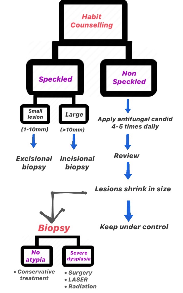
LEUKOPLAKIA

3) Surgical Approach
References: Ghoms, Textbook of Oral Medicine
Dr. Mehnaz Memon🖊

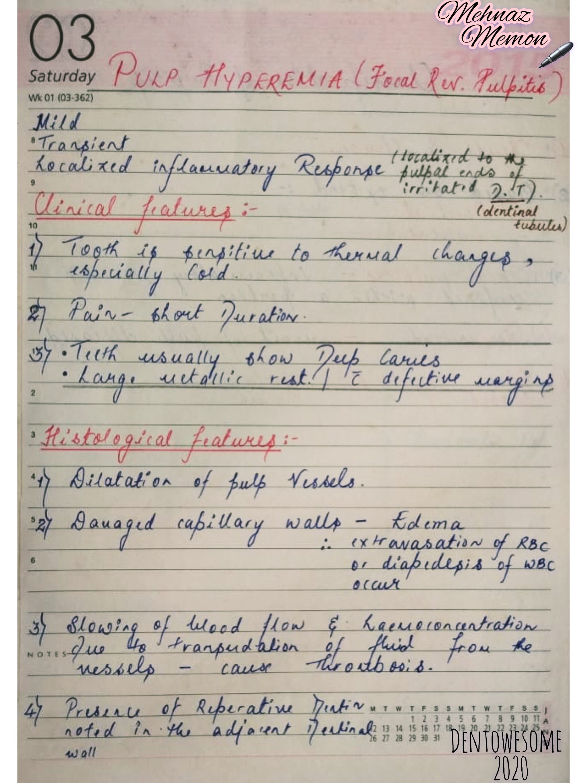
1) Pulp Hyperemia (Focal Reversible Pulpitis)
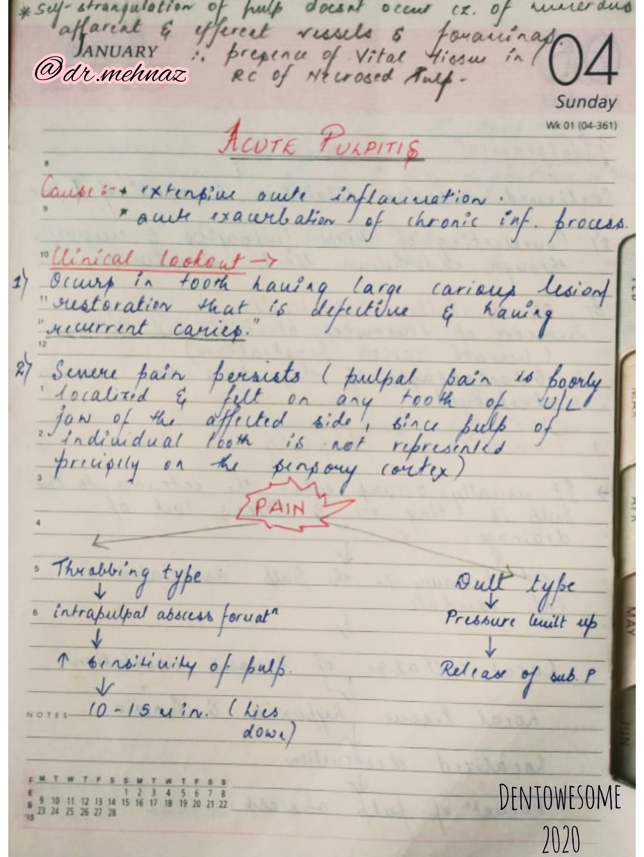
2) Acute Pulpitis


3) Chronic Pulpitis
References: Shafer’sTextbook Of Oral Pathology
Reference:-
1.Shafers 8th e
2. Neville 3rd e
3.Purkit’s 3rd e
