BULLOUS PEMPHIGOID

A chronic, autoimmune, sub-epidermal blistering skin disease that rarely involves mucous membrane.

🔹Clinical Features:

Age: Elderly (>60 years)

Skin Lesions:

  • Generalised non-specific rash, commonly on Limbs.
  • Appears urticarial/eczematous; persist for several weeks to months.
  • Vesicles & bullae arise in prodromal skin lesion as well as Normal skin.
  • The blisters are thick walled and don’t rupture easily.
  • ruptured blisters are usually sensitive and painful, have raw eroded area which heals rapidly.

Oral Manifestations:

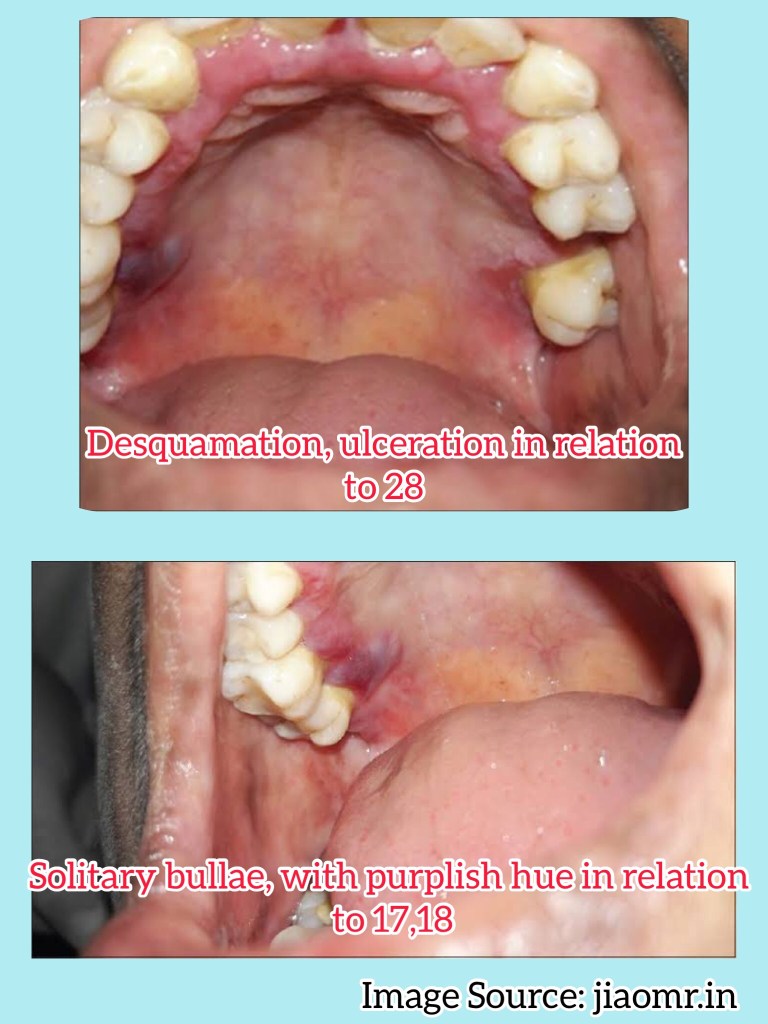
Vesicles appear gingivally👇🏻

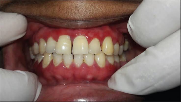
Erythematous & desquamate as result of minor frictional trauma

👉🏻 Oral lesions comprise of bullae/vesicle that rupture to form erosions and ultimately leave out ulcerations

👉🏻 Other sites:

  • Buccal Mucosa
  • Tongue
  • Floor of the mouth
  • Palate

🔹Diagnosis:

Apart from evaluating history, clinical presentation, histopathological analysis is carried out followed by direct immunofluorescence study for the differential diagnosis and confirmation of the condition.
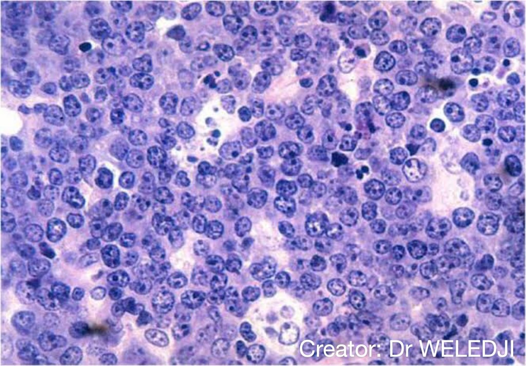
👉🏻Histopathology:

  • Acanthotic mucosa
  • Subepidermal non-specific vesicles with fibrous exudate

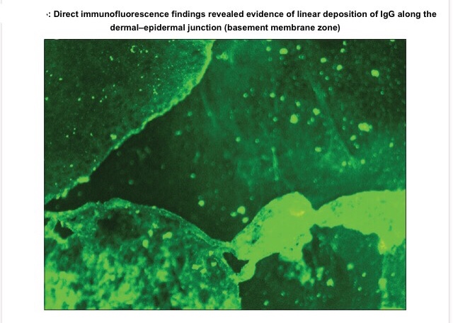
👉🏻Direct immunofluorescence is found to be the gold standard test. Deposition pattern of different types of immunoreactants differentiates the various immune-mediated diseases. Direct immunofluorescence shows presence of IgG and C3 deposits along the basement membrane zone.

©️jiaomr.in
👉🏻Electron Microscopy: In bullous pemphigoid (BP), the 180 kD antigen (BPAG2) was shown by immuno-EM to be a transmembrane molecule and to possess an autoantibody binding site outside the cell, suggesting a major pathogenic role for the BPAG2 in blister formation.

🔹Differential Diagnosis:

• Mucous membrane pemphigoid can be differentiated from BP by its predominant involvement of mucosal surfaces and positive Nikolsky’s sign.

• Lichen planus pemphigoides is clinically differentiated by the presence of lichen planus lesions in addition to tense blisters.

• Nikolsky’s sign is present in case of pemphigus and cicatricial pemphigoid, but not in the case of BP.

🔹 Treatment:

👉🏻Treatment is based on the degree of cutaneous and oral involvement. Mostly, topical steroid (clobetasol propionate) gives satisfactory result in case of smaller area of skin involvement, whereas larger area of skin involvement and recurrent cases are treated satisfactorily with systemic steroids and immunosuppressive agents.

👉🏻Recommended dosage for oral prednisolone is 0.3–1.25 mg/kg body weight/day, controls disease within 1–2 weeks, followed by which the dose is tapered. Dexamethasone (100 mg in 500 mL 5% dextrose i.v. over 2–3 h for three consecutive days) is the preferred steroid for pulse therapy, either administered alone or in combination with cyclophosphamide.

Other drugs for treating BP include new antibody modulators, rituximab 375mg/m2weekly over 4 weeks and omalizumab subcutaneously 300–375 mg for every 6 weeks.

👉🏻Higher doses of systemic corticosteroids seem to be associated with higher mortality rates, which led to the addition of corticosteroid-sparing agents to the treatment of BP. The most frequently used immunosuppressive agent is azathioprine (0.5–2.5 mg/kg body weight/day). Others being cyclophosphamide, methotrexate, cyclosporine A, combination tetracycline/minocycline along with nicotinamide, and more recently, mycophenolate mofetil, a DNA synthesis inhibitor, and methotrexate, a folate antagonist.

👉🏻IVIg – A dose of 1–2 g/kg for five consecutive day cycle of 0.4 g/kg/day, although a 3-day cycle may be used in cases that are nonresponsive to conventional therapy.

Dr. Mehnaz Memon🖊


References:

  1. http://www.jiaomr.in/article.asp?issn=0972-1363;year=2018;volume=30;issue=4;spage=432;epage=435;aulast=Aparna
  2. https://www.cidjournal.com/article/S0738-081X(00)00178-4/abstract
  3. Shafer’s Textbook of Oral Pathology, 7th edition
Read More »

APERT SYNDROME

🔖 Acrocephalosyndactyly. The condition is autosomal dominant i.e, one copy (out of 2) of the defective gene is sufficient to cause the abnormality in the offspring.

🔹Characteristics:

  • Craniosynostosis (premature fusion of the skull bones)
  • Craniofacial anomalies
  • Syndactyly (Fusion of fingers and toes)

🔹Etiology:

🔹What are the Symptoms and Signs of Apert Syndrome?

The various clinical features include:

  1. Asians affected
  2. Acrocephaly, Brachycephaly, flat occiput & prominent forehead.
  3. Late closing fontanels
  4. Low set ears, hearing loss
  5. Eyes: Down slanting of palpebral fissures, Widely spaced eyes(Hypertelorism), Shallow orbits, Abnormally bulging eyes (Exophthalmos)
  6. Nose: Depressed Nasal bridge, short, wide with bulbous tip, Parrot beaked appearance, Atresia
  7. Jaw:
  • Prominent Mandible
  • Maxillary hypoplasia
  • Drooping angles of mouth
  • High arched palate
  • Bifid uvula
  • Cleft palate
  • Crowded upper teeth
  • Malocclusion
  • Delayed & ectopic eruption
  • Shovel shaped incisors
  • Supernumerary teeth
  • V-shaped maxillary dental arch
  • Bulging alveolar ridges

8. Partial to complete fusion of digits: 2-4th digits – MITTEN HANDS & SOCK FEET; Sole – supinated

9. Intelligence – Normal

10. ⬆️ intracranial pressure – optic atrophy, papilledema

11. Hyperhidrosis

12. Cardiovascular system: Atrial Septal defect, Ventricular septal defect, Patent ductus Arteriosus

🔹How do you Treat Apert Syndrome?

Standard Therapies

The treatment of Apert syndrome aims at addressing the specific symptoms that may be present in the particular individual. Treatment is usually symptomatic and supportive.

  • Craniosynostosis and associated hydrocephalus in some cases may give rise to an abnormally increased pressure within the skull (intracranial pressure) and on the brain. In these cases, early surgery (within 2 to 4 months after birth) becomes necessary to correct the defects in the skull and facial bones.
  • Insertion of a tube (shunt) to drain excess cerebrospinal fluid (CSF) away from the brain and into another part of the body like the abdomen where the CSF can be absorbed can be done to relieve associated hydrocephalus (fluid accumulation in the brain).
  • Early repair and reconstructive surgery may also be done in some infants with Apert syndrome to address craniofacial abnormalities.
  • Other defects such as those of heart, eye and ear defects may also need correction.

Dr. Mehnaz Memon🖊


References:

  1. https://www.medindia.net/amp/patientinfo/apert-syndrome.htm
  2. Shafer’s textbook of Oral Pathology – 7th Ed.

PAGET’S DISEASE

Introduction & Etiology👆🏻
Clinical Features & Phases of PDB..👆🏻
Clinical Features contd…👆🏻
O/M of PDB..👆🏻
Radiographic Features of PDB..👆🏻
Histological Features of PDB…👆🏻
Lab. Findings & Treatment of PDB..👆🏻

References: Shafer’s Textbook of Oral Pathology 7th Edition


Dr. Mehnaz Memon🖊

BURKITT’S LYMPHOMA

🌏 African Jaw Lymphoma

🌏 The endemic form is linked to malaria and to the Epstein-Barr virus (EBV), a common virus that also causes glandular fever.

🌏 A tumour peculiar to children of central Africa was reported by Denis Burkitt in 1950. It is a lymphoreticular cell malignancy.

▪️It is a high grade B-cell neoplasm & has 2 major forms:

🔷 Clinical Features:

1. Age – between 6 & 9 years

2. Sex – M:F – 2:1

3. Site: In African form (Endemic),

  • Maxilla > Mandible
  • Spreads to floor of orbit
  • Molar area
  • More than one quadrant is involved

American form (Non-endemic) – Oral: only 1 quadrant involved. Other: Mainly involves Abdomen.

4. Onset & progress – fast growth with tumor doubling time of <24 hours.

5. Symptoms –

  • Swelling of jaws, abdomen & paraplegia
  • Loosening of teeth
  • Abdominal tumors
  • Bowel obstruction

6. Sign – Lymphadenopathy (Non-tender)

🔷 Oral Manifestations:

  • Gingiva and mucosa – swollen, ulcerated, necrotic
  • Facial asymmetry
  • Teeth are pushed out of their sockets

🔷 Radiographic Features:

🔷 Histological Features:

1. Monoclonal proliferation of B-lymphocytes characterized by small non-cleaved cells.

2. Burkitt cells are homogenous in size & shape with –

  • round to oval nuclei
  • coarse chromatin
  • Multiple nucleoli
  • Basophilic vacuolated cytoplasm with neutral fat

🔷 Differential Diagnosis:

  • Non-hodgkins lymphoma
  • Cherubism
  • Osteosarcoma

🔷 Treatment: Intrathecal Chemotherapy

References: Shafer’sTextbook Of Oral Pathology


Dr. Mehnaz Memon🖊


Ann Arbor Staging System for Lymphoma: https://dentowesome.wordpress.com/2020/06/15/ann-arbor-staging/