Rhinosporidiosis

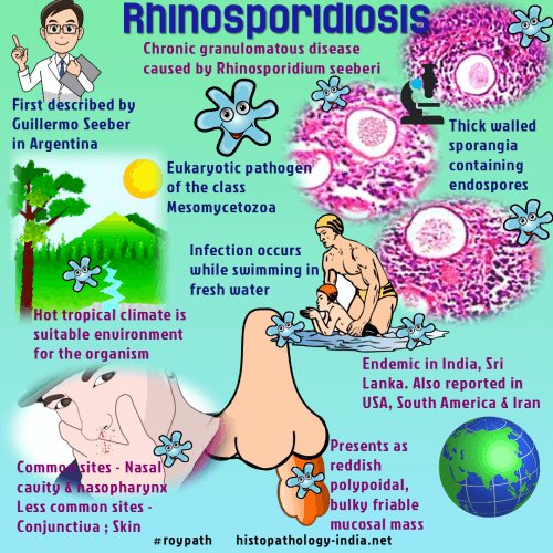
(i) Rhinosporidiosis is a chronic granulomatous disease characterised by formation of friable polyps, usually confined to the nose, mouth or eye.

Nasal polyp

(i) Causative agent is Rhinosporidium seeberi.

Rhinosporidium seeberi

(iii) More than 80% cases are reported in India and Sri Lanka.

(iv) The mode of infection is not known but most infections occur in males who have frequent contact with stagnant water or aquatic life.

ORAL MANIFESTATIONS

Oronasopharyngeal lesions appear as soft red polypoid growth which spread to pharynx and larynx.

• These lesions often contain mucoid discharge and are vascular.

LAB DIAGNOSIS

• The fungus has not been cultivated.

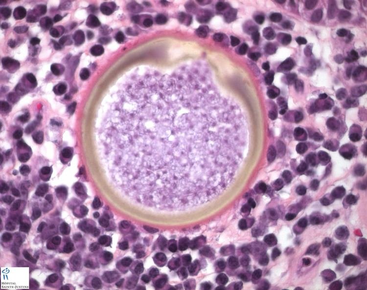
• Diagnosis depends on the demonstration of sporangia

•Tissue sections stained with H & E stain show large number of endospores within the sporangia embedded in a stroma of connective tissue and capillaries.

The sporangium (10-200 um) contains thousands of endospores (6-7 µm in diameter)

Source – textbook of microbiology for dental students c p baveja and Google images

DERMATOPHYTES

(1) Dermatophytes are a group of fungi that infect only superficial keratinised tissue (skin, hair and nails) without involving the living tissue.

(ii) They break down and utilise keratin.

(iii) They are incapable of penetrating subcutaneous tissue.

(iv) They cause dermatophytosis, also known as tinea or ringworm

Ring worm

Classification-

Dermatophytes are classified into three genera as follows:

Genus. Infection of

  1. Trichophyton- hair,skin,nail
  2. Microsporum- hair ,skin
  3. Epidermophyton- skin, nail

Clinical types

Clinically, ringworm can be classified depending on the site involved. These include

1.Tinea capitis (scalp)

2.Tinea corporis (non-hairy skin of the body)

3. Tinea cruris (groin)

4.Tinea pedis (foot) or athlete’s foot

5. Tinea barbae or barber’s itch (bearded areas of the face and neck).

🔶Favus- is a chronic type of ringworm involving the hair follicles. It leads to alopecia and scarring.

• In favus, there is sparse hyphal growth and formation of air spaces within the hair shaft.

Two types of hair infection may be present, ectothrix and endothrix.

🛑Ectothrix

In ectothrix, a sheath of arthrospores is present on the surface of hair shaft.

🛑Endothrix

In endothrix the arthrospore formation occurs entirely within the hair shaft.

Source – slide share

Lab diagnosis-

🔻Specimens

• Skin scrapings

• Hair clippings

• Nail

🔻Direct microscopy

• Direct 10% KOH mount may show fungal hyphae.

🔻Culture

SDA and SDA with antibiotics are used.

• Culture media are incubated at 25-30°C for three weeks.

• Identification of dermatophytes is based on

🔶colony morphology

🔶pigment production

🔶microconidia and macroconidia

🔻Colony characters

🔶Reverse of media is red in T. rubrum.

🔶White to creamy, cottony growth

Dermatophytes On SDA media

🔻Microscopy

🔶 Lactophenol cotton blue preparation from colony reveals microconidia, macroconidia or both. The following are the characteristics of three genera:

🔶Genus TrichophytonMore microconidia, very few macroconidia

🔶Genus Microsporum- Predominant macroconidia

🔶Genus Epidermophyton-macroconidia

🔻Treatment of dermatophytoses

🔶Topical antifungal agents are generally used for treatment.

🔶Oral griseofulvin is the drug of choice.

Source – textbook of microbiology for dental students c p baveja