Types Of Caries

• Clinical Classification of Caries:

1️⃣ According to Anatomical Site –

  • Pit & fissure caries
  • Smooth Surface Caries
  • Cervical
  • Root caries

2️⃣ According to rate of caries progression –

  • Acute dental caries
  • Chronic dental caries

3️⃣ According to nature of attack-

  • Primary
  • Secondary

4️⃣ Based on chronology –

  • Infancy caries
  • Adolescent caries

A. Pit & Fissure Caries:

https://dentowesome.wordpress.com/2020/05/11/pit-fissure-caries/

B. Smooth surface caries:

  • On proximal surface of teeth or gingival 3rd of buccal & lingual preceded by formation of plaque.
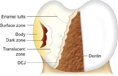
  • Early while chalky spot – decalcification of enamel.

C. Linear Enamel Caries:

  • Atypical form
  • Found in primary dentition
  • Gross destruction of labial surface of incisor teeth

https://dentowesome.wordpress.com/2020/05/07/dental-caries/
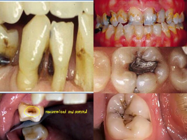
D. Root caries:

  • Soft progressive lesion that is found everywhere on root surface that has least connective tissue attachment & is exposed to oral enviornment.
  • Older age group & gingival recession

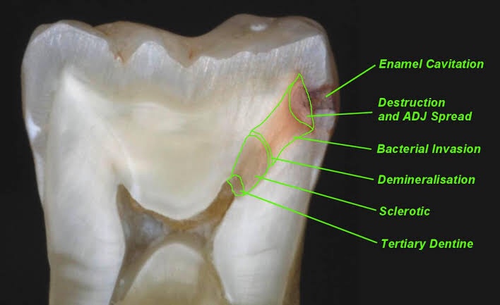
E. Acute Dentinal Caries:

  • Rapid clinical course
  • Early pulp involvement
  • Initial lesion is small, while rapid spread of process at DEJ & diffuse involvement of dentin produce large internal excavation.

F. Rampant Caries:

Sudden, rapid & almost uncontrolled destruction of teeth affecting surface that are relatively caries free.

G. Nursing bottle caries (Baby bottle syndrome)

Affect deciduous teeth due to prolonged use of nursing bottle containing milk, sugar or honey.

💬 What is 👶 bottle decay? What causes it and how to prevent it? 👇🏻

H. Chronic dental caries: (Slower progress)

I. Recurrent caries: (Presence of leaky margins)

J. Arrested caries:

  • No tendency of future progression, caries become static.
  • Brown pigmentation in the hard tissue.

Dentowesome|@drmehnaz🖊


Image Source: Google.com

Histopathology of dentinal caries

Zone I: Zone of fatty degeneration of odontoblast process
Zone 2: Zone of dentinal sclerosis characterized by deposition of cal- cium salts in dentinal tubules
Zone 3: Zone of decalcification of dentin, a narrow zone, preceding bacterial invasion
Zone 4: Zone of bacterial invasion of decalcified but intact dentin Zone 5: Zone of decomposed dentin

Earldentinal caries

Fatty degeneration oodontob/ast process

>Disposition of fat globules – precedes early sclerotic changes  >Special stains – Sudan red
>Significance-
1.Fat contributes to impermeability 

2.Predisposing factor for dental sclerosis

Sclerotic dentin

>Reaction of vital pulp – calcification of dentinal tubules (DT)

>Seals off DT from further penetration of microorganisms

>Minimal in rapidly advancing caries

>Prominent in slow caries

>Sclerotic dentin – appear white in transmitted light

Decalcification odentinatubules

>Above dentinal sclerosis – zone of decalcification

>Occurs in advance of bacterial invasion of DT 

>Pioneer bacteria
>The initial decalcification – only the walls of DT 

>Study of tubules- pure form of microorganisms

Zone omicrobial invasion

>Proteolytic organisms – predominantly in deeper layers Acidogenic microorganisms – more in early caries
>Supporting the hypothesis that initiation and progression are two distinct processes and must be differentiated

Advanced dentinacaries

>Decalcification of the walls of DT – confluence

>Thickening of sheath of Neumann – along its course • Increase in the diameter of DT – microorganisms

>Focal coalescence of adjacent tubules and ovoid area of destruction- liquefaction foci
>Acidogenic organisms – initial decalcification

>Proteolytic organisms – matrix destruction

>Multiple areas of destruction>Necrotic mass of dentin (leathery consistency)

>Formation of transverse cleftsExtend at right angles to DT and parallel contour line

>Peeling away of carious dentin

REFERENCE- Shafers textbook of oral pathology 8th edition

Histopathology of enamel caries

Four zones are clearly distinguishable, starting from the inner advancing front of the lesion. These are the (1) translucent zone, (2) dark zone, (3) body of the lesion and (4) surface layer.

Zones of enamecarieTranslucenzone {TZ)

First recognizable zone of alteration

Advancing front of the lesion

Half the lesions demonstrate TZ, not always present

Seen in longitudinal ground sections in clearing (quinoline – RI – 1.62)

TZ appears structureless

Pore volume – I% (compared to 0.1 % of sound enamel)

Dark zone

Lies adjacent and superficial to the translucent zone Positive zone

Shows positive birefringence (in contrast to sound enamel.

Pore volume of 2-4% (polarized light)
Presence of small pores; large molecules of quinoline are unable to penetrate
Micropore system – gets filled with air and becomes dark
Medium like water may penetrate

Body othlesion

Between unaffected, surface and dark zone
Area of greatest demineralization
Pore volume – 5% in periphery and 25in centre
Quinoline imbibition – body appears transparent
Water imbibition – positive birefringence compared to sound enamel Striae of Retzius – prominent

Surfaczone

Quantitative studies – partial demineralization of 1-10% • Pore volume – less than 5% of the spaces

Negative birefringence – water imbibition

Positive birefringence – porous subsurface
All the four zones of enamel caries cannot be seen with same immersion medium.

REFERENCE – Shafers textbook of oral pathology 8th edition