- Solitary Bone Cyst
- Hemorrhagic Cyst
- Extravasation Cyst
🔷 Etiology: Trauma Hemorrhage Theory
Origin from traumatic injury
⬇️
Intramedullary Hemorrhage
⬇️
Clots break down and leave empty space within bone
🔷 Clinical Features:
- Age: 18 years
- Sex: Male predominance
- Site: Posterior region of mandible ( More common). Also due to presence of hemopoietic marrow – incisor region also reported.
- In majority of cases, pulp of involved teeth is vital.
- Cavity contains Sero-sanguinous fluid, shreds of necrotic blood clot, fragments of Connective tissue.
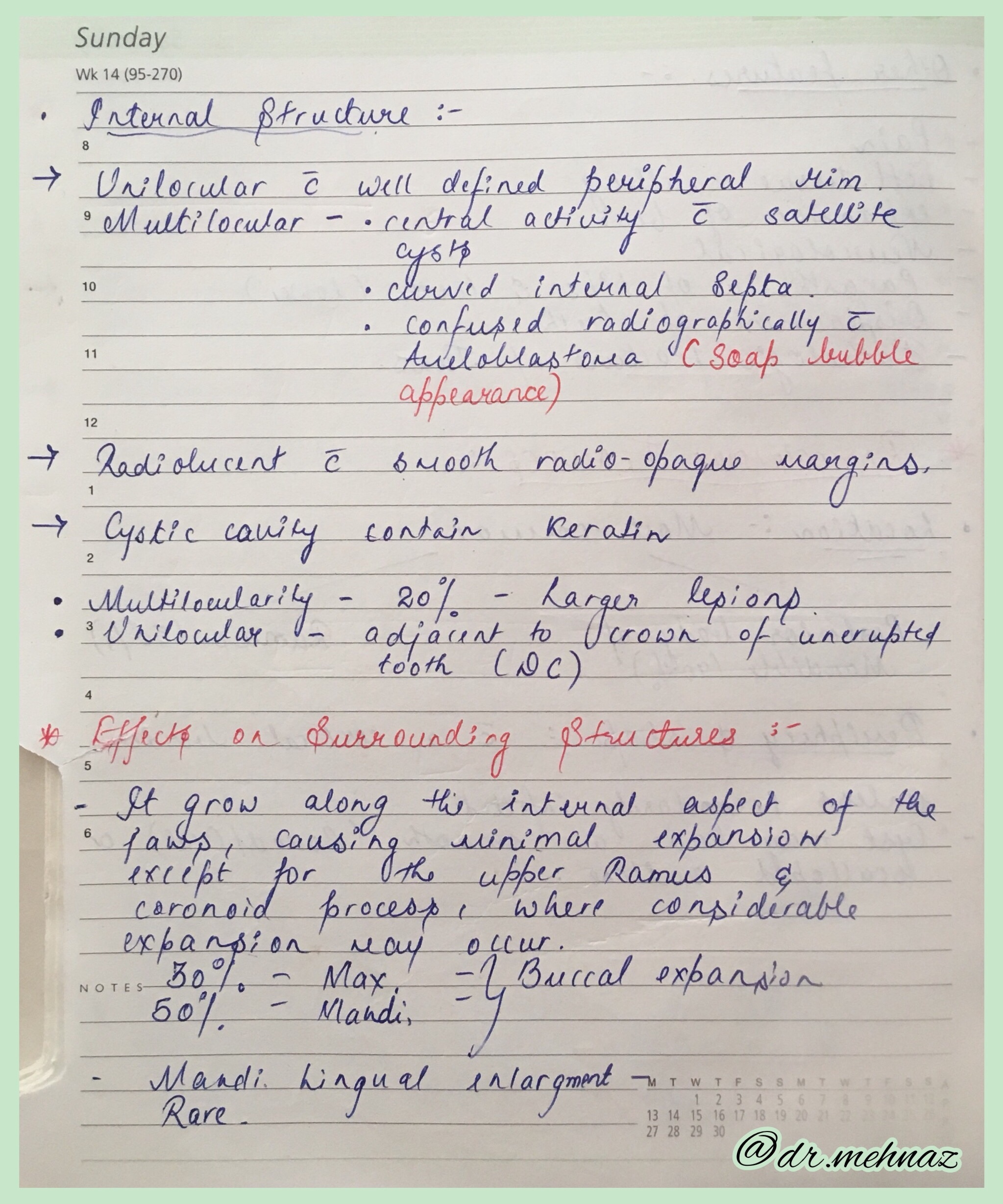
🔷 Radiographic Features:
- Smoothly outlined radiolucent area of variable size, sometimes with thin sclerotic borders. (D/D – Lingual salivary gland depression of mandible)
- However the latter lesion is usually located below the mandibular canal, whereas Traumatic Cyst usually lies above it.
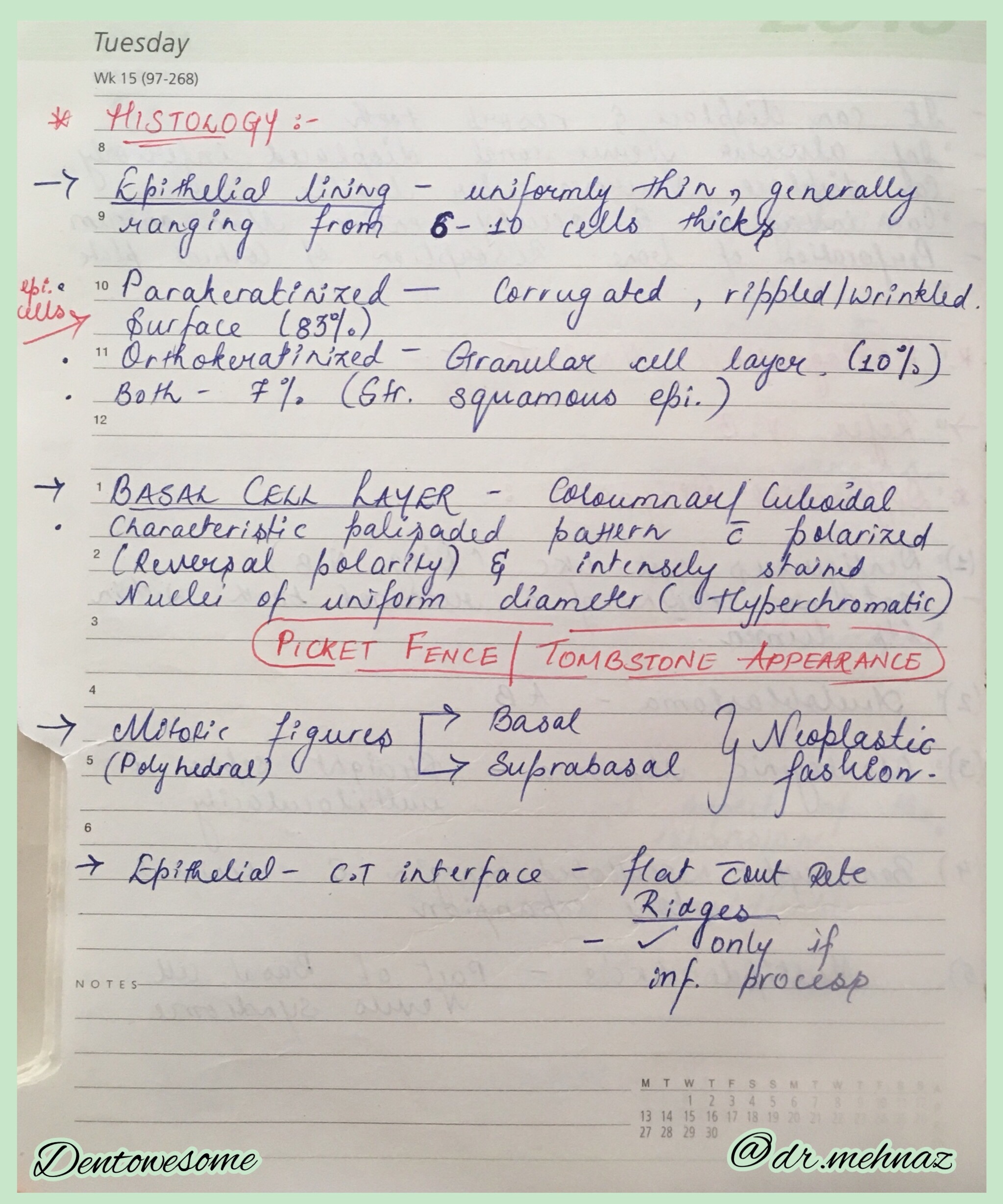
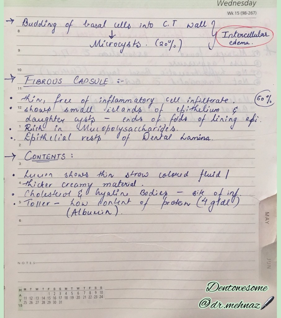
🔷 Histological Features:
- Bone cyst lined by thin Connective tissue membrane.
- On outer surface of cortical plate – Osteophytic Reaction
🔷 Treatment and prognosis:
➡️ Surgical exploration is often undertaken. The bony cavity is scraped to generate bleeding, which is considered the treatment of choice for this condition. Other methods of treatment have been tried, such as packing the curetted cavity with autologous blood, autologous bone, and hydroxyapatite. Exploration surgery usually leads to healing. Recurrence is rare.
References: Shafer’s Textbook of Oral Pathology 7th Edition, Internet
Dr. Mehnaz Memon🖊