Biomechanics of edentulous state

What is biomechanics?

It’s the interaction between the biological environment and the mechanical device that is fixed or removable prosthesis is called as biomechanics.

• They are 2 types of forces that is horizontal and vertical.

• The forces are directed in the vertical direction to the long axis of the tooth. Forces transferred by vertical direction are during mastication, clenching and swallowing.

• The forces transferred by the vertical direction is much more than the horizontal direction.

• The forces transferred by horizontal direction are mainly by the tongue and by perioral musculature.

• The factors for edentulism are trauma, patient’s oral hygiene, ageing , attitude of the patient , diet and financial status.

• If the forces are directed obliquely then the bone remodelling takes place and bone deposition will occur.

• In natural dentition , masticatory forces will be transferred through the periodontal ligament to the bone. The arrangement fibres in periodontal ligament also plays a major role.

• The periodontal ligament acts like a cushion and distributes the masticatory forces.

• The tooth is supported by the periodonium. Hard tissues supporting are cementum and bone and soft tissues are PDL and lamina propria of gingiva covered by the epithelium.

• The load taken up by the natural dentition is 20 kg and that in complete denture wearers is 8 kg.

• The major difference between the natural dentition is the compressibility of PDL and in the complete denture patient the oral mucous membrane has less compressibility to deliver forces.

• Normal mastication – 30 sec. Act of deglutution – 18 sec.

• We swallow about 500 times in a day and masticate about 17 minutes approximately in a day.

Reference :- deepak nallaswamy

Prostho Case 10

LEARNING GOALS AND OBJECTIVES
􏰀 Identify the consequences of untreated ectodermal dysplasia.
􏰀 Discuss the importance of diagnostic mock-up procedures to assess the degree of an underdeveloped maxilla and mandible.
􏰀 Understand the determining factors in assessing desired vertical dimension of occlusion.
􏰀 Understand the determining factors affecting the facial profile.

LINK: https://drive.google.com/file/d/1L4Y19Ah6HnYvHoaMmIpZ13-Xsv8vCgL8/view?usp=drivesdk

Prostho Case 9

LEARNING GOALS AND OBJECTIVES
􏰀 Understand the growth pattern of the maxilla and mandible in the growing patient.
􏰀 Understand the effects of anodontia and hypodontia.
􏰀 Understand the implication of implant prostheses in a growing child.
􏰀 Describe the importance of a maintenance program for young patients with ectodermal dysplasia with implant prostheses.

Link: https://drive.google.com/file/d/1trjQcK-3Db4eRXziOH8rWYsL0LMydsed/view?usp=drivesdk

Prostho Case 7

Brief Summary

A 72 – year – old Caucasian female presents with a chief complaint of: “ I need new partial dentures and a new crown, so I can chew better. ”

She is partially edentulous in the maxilla and the mandible and has multiple fixed partial dentures.

LEARNING GOALS AND OBJECTIVES
– Sequence treatment of a patient requiring a combination of fixed and removable prostheses.

– Discuss critical design elements for/removable partial dentures.
– Use a surveying instrument.
– Understand parameters for abutment selection for removable partial dentures.
– Recognize and treat common postinsertion
sequelae.

LINK: https://drive.google.com/file/d/1KDMmuWUNZ9KceBCwAp1jLWlMjGmE46PL/view?usp=drivesdk

Prostho Case 6

*Brief Summary* ‍
– Patient has come with complaint of loose RPD on left side which is due to little retention. Dental History of mutliple extractions, endodontic therapy, FPD and RPD.

*What may be the reason for compromised RPD?* READ THE PDF. Nicely Explained

*Treatment Options:*
Extractions and CD
Conventional RPD
Implant retained RPD

Since Patient has expressed the desire to maintain his remaining teeth. We go with Implant retained RPD

LINK: https://drive.google.com/file/d/1JE_r_zyBfQYHYzZ3SGOFpU_S9ZXWhNBN/view?usp=drivesdk

Residual Ridge Resorption

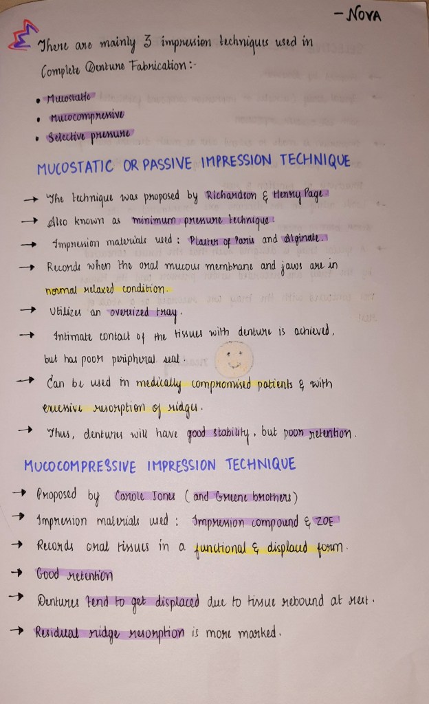
Ridge = Soft tissue + bone

• Residual ridge resorption is a life long process. Residual ridge resorption is maximum upto 3 to 6 months and after that it tappers off.

Alveolar bone :- defined as a bony portion of maxilla and mandible held by the fibres of PDL.

Bone ( Dynamic Process)

Depending on the type of bone the resorption pattern varies like for spongy bone replaced 3-4 yrs and compact bone replaced 10yrs

Which is the process coupled that is bone deposition by osteoblast and bone resorption by osteoclast.

• Residual ridge resorption pattern varies in different individual

According to American college of prosthodontics

Based on bone ht

Type 1 :- residual bone height 21 mm

Type 2 :- residual bone height 16mm

Type 3:- residual bone height 11 – 13mm

Type 4 :- residual bone height 10mm

Type 1 is having good prognosis and type 4 is having poor prognosis that means it’s difficult to gain stability , retention and support

Epidemiology of residual ridge resorption

•It occurs world wide . RRR is accelerated in 1st 6 months with more loss in mandible than maxilla.


• After menopause that is due to harmonal disturbances osteoblastic activity is very less and it’s dominated by osteoclastic activity

” RESORPTION OF MANDIBLE >>>> RESORPTION OF MAXILLA

Direction of bone resorption :-

• Maxilla resorbs upwards and inwards ( centripetal) to become progressively small

• Mandible resorbs outwards and progressively wider

Etiology of residual ridge resorption :-

• Anatomical factors that is quality and quantity of bone of residual ridge RRR is directly proportional to anatomical factors

• Metabolic process :- RRR is directly proportional to bone resorption factors and inversely proportional to formation factors

• Mechanical factors :- If there is excess stimulus or no stimulus resorption takes place

Consequences of residual ridge resorption :-

– loss of sulcus width and depth

– Displacement of muscle attachment to the ridge

– loss of vertical dimension of occlusion. Reduction of lower facial height

– Increase in relative prognathism. Changes in inter alveolar relation. Change in the location of mental foramen.

Treatment :-

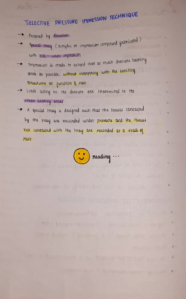
– Prevention of loss of natural tooth. Change in the design of denture like impression technique by using minimal pressure impression and selective pressure impression techniques.

• Provide adequate relief on relief are areas. Avoidance of inclined planes. Centralization of occlusal contacts to increase stability and maximise compressive forces

• Adequate interocclusal distance that is by providing enough free way space . Occlusal table should be narrow .

Source :- Deepak nallaswamy and rangarajan.

Examples of Kennedy’s Classification

Kennedy’s class lV

* A single bilateral edentulous area crossing the midline.

Kennedy’s class l – mod 2

* Class I :Bilateral edentulous area most posteriorly . Mod 2 : two extra edentulous areas

Kennedy’s Class III- mod I

* Class III- Edentulous area present between anterior and posterior natural teeth posteriorly. Mod I : one extra edentulous space

Kennedy’s Class II

* Unilateral Edentulous area

Kennedy’s class I

* Bilateral Edentulous area

Kennedy’s Class III

* Edentulous area between anterior and posterior natural teeth

Kennedy’s Class II- mod 2

* Class II : unilateral edentulous area present from most posteriorly. Mod 2: Extra 2 edentulous space

Kennedy’s Class III- Mod 1

* Class III- Edentulous area present between anterior and posterior natural teeth. Mod 1 – one extra edentulous space present.

Reference : Google pictures