An In-Vitro Evaluation of Occlusal Fissure Morphology in Primary Molars

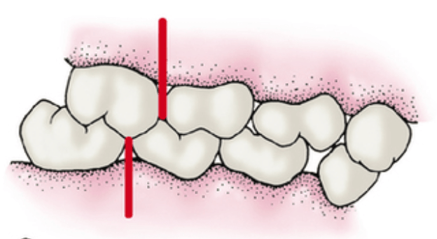
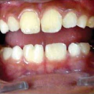
The occlusal surfaces of molars, particularly the permanent first molars, are commonly affected by caries (Demirci et al., 2010). This is consistent with the findings of a study that reported over two-thirds of permanent first molars being affected by caries (Demirci et al., 2010). Additionally, the occlusal surfaces of permanent second molars also contribute significantly to caries development (Demirci et al., 2010). The high caries rates in the permanent dentition are primarily limited to the pit and fissure surfaces of molars (Demirci et al., 2010).

The pattern of caries in primary molars, specifically the maxillary incisors and first molars, is thought to be related to the eruption times of primary teeth and the acquisition of cariogenic bacteria (Xu et al., 2014). Children who develop caries before 2.5 years of age typically exhibit decay on the smooth surfaces of maxillary incisors and occlusal fissures of the first molar teeth (Xu et al., 2014).

The progression of caries lesions in primary molars is more frequent in second molars compared to first molars (Guedes et al., 2015). This is consistent with previous studies that have reported a higher probability of progression in occlusal surfaces, both in primary and permanent teeth (Guedes et al., 2015).

Sealants are effective in preventing and arresting pit-and-fissure occlusal caries lesions in primary and permanent molars in children and adolescents (Prabakar et al., 2018). A systematic review conducted by the American Dental Association Council on Scientific Affairs and the American Academy of Pediatric Dentistry recommended the use of pit-and-fissure sealants on the occlusal surfaces of primary and permanent molars (Prabakar et al., 2018).

The morphology of occlusal fissures in molars plays a significant role in caries prevention (Dindar & Atay, 2022). Understanding the types of fissure morphology is important for preventing caries on the occlusal surfaces of molars and premolars (Dindar & Atay, 2022). The presence of deep occlusal pits and fissures increases the risk of plaque accumulation and subsequent decay (Alqahtani et al., 2022).

The retention of sealants on primary molars is generally lower compared to permanent molars (AL-Sultani et al., 2020). However, there is conflicting evidence regarding the effectiveness of sealants on primary molars compared to permanent molars (AL-Sultani et al., 2020). Some studies have reported that sealants are more effective in permanent molars, while others have found no significant difference in retention between primary and permanent molars (AL-Sultani et al., 2020).

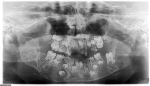
The morphology of the pulp chamber in primary molars can be evaluated using spiral computed tomography (SCT) (Selvakumar et al., 2014). SCT can provide information on the distance between the central fissure and furcation, the distance between the central fissure and the floor of the pulp chamber, the height of the pulp chamber, and the distance between the floor of the pulp chamber and the furcation (Selvakumar et al., 2014).

The use of self-etching self-adhesive flowable composite for fissure sealing in primary molars has been studied (Шхагошева et al., 2021). This material has shown efficiency in sealing fissures and preventing caries in primary molars (Шхагошева et al., 2021).

The impact of occlusal tooth morphology on the penetration of fissure sealants has been investigated (Petrovic et al., 2006). The study found that the type of occlusal tooth morphology can affect the penetrating abilities of different types of sealants (Petrovic et al., 2006).

The retention of fissure sealants in primary molars can be influenced by various factors, including etching time (Duggal et al., 2009). Different etching times have been shown to affect the retention of sealants in primary molars (Duggal et al., 2009).

The efficacy of glass ionomer sealant (GIS) and fluoride varnish (NaFV) in preventing occlusal caries in primary molars has been compared (Lam et al., 2021). Both GIS and NaFV have shown effectiveness in preventing and arresting occlusal caries in primary second molars among preschool children (Lam et al., 2021).

The microtensile bond strength of bioactive pit and fissure sealants bonded to primary and permanent teeth has been evaluated (Alqahtani et al., 2022). The study found that the bond strength of sealants can vary between primary and permanent teeth (Alqahtani et al., 2022).

In conclusion, the occlusal surfaces of molars, particularly the permanent first molars, are commonly affected by caries. The pattern of caries in primary molars is related to the eruption times of primary teeth and the acquisition of cariogenic bacteria. The progression of caries lesions is more frequent in second molars compared to first molars. Sealants are effective in preventing and arresting pit-and-fissure occlusal caries lesions in primary and permanent molars. The morphology of occlusal fissures and the retention of sealants can be influenced by various factors. Understanding the occlusal morphology and evaluating the pulp chamber morphology in primary molars can provide valuable information for caries prevention and treatment.

References:

AL-Sultani, H., Al-Janabi, W., Hasan, H., Al-Murib, N., Alam, M. (2020). Clinical Evaluation Of Pit and Fissure Sealants Placed By Undergraduate Dental Students In 5-15 Years-old Children In Iraq. Pesqui. Bras. Odontopediatria Clín. Integr., (20). https://doi.org/10.1590/pboci.2020.004 Alqahtani, A., Al-Dlaigan, Y., Almahdy, A. (2022). Microtensile Bond Strength Of Bioactive Pit and Fissure Sealants Bonded To Primary And Permanent Teeth. Materials, 4(15), 1369. https://doi.org/10.3390/ma15041369 Demirci, M., Tuncer, S., Yuceokur, A. (2010). Prevalence Of Caries On Individual Tooth Surfaces and Its Distribution By Age And Gender In University Clinic Patients. Eur J Dent, 03(04), 270-279. https://doi.org/10.1055/s-0039-1697839 Dindar, M., Atay, M. (2022). Microscopic Evaluation Of Fissure Patterns Of Posterior Permanent Teeth: An In Vitro Study. Int Dent Res, 3(12), 107-111. https://doi.org/10.5577/intdentres.2022.vol12.no3.1 Duggal, M., Tahmassebi, J., Toumba, K., Mavromati, C. (2009). The Effect Of Different Etching Times On the Retention Of Fissure Sealants In Second Primary And First Permanent Molars. International Journal of Paediatric Dentistry, 2(7), 81-86. https://doi.org/10.1111/j.1365-263x.1997.tb00283.x Guedes, R., Piovesan, C., Floriano, I., Emmanuelli, B., Braga, M., Ekstrand, K., … & Mendes, F. (2015). Risk Of Initial and Moderate Caries Lesions In Primary Teeth To Progress To Dentine Cavitation: A 2-year Cohort Study. Int J Paediatr Dent, 2(26), 116-124. https://doi.org/10.1111/ipd.12166 Lam, P., Sardana, D., Luo, W., Ekambaram, M., Lee, G., Chu, C., … & Yiu, C. (2021). Glass Ionomer Sealant Versus Fluoride Varnish Application To Prevent Occlusal Caries In Primary Second Molars Among Preschool Children: a Randomized Controlled Trial. Caries Res, 4(55), 322-332. https://doi.org/10.1159/000517390 Petrovic, B., Markovic, D., Blagojevic, D. (2006). The Impact Of Occlusal Morphology On Fissure Sealant Penetration. SERBIAN DENT J, 2(53), 87-94. https://doi.org/10.2298/sgs0602087p Prabakar, J., John, J., Arumugham, I., Kumar, R., Srisakthi, D. (2018). Comparative Evaluation Of Retention, Cariostatic Effect and Discoloration Of Conventional And Hydrophilic Sealants – A Single Blinded Randomized Split Mouth Clinical Trial. Contemp Clin Dent, 6(9), 233. https://doi.org/10.4103/ccd.ccd_132_18 Selvakumar, H., Kavitha, S., Vijayakumar, R., Eapen, T., Bharathan, R. (2014). Study Of Pulp Chamber Morphology Of Primary Mandibular Molars Using Spiral Computed Tomography. The Journal of Contemporary Dental Practice, 6(15), 726-729. https://doi.org/10.5005/jp-journals-10024-1606 Xu, H., Hao, W., Zhou, Q., Wang, W., Xia, Z., Liu, C., … & Chen, F. (2014). Plaque Bacterial Microbiome Diversity In Children Younger Than 30 Months With or Without Caries Prior To Eruption Of Second Primary Molars. PLoS ONE, 2(9), e89269. https://doi.org/10.1371/journal.pone.0089269 Шхагошева, А., Маслак, Е., Фурсик, Д. (2021). The Results Of Self-etching Self-adhesive Flowable Composite Application For Primary Molars Fissure Sealing In Children. jour, 2(21), 113-117. https://doi.org/10.33925/1683-3031-2021-21-2-113-117

Pictorial Puzzles – Pedodontics – Module 2

Veau Classification of Cleft Palate – CLASS 1 – Only soft palate is involved

Distal Molar relationship transforms into Class 2

Edge to edge molar relationship transforms into Class 1 or Class 3

Transpalatal arch = In cases of unilateral loss

Nance Palatal arch = In cases of bilateral loss

Maxillary Anterior bite plane = correction of deep bite

Split labial bow – correction of midline diastema

Crouzon syndrome – Obliteration of saggital sutures, multiple marks in the skull region

This is Treacher collins syndrome = Autosomal Dominant

Autosomal Recessive – Cystic fibrosis and sickle cell anaemia

X Linked recessive = Hemophilla

X linked dominant = Double cortex syndrome

Red Line = Genital growth

Pictorial Puzzles – Pedodontics – Module 1

Internal Resorption

First permanent molar is about to erupt, so the age = is 5 years

Number of teeth = 19, FUSION

LEEWAY SPACE – Maxillary = 0.9 mm and Mandibular = 1.7 mm

OPG of 10 year old child – permanent CI, LI and first molar has erupted. The primary canine, first molar and second molar are also present.

8 year old male patient comes to your clinic with bilateral swelling in the jaw, upturned eyes with excessive sclera visible beneath the iris = CHERUBISM

AMELOBLASTOMA = Multilocular radiolucency = Honeycomb appearance

FIBROUS DYSPLASIA = mc seen in maxillary bone

Thumb sucking appliance – CRIBS are given

Mouth breathing appliance – ORAL SCREEN

Endodontics and Pulp Therapy – NEET and INICET pearls from Dentest and Pulse

  1. Pulp of all primary teeth is active for a period of = 8.3 years/9.6 years
  2. Infected dentin (TRUE STATEMENTS)
    1. Loss of organic network 
    2. Lacking sensation
    3. Irreversible demineralization 
    4. Excavated during cavity preparation 
  3. Affected dentin (TRUE STATEMENTS)
    1. Demineralised but can be mineralized
    2. No loss of organic network 
    3. Sensitive
    4. Should be left out for remineralisation during cavity preparation
  4. The dye used for the detection of dentin caries is = Acid red system
  5. The greatest problem in pulpal diagnosis is estimating the amount of = INFLAMMATION
  6. Radiographs of permanent molar 
    1. Acute apical abscess shows
      1. Early stages – no radiographic changes
      2. Later stages – little change from the normal structure
    2. Acute alveolar abscess = thickened periodontal membrane
    3. Chronic pulpal infection = rarefaction at bony furcation
    4. Carious involvement = Radiolucency in the furcation area 
  7. ACUTE ALVEOLAR ABSCESS (TRUE STATEMENTS)
    1. Primary tooth = more diffuse 
    2. A localized collection of pus at root apex in alveolar bone
    3. There is swelling, mobility and elevation of a tooth from its socket 
  8. Pulse oximetry 
    1. Measures the pulpal circulation directly 
    2. TRUE STATEMENTS
      1. Objective test
      2. Measures and compares amplitudes of ratios of transmitted infrared light with red light 
      3. Capable of evaluating the stature of blood vasculature
      4. Depends on pulsatile blood flow
      5.  
  9. Laser doppler flowmetry
    1. Based on the velocity of RBC cells in capillaries 
    2. A non-invasive method to measure blood flow
    3. GAZELIUS
  10. Indirect Pulp Therapy 
    1. Objective = Eliminate the removal of deep caries
    2. Indicated (imp)
      1. in primary molars when the carious lesion is suspected of producing an exposure of the pulp
      2. when there is no history of spontaneous pain  = Tooth must be asymptomatic 
    3. IPT = It is dependent on the remineralisation of affected dentin and reparative dentin formation  
    4. ​​
    5. The success of IPT = placement of temporary restoration with excellent sealing properties
    6. Most ideal material = Calcium hydroxide
      1. Introduced by FAUCHARD
      2. When applied to residual carious dentin = it remains for 6 – 8 weeks 
      3. Following amputation of the coronal portion of the pulp of an immature permanent first molar = the stump is capped with CaOH
      4. The success of CaOH pulpotomy is determined by = Continuation of root formation and apexogenesis
      5. Dentin bridge is formed = at a level slightly below the amputation
  11. Direct Pulp Therapy 
    1. Amount of repartive/3* dentin formed after 30 days = 1.5 microns/day and then slows down after 48 days 
    2. Contraindicates in primary teeth 
    3. Prognosis is best = mechanically exposed permanent tooth
    4. Indicated in cases of little (Pinpoint exposure = less than 1 mm) or no haemorrhage from the exposure site
    5. The pulp should be vital
    6. Done in traumatic or iatrogenic cases = Reported within 24 hours
    7. High rate of failure = high cellular content of pulp
  12. Pulpotomy
    1. The simplest form of pulp therapy 
    2. Contraindicated = If there is Inflammation of radicular pulp and pain
    3. Mechanical exposure of mesiobuccal pulpal horns in primary maxillary 1st molar with moderate caries lesion on the mesial-distal surface = formocresol PULPOTOMY + stainless steel CROWNS
    4. Pulp preservative material and best   = MTA
    5. The primary function of MTA = Apexification of immature traumatised pulpless tooth
    6. Important criteria of success = Root end completion 
  1. Formocresol Pulpotomy 
    1. SWEET
    2. Composition of Buckley Solution
      1. 1:2 parts F: C
      2. 19% formaldehyde 
      3. 13% cresol
      4. 15% glycerine 
    3. Successful treatment for vital primary second molar with a large carious and pulpal exposure 
    4. The success of the technique depends on vital root pulp 
    5. Cotton pellet applied to the pulpal stumps after formocresol pulpotomy should be = Dampened with formocresol and placed for 5 mins
    6. Amount of pulp to be removed in formocresol technique= entire coronal pulp down to the cervical constriction of each root canal
    7. FERRIC SULPHATE = material with haemostatic effect, used as a substitute for formocresol pulpotomy
    8. EFFECT = Surface fixation of pulpal tissue accompanied by degeneration of odontoblasts 
    9. The first sign of formocresol technique failure = Internal resorption
  2. Glutaraldehyde Pulpotomy 
    1. Superior fixative properties 
    2. Conc as pulpotomy agent = 2 to 5 % concentration 
    3. The cotton pellet is kept on pulp stumps = for 3-4 mins
    4. KOPEL
  3. Ferric Sulphate Pulpotomy 
    1. 2 – 5 seconds 
    2. 15.5% concentration
    3. 100% clinical success rate
  4. CaOH Pulpotomy
    1. contraindicated in primary teeth due to internal resorption – odontoclastic reaction
    2. Immediate reaction = Severe inflammatory reaction
    3. The calcified bridge is evident within = 1 month
    4. Increases in thickness – next 12 months 
    5. Failure of technique = 
  5. Cvek Pulpotomy
    1. Partial technique
    2. Important criteria of success = Root end completion 
  6. Pulpectomy
    1. The entire pulpal tissue is removed
    2. Complete Pulpectomy technique for primary molars is developed by = STARKEY
    3. Pulp extirpation in primary teeth is difficult because = Tortuous anatomy and branching of canals 
    4. The major problem with this technique = is limited knowledge of pulpal anatomy
    5. KRI paste used for obturation in case of pulpectomy is primarily a mixture of = Iodoform and CMCP
    6. MC filling material, traditionally = Zinc Oxide Eugenol 
    7. ROOT CANAL SPREADER = It is not used in the widening of root canals 
  7. Pulp Devitalization 
    1. PARAFORMALDEHYDE = common medicament in all types of devitalization pulpotomy 
    2. Laser = Nd: YAG
  8. Pulp mummification = Indicated in = traumatic exposure of a vital primary tooth 
  9. Apexification
    1. Rx of pulpless/nonvital immature young permanent tooth
    2. A condition that leads to apexification = Pulpectomy of the young permanent tooth with incomplete root formation
    3.  Any excess CaOH periapically = will be removed by multinucleated giant cells 
    4. Increase success rate = Increased blood supply through the wide apex
    5. Best obturated by = OBTURA – thermoplasticized gutta percha system
    6. FRANK technique 
      1. Uses CaOH and CMCP (Camphorated monochlorophenol) 
      2. To stimulate root closure/ apexification in incompletely developed young permanent tooth 
      3. CMCP = 6 months
  10. Rate of resorption in pulpless primary teeth compared to vital teeth = SIMILAR
  11. In the absence of second premolar roots of primary second molar will most likely = resorb more slowly than normal
  12. Ideal Root Canal filling = CaOH
  13. Rx choice in poor prognosis cases, alveolar abscess and necrotic pulp cases = extraction 
  14. Apexogenesis
    1. TRUE STATEMENTS
      1. More of a physiological process
      2. Root development continues
      3. Indicates in pulp tissue with mild inflammation 
    2. Done in the vital and infected tooth
  15. In regenerative endo therapy, Metronidazole is replaced by = Ornidazole 
  16. Reattachment of fractured tooth fragments using the resin bonding technique is called = Fragment Restoration 
  17. STAINLESS STEEL CROWN
    1. Rx of choice for Ankylosis of a primary molar with the absence of a permanent successor
    2. Rx of choice for amelogenesis imperfecta in primary dentition = SSC
    3. HUMPHREY
    4. SSC is a = Semi-permanent restoration 
    5. Contraindicated in Medically compromised patients – Heart problems – VSD, ASD
    6. INDICATIONS 
      1. Restoration of hypoplastic teeth 
      2. Following pulpotomy or pulpectomy 
      3. As an abutment for space maintainers
      4. NOT USED = teeth that are not restorable 
    7. Iron content in 3M SCC = 10%
    8. CHENG CROWNS = pre-veneered crown which is stain resistant and pre-crimped
    9. Countering of stainless steel crown is done at middle 1/3rd of the crown to produce = BELLING EFFECT
    10. MC complication while tooth prep = LEDGE Formation
    11. PREVENTION OF LEDGING = most important for using burs in opening proximal contacts to receive SSC
    12. SCC should extend
      1. intra gingivally or below the gingival crest = 0.5 – 1mm 
      2. Occlusal = 1.5-2mm 
      3. Buccal and lingual = 0.5mm 
      4. Proximal = 1-1.5mm 
    13. Basket Crown technique
      1. Temporary crowns 
      2. Placed in the anterior tooth 
      3. 3/4th crown
      4. The window is prepared on the LABIAL aspect for aesthetic

    14. Finish Line of SSC = FEATHER edge
    15. Difficulty in adopting an SSC to a primary mandibular first molar = Buccal cervical ridge showing constriction
    16. Surface requiring the least amount of reduction = BUCCAL and LINGUAL 
    17. Retention is achieved primarily by = PARALLEL distal and mesial walls
  18. In a con-compliant pulp chamber, average intrapulpal pressure = 10 mm HG and varies with each arterial pulse
  19. PREPOMETER = device to measure the thickness of the dentin layer above the pulp chamber
  20. PULPDENT = most capable of stimulating early dentinal bridge formation
  21. Obturation of deciduous teeth can be done with = Iodoform paste

Childhood Diseases – NEET Pearls from Dentest and Pulse – 2023

  1. Normal colour of
    1. Primary teeth = China white
    2. Permanent teeth = Ivory white
  2. In primary dentition =
    1. dentine is uniformly calcified 
    2. Enamel and dentin is less mineralized
    3. Enamel and dentin thickness is less
    4. DEJ is less scalloped
  3. Main difference between primary and permanent tooth = Mineral content 
  4. Cleft of secondary palate causes = cleft lip
  5. Ectodermal dysplasia in 3-year-old child with only primary molars and canine and overall appearance of an older person
  6. Treatment of geographic tongue = No treatment
  7. Dentinogenesis Imperfecta 
    1. Grey in colour
    2. Exhibit extensive occlusal and incisal wear
    3. r/g = secondary dentin deposition
    1. Transillumination of soft tissue is useful in detection of = Sialolithiasis 
  8. KOPLIK SPOTS
    1. Small irregular bright red spots with white specks in centre 
    2. Indicate the onset of RUBEOLA/MEASLES
  9. MC cause of acute generalised inflammation 
    1. in pre-school children = Acute herpetic gingivostomatitis/ herpes
      1. Child with fever of 102*C and vesicles in oral cavity
      2. Rx = symptomatic rx for fever and prevent secondary infection and dehydration
    1. In adolescents = ANUG
  10. Apthous ulcer should be treated by = Palliation and Patience
  11. Rx of severe intraoral infection differs from that in adult because = dehydration occurs more rapidly and severely in children
  12. The purulent lesion in the oral vestibule of an 8-year-old child = ODONTOGENIC FISTULA
  13. Bluish dome shaped lesion on the inside of a lip = Mucocele
  14. MC in children = gingivitis
    1. Reaches in its severity at peak of = 11 – 13 years
  15. Gingiva in children = less keratinized, less stippled
  16. NOT seen on attached gingiva = interdental clefts
  17. Gingival stripping in children = due to narrow attached gingiva 
  18. Periodontosis or Juvenile Periodontitis
    1. Vertical pockets around incisors and first molars
    2. Presence of plaque isn’t consistent with inflammatory findings 
  19. NOT related to gingival inflammation = Spirochetal infection
  20. Pulp starts reacting to caries when carious lesion reaches = Dentinoenamel Junction 
  21. Periapical abscess more diffuse in primary teeth because = Alveolar bone surrounding the teeth is less dense
  22. Gingival abscess is common in primary teeth because = More accessory canals are present on buccal surface with porous floor and thin bony surface
  23. MCC of draining sinuses in oral cavity of children = chronic periapical abscess
  24. Hypoplasia of primary enamel that forms before birth is = RARE
  25. Eruptive cysts are best treated by = No treatment, only observation
  26. A disease that only affects formation and eruption of tooth but doesn’t cause hypoplasia = RICKETS
  27. OSTEOGENESIS IMPERFECTA = doesn’t cause delayed eruption
  28. MANDIBULAR second premolar = shows greatest variation relative to the onset of mineralization of crowns
  29. Child with Down syndrome (all are important points)
    1. Affectionate and cooperative
    2. Capable of understanding operative procedures
    3. Fearful of quick movements
    4. Simian crease in hand
    5. Head shape = brachycephalic 
    6. Incidence of caries = less as compared to general population 
    7. High incidence of periodontal disease
    8. Retarded eruption
    9. Delayed exfoliation**
    10. Retained deciduous teeth and microdontia
    11. Typical face = hypoplastic maxilla
    12. IQ = 120 – 139
    13. Syndrome associated with congenital heart lesions
  30. Fluoride is contraindicated in = Chronic renal failure 
  31. DIABETES
    1. Serious complication of juvenile diabetes/ DM – 1 = blindness
    2. MC type of diabetes = DM – type 2
    3. Level of glucose for diabetic ketoacidosis = 300 to 600 mg/dl 
  32. LEUKAEMIA 
    1. Child suffering from acute leukaemia is more susceptible to = Oral infections
    2. Primary cause of death in patients = Infections 
    3. TRUE STATEMENTS 
      1. May be manifested by mucosal pallor
      2. Cause Obvious Purpura 
      3. Lymphoblastic variety 
    4. Line of Rx for periapical abscess = Obtain medical consultation before treatment 
    5. Down Syndrome = 10 – 20 fold increased risk of leukaemia pt  
  33. HEMOPHILIA
    1. Epsilon aminocaproic acid = given to haemophilic child before surgical procedures to control bleeding
    2. Line of Rx for periapical abscess = Obtain blood count before extraction, determine the amount and duration of factor concentrate replacement 
    3. In extraction cases, Minimal level of Factor VIII = 50%
    4. Minimal level needed for adequate hemostasis = over 25%
    5. 5 years old, primary second molar with non vital pulp is treated by = conventional pulpectomy
    6. Mode of anaesthesia contraindicated = INTRAMUSCULAR
    7. MC type = Hemophilia A
  34. CYSTIC FIBROSIS (all points imp)
    1. Maldigestion and malnutrition
    2. Chronic respiratory infection
    3. Thyroid deficiency 
    4. Water and electrolyte imbalance
    5. Staining of teeth = yellow in colour  
  35. Yellowish primary tooth due to trauma indicates = calcific reaction of pulp
  36. Pink primary tooth indicates = Internal resorption
  37. TETRACYCLINE PIGMENTATION
    1. Yellow primary tooth, under UV light – faint overall yellow green autofluorescence = Tetracycline pigmentation
    2. TS occurs in primary tooth during the period of = mineralization of first millimetre of dentin at DEJ
    3. Tetracycline administration causes primary tooth staining = upto 9th month of life
  38. Sensitive period for tetracycline discolouration 
    1. PRIMARY
      1. Max and mand incisors = 4 months in utero to 3 months postpartum 
      2. Max and mand canines = 5 months in utero to 9 months postpartum 
    2. PERMANENT
      1. Max and mand incisors and canines = 3 months postpartum to seventh year of life
  39. SEIZURES
    1. Rx of Petit mal seizure in dental office = Watch until episode passes away
    2. Occurring in children several times in a day with no involvement of aura = Petit Mal 
    3. MC type of epilepsy seen in children = Petit Mal 
  40. Following are associated with slowness of mental retardation in a child
    1. Family history
    2. Metabolic disease
    3. Pregnancy and delivery history 
    4. Phenylketonuria
    5. Galactosaemia 
    6. Non trainable type of mental retardation = IQ level of below 20 
  41. CEREBRAL PALSY
    1. Features seen
      1. Increases Caries
      2. Increased salivation
      3. NOT SEEN = fluorosis 
    2. Most of CP children = SPASTIC
    3. MC type of CP = Spastic and Athetosis
    4. SPASTICITY = characterised by sudden violent involuntary contraction of a muscles
    5. TYPES
      1. Spasticity = muscular contractions which are irregularly spaced and have no purpose 
      2. Athetosis = slow, worm like, constant involuntary uncontrollable purposeless movements 
      3. Ataxia = disturbances of balance and equilibrium
      4. Rigidity = marked resistance to passive motion
    6. MC classifications = physiological and topographic 
    7. Oral Manifestations 
      1. Periodontal diseases
      2. High incidence of caries
      3. Attrition of teeth due to bruxism
    8. Class II Divison 2 = mc malocclusion observed in pt with spastic type 
  42. AUTISM
    1. Delayed milestones, playing with herself, unable to make friends, and difficulty in learning 
    2. It is a severely incapacitating disturbance of mental and emotional development that causes problem in learning, communication and relating to others 
    3. Incapacitance of emotional and mental disturbance
    4. Parrot like repetition speech 
    5. Also known as Kanner’s syndrome
  43. RESTRAINTS
    1. In uncooperative child, last resort is = Physical restraints
    2. TRUE STATEMENTS
      1. Papoose board is for restraining body
      2. Use of restraining device is a passive method
      3. Posey straps are used for restraining extremities
    3. MC restraints in children with neuromuscular diseases = Papoose Board
    4. Mouth props are used as = Restrainers
    5. Bean body is used in dental chair for = Restraining body
  44. BACTERIAL ENDOCARDITIS
    1. Antibiotic prophylaxis = 1 hour before
    2. Prophylactic antibiotic coverage in a child with history of rheumatic fever is precaution against developing = subacute bacterial endocarditis
  45. MOUTH BREATHING
    1. Reliable method for quantifying the extent of mouth breathing = Rhinomanometry
    2. TRUE STATEMENTS
      1. Increase incidence of caries in open bite cases
      2. Decrease in masticator scouring action in the area of open bite
  46. GENOME
    1. Albino mother, normal father, chances of children being carriers = 50% carriers
    2. ALBINISM = Autosomal Recessive, alternate generations, not affected by sex
    3. Normal parents, affected male infant = Polygenic disorder
    4. Affected father, one child affected out of three = Autosomal Dominant 
    5. Affected fathers, affected grandsons = X linked recessive
    6. Affected fathers, affected grand-daughters = X linked dominant
  47. HANDICAPPED CHILDREN
    1. Radiograph of choice = panoramic 
    2. Mc tooth brushing technique = Horizontal scrub
  48. Use of penicillin on a patient with asthma = CONTRAINDICATED
  49. Chicken wire appearance of alveolar bone = THALASSAEMIA 
  50. Virus responsible for causation of AIDS = HTLV 3
  51. Denture bearing mucosa in children as compared to adults = thinner and well-circulated 
  52. Impression taking order in paediatric prosthetic procedures = upper first and then of lower jaw 
  53. Green stains frequently seen in children’s teeth = chromogenic bacteria 
  54. Tender, painful, unilateral or bilateral swelling of salivary glands = Parotitis or Mumps
  55. Dentinogenesis Imperfecta = dark brown coloured tooth, frequently broken bones associated with blue sclera
  56. Delayed eruption of permanent teeth and large tongue = Hypothyroidism 
  57. Premature eruption of permanent teeth and large tongue = Hyperthyroidism 
  58. Fever, acute painful ulcers, lymphadenopathy = Acute herpetic gingivostomatitis
  59. Exanthematous fever and strawberry tongue = Scarlet fever
  60. Congenital anodontia and perspiration = Ectodermal dysplasia
  61. Brown discoloration of teeth, malnutrition, steatorrhoea, disturbances in function of exocrine glands = Cystic Fibrosis 
  62. Multiple supernumerary teeth unerupted with open fontanelle and abnormal development of clavicles = Cleidocranial dysostosis
  63. Causes both hypocalcified as well as irregular but well calcified enamel = Amelogenesis Imperfecta
  64. Flabby white lesions occur bilateral and seen in few other members of family = White sponge nevus
  65. DRUGS
    1. MC used antibiotic in child allergic to penicillin = Erythromycin
    2. Common antagonist of meperidine = NALOXONE
    3. Moniliasis/Candidiasis 
      1. Unfavourable oral sequel with prolonged use of antibiotics in children 
      2. Thick curd like white patch, on rubbing leaves erythematous patch
  66. PERCENTAGES
    1. Gingivitis at age of 1 – 13 years = 90%
    2. Periodontitis by age 18 years = 25%
  67. Dentitia Tarda = Retaded eruption of deciduous dentition 
  68. MC malignant tumour = Osteosarcoma
  69. Chronic periapical infection in primary molars, first noted as = rarefaction of bony furcation
  70. MC cause for gingival fistula = chronic apical lesion
  71. CLEFT PALATE
    1. Alveolar grafting in a pt of cleft should be ideally placed = after maxillary expansion, cross bite correction and before cuspid eruption
    2. After treating the cleft palate, the occlusion is = Unilateral and bilateral crossbite

Histopathology of Dentinal caries

  1. As the carious lesion invades dentin, dentinal tubules become involved.
  2. As dentin is part of the dentin pulp complex, it leads to reparative dentine formation.

FIVE PATHOLOGICAL ZONES

  1. Zone 1: Zone of fatty degeneration of Tomes fibers.
    1. Starts at pulp
    2. No fat degeneration (misnomer name)
    3. Alteration by dentin by fat deposition leads to impermeability of tubules which leads to sclerosis 
    4. Sclerosis is reaction between vital pulp and vital dentin
    5. It is the calcification of tubules against further penetration of microorganisms.
  2. Zone 2: Zone of dentinal sclerosis characterized by deposition of calcium salts in dentinal tubules.
    1. Sclerotic zone is minimum in rapidly advancing caries and prominent in slow/chronic caries.
    2. This is a translucent zone due to the vital reaction of odontoblasts
    3. High mineral content
    4. Reflected Light: Appears Dark
    5. Transparent Light: Appears White
  3. Zone 3: Zone of Decalcification of dentin
    1. Tubules are made of pure microorganisms such as pioneer bacteria (Cocci and Bacilli etc)
    2. Intertubular matrix is mainly affected by waves of acid produced by bacteria in the zone of bacterial invasion
    3. Initial decalcification occurs in the wall of tubules. 
    4. Softened infected dentin cannot be differentiated with sterile soft dentine, clinically.
    5. Appears yellow-brown
  4. Zone 4: Zone of bacterial invasion of decalcified but intact dentin.
    1. Bacteria multiply within dentinal tubules.
    2. Acidogenic microorganisms: Seen in early caries
    3. Proteolytic organisms—predominate in deeper layers
    4. It supports the hypothesis that initiation and progression are two distinct processes and must be differentiated
  5. Zone 5: Zone of decomposed dentin.
    1. Bacteria invade both peritubular and intertubular dentin. Hence, Little architecture of dentin remains
    2. Liquefaction foci of miller enlarge and increase in number.
    3. Transverse Clefts: Perpendicular to tubules
    4. Acute Caries: necrotic dentin is very soft and yellowish-white
    5. Chronic Cases: Dentin is leathery and brownish-black

SUGAR SUBSTITUTES

  1. Xylitol
    1. its 5 carbon sugar
    2. comes from birch trees. 
    3. preferential binding to Mutans strep but it cant ferment xylitol to acid
    4. Hence, no decrease in pH
  2. Sorbitol = caloric sweetness
  3. Aspartans = non caloric sweetener = 4Kcal/g = 200 times sweeter than sugar 

Caries activity tests

  1. Lactobacillus Count Test
    1. It was introduced by Hadley in 1933.
    2. It estimates the number of bacteria in the patient’s saliva by counting the number of colonies appearing on tomato Peptone Agar at ph 5
    3. Interpretation of caries activity
      1. Immune: less than 1000 
      2. Slight: 1000 – 5000
      3. Medium: I5000 – 10,000
      4. High: more than 10000.
  2. Snyder Test 2m
    1. The rapidity of acid formation by cariogenic bacteria
    2. Stimulated saliva + Glucose in Agar Medium containing bacto peptone, sodium chloride, and bromocresol green 
    3. Dye changes from blue-green to yellow
    4. Interpretation of color change with caries activity 
  • High—24 hours
  • Medium— 48 hours
  • Slight—72 hours
  • Immune—no color change
  1. Streptococcus mutans Level in Saliva
    1. The number S.Mutans colonies per ml of saliva is indicative of caries activity.
    2. Saliva samples obtained by using tongue blades are incubated on MSB agar (Mitis Salivarius Bacitracin Agar).
  2. Buffer Capacity Test
    1. The test evaluated the quantity of acid required to lower the pH of saliva using an arbitrary pH interval
    2. Buffering Capacity is inversely proportional to the process of caries
  3. Swab Test
    1. Oral swab- the buccal surface of teeth and placing it in Snyder media. 
    2. This is incubated for 48 hours and the pH changes are read and correlated with caries activity.
  4. Fosdick Calcium Dissolution Test
    1. Patient saliva is mixed with glucose and powdered enamel
    2. Measuring of powdered enamel dissolved in 4 hours by acid formed
    3. This is not a single test and requires trained personnel
  5. Reductase Test
    1. Measures the activity of salivary enzyme reductase. 
    2. Stimulated Saliva + diazo resorcinol, which colors the saliva blue. 
    3. The change in color from blue to red is measured after 30 seconds – 15 minutes and this is taken as a measure of caries activity.
    4. Interpretation
      1. Non-Conducive: Remains blue after 15 minutes
      2. Slightly conducive: Orchid after 15 minutes
      3. Moderately conducive: Red after 15 minutes
      4. Highly conducive: immediately to red
      5. Extremely conducive: Changes to pink or white

EARLY CHILDHOOD CARIES

DEFINITION = American Academy of Paediatric Dentistry (AAPD) defines early childhood caries as ‘the presence of one or more decayed (non-cavitated or cavitated lesions), missing (due to caries) or filled tooth surfaces in any primary tooth in a child 71 months of age or younger.

A window of infectivity 2M**:

  1. Caufield (1996) stated that there is a window of infectivity between 19 and 33 months during which teeth get infected with S. mutans 
  2. The most beneficial time for vaccination against dental caries would be in infancy before the eruption of teeth. 
  3. This would promote the induction of adherence inhibiting salivary IgA thus delaying colonization of S. mutans
  4. With the establishment of early colonizers, there would be a synergistic effect of suppressing the colonization of S.mutans during the period of the window of infectivity. 
  5. A booster dose of vaccination may be required at the time of eruption of first permanent molars 

ETIOLOGY AND PATHOGENESIS 3M

  1. Early colonization of MS is the most imp risk factor for developing ECC – MS transmission can be through the mother or from peers [ other kids] 
  2. MS Colonization of pre dentate children is mostly associated with maternal factors [ high level of MS in the mother, poor OH, and active caries ] 
  3. How is nocturnal bottle feeding/breastfeeding related to ECC? 
    1. When a child is laid to rest, the bottle or breast nipple rests against the palate and the tongue covers the lower incisors [ that’s why they are not affected] – 
    2. As the child becomes sleepy, saliva flow and swallow reflex are reduced
    3. Sugar remains stagnant around the neck of the teeth 
  4. what practices increase the chance of developing ECC?
    1. Prolonged nighttime bottle feeding
    2. On-demand breastfeeding after the age of 1
    3. Frequent snacking with sugary foods
    4. Frequent sipping of sugary drinks throughout the day

CLINICAL FEATURES 2M

  1. ECC = also known as nursing bottle caries, baby bottle tooth decay 
  2. Seen in infants and preschool children [ below the age of 6
  3. Demineralization at the necks of the upper incisors – mandibular incisors are not affected
  4. Decay pattern:
  1. Maxilla: incisors, canines, first molars 
  2. Mandible: canines, first molars
  1. Lesion progresses to the necks of the teeth and in advanced cases, only a root stump is left
  2. Why does ECC follow this specific pattern? 
    1. Chronology of primary tooth eruption
    2. Duration of the deleterious habit [ bottle feeding]
    3. A muscular pattern of infant sucking
  3. STAGES
    1. Very mild: slight demineralization usually at the gingival crest and no cavitation.
    2. Mild: demineralization in a gingival third of tooth and moderate cavitation.
    3. Moderate: frank cavitation on multiple tooth surfaces.
    4. Severe: Widespread destruction of tooth and loss of the clinical crown 

MANAGEMENT 6M

  1. Identify the cause and stop the habit
  2. Give parental instruction on proper oral hygiene measures + diet counseling
  3. Decide if the case can be managed in the clinic [ with regular LA or nitrous sedation ] or the child needs GA
  4. If the case is treated in the clinic: full assessment of all affected teeth to know 
    1. which teeth can be restored
    2. which need pulp therapy
    3. which need extractions
First visitSecond visitThird visit
Immediate excavation of caries followed by temporization Dietary chart Caries activity doneTopical fluoride application doneParent counsellingExamine diet chart Caries activity done again Replace any temporary restoration with permanent restorationPulpul procedures Extractions Space maintersRecall the pt every 3 months

What instructions would you give the parents to a child with ECC? 

  1. STOP NIGHT TIME BOTTLE FEEDING / stop breastfeeding at will after the first tooth erupts
  2. Feed the child while being held + burp the infant after feeding
  3. Clean the teeth after each feeding [ wipe the teeth with a wet gauze]
  4. regularly lift the upper lip to check for signs of demineralization of the upper Interiors
  5. OH should start with the eruption of the first tooth – wipe the teeth with gauze and for ages 2- 6 brush with low fluoride tooth past [ 400-500 ppm] – parental supervision until the child can properly spit
  6. Children are encouraged to drink from a cup as they become 1 year old
  7. Avoid frequent snacking and have regular meals instead
  8. First dental visit should be combined with immunization dates [ at or before 6 months]

Prevention of ECC ideally begins prenatally: 

  1. give the mother information about diet and OH
  2. treat the mother’s own oral diseases and lower MS count by mouth rinses and restorative care
  3. educate the mother on modes of transmission of MS [ don’t lick spoons or pacifiers etc..]