Osteomyelitis

Definition: Osteomyelitis is defined as inflammation of the bone and marrow.

Similar Images, Stock Photos & Vectors of Osteoporosis Labeled ...
source -google

Classification

🔻Primary: Where bone is the primary solitary focus of disease .

🔻Secondary: Develops as a complication of any systemic infection.

Any infection (bacteria, viruses, parasites, fungi) may cause osteomyelitis but infections certain pyogenic bacteria and mycobacteria are the most common
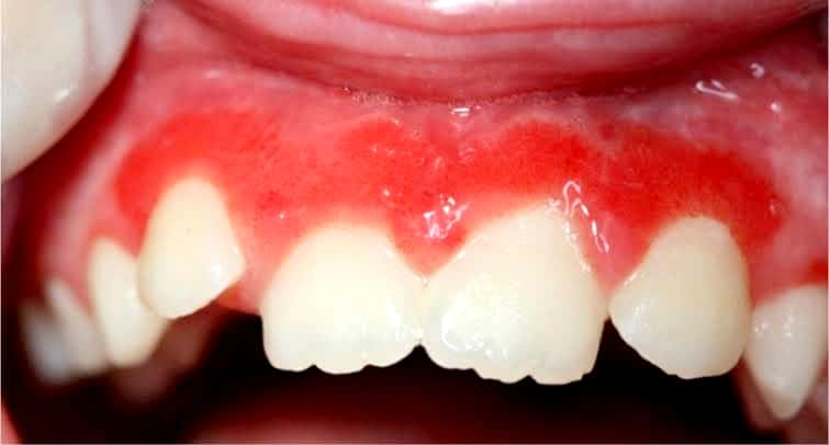
Etiology

It is usually caused by bacteria.

• Most common pathogens are Staphylococcus species (aureus in 80-90% of the cases).

🔻Other organisms: Klebsiella, Neisseria gonorrhoeae, Haemophilus influenzae Escherichia coli, Pseudomonas, and Salmonella species

🔻Escherichia coli in patients with genitourinary tract infections or intravenous drug abusers.

Neonatal period: Haemophilus Influenzae and group B streptococci.

Patients with sickle cell disease: Salmonella infection.

Mixed bacterial infections: It is due to direct spread or surgery or open fractures.

• In about 50%, no organisms can be isolated.

Portal entry of organisms

Causative organisms may reach the bone through the bloodstream, directly or extend from a contiguous site.

  1. Hematogenous spread:

• Source of organisms may be a focus of infection anywhere in the body (e.g skin pustule or infected teeth and gums, intestinal mucosa).

Minor injuries to the mucosa (vigorous chewing of hard foods, brushing of teeth), or minor infections of the skin, release these organisms into the blood → causing temporary bacteremia → reach the bone.

In children (5 to 15 years) and drug addicts (infected needles), it develops in the long bones.

  1. Direct implantation: Organisms may enter into bone by penetrating wounds, open fractures, or surgical procedures (staphylococci, streptococci, anaerobic organisms).
  2. Spread from adjacent (contiguous) site: e.g. infections of the feet may spread into the bone in diabetics.

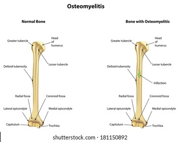
Location of infection: It varies with age.

Neonate: Metaphysis, epiphysis, or both.

Adult: Epiphysis and subchondral regions

Children with hematogenous spread: Metaphysis of long bones (knee, ankle, and hip).

Pathogenesis and Morphology

The sequence of events and morphological features in osteomyelitis are described together.

Transient bacteremia: Mild injury or trauma can initiate bacteremia by organisms (eg.Staphylococcus aureus).

Infection reaches metaphysis long bone: Because in the metaphysis capillaries form loop – which slows the blood flow provides time for bacteria to penetrate blood vessel walls and establish infective foci within the marrow.

Inflammatory Reaction: Once in bone, the bacteria grow and induce an acute inflammatory reaction with exudates.

Necrosis of bone: Exudate increases the pressure on the adjacent vessels and further decreases the blood supply produces bone necrosis. The necrotic areas coalesce and allow further bacterial proliferation.

Formation of sequestrum: Bacterial infections and pus spreads into the cortex and collects beneath the periosteum and may lift the periosteum and reduce the blood supply to the affected region.

The fragment of dead necrotic piece of bone, which is embedded in the pus, is known as a sequestrum.

abscess infection - pictures, photos
source -google

What is a sequestrum ?

🔻The pus penetrates the periosteum and leads to a soft-tissue abscess may penetrate the skin form a draining sinus.

Hole formed in the bone during the formation of a draining sinus is known as cloaca

🔻The sinus tract may become epithelialized and may remain open, continually draining pus, necrotic bone, and bacteria.

Involucrum

After first week, chronic inflammatory cells become more numerous and the cytokines released stimulates osteoclastic bone resorption and deposition of reactive bone in the periphery.

Reactive new bone forms a sheath around the necrotic (segment of devitalized infected bone) sequestrum. This reactive new bone formed is known as involucrum.

A sequestrum is usually a complication of osteomyelitis and ...

Variants of osteomyelitis

Brodie abscess: It is a distinctive form of subacute pyogenic osteomyelitis.

  • It appears as a small, solitary, intraosseous abscess localized to the metaphysis and is surrounded by reactive bone.

It may be due to inadequate treatment of infection by less virulent organisms.

Brodie's abscess | Development, Videos tutorial, Learning
source -google

Sclerosing osteomyelitis of Garre – characterized by extensive newborn formation which obscures the underlying structure of the bone and typically develops in the jaw.

Management of Garré's sclerosing osteomyelitis by endodontic ...
source -google

Complications

Septicemia: From infection in the bone, organisms may disseminate through the blood stream and cause septicemia.

Acute suppurative arthritis: Infection may spread through the articular surface into a joint producing suppurative arthritis may lead to destruction of the articular cartilage and permanent disability. It is more common in infants.

Pathologic fractures.

Squamous cell carcinoma: It may arise from the epithelialized sinus tract, rarely sarcoma of bone may develop.

Secondary amyloidosis.

Chronic osteomyelitis: It may develop due to delay in diagnosis, extensive bone necrosis, and inadequate therapy.

Clinical therapy

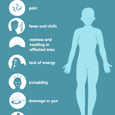
🔻 present with malaise, fever, chills , leukocytosis and throbbing pain over the affected area

Osteomyelitis: Symptoms, Causes, Diagnosis and More
source -google

🔻 Diagnosis-

Radiography- lytic focus of bone destruction

Blood cultures are positive

Biopsy and bone culture

source -textbook of pathology for dental students Harsh mohan

Osteoporosis

Osteoporosis is a disease of bone which characterized by low bone mass that leads to porosity of bones.

🔻characterised by porus bone – thinning of the cortex and trabaculae

🔻 increase in the risk of fractures – hip ,wrist and spine

Types:

Osteoporosis may be:

Localised : It is usually due to disuse and is seen as a complication of some other disease.

For example, local immobilization following fracture due to other causes.

Generalized: Involves the entire skeleton. It may be primary or secondary.

Primary osteoporosis occurs without any known cause.

These include senile postmenopausal.

-Secondary osteoporosis develops due to a large variety of conditions.

These include endocrine disorders (hyperparathyroidism, hyperthyroidism, etc.),

gastrointestinal disorders (e.g. malabsorption, vitamin D deficiency, malnutrition),

drugs (e.g. corticosteroids),

tumors (multiple myeloma)

reduced physical activity,

hormonal disturbances

and other causes (e.g. immobilization).

Etiology

Age : Peak bone mass is achieved between 25 and 35 years of age and gradually declines from the age of 50 years onwards.

Sex: It is generally seen in elderly and women. Genetic/hereditary factors also play an important role .

Pathogenesis

🔸Ageing-due to decrease in the osteoblastic activity and decrease in the growth factors.

🔸Hormonesdecreased estrogen, cytokines like IL-1 and IL-6,which increase the osteoclastic activity.

🔸Decrease in the physical activity

🔸Genetic factors

🔸Nutritional status- low calcium intake

Morphology

Entire skeleton is involved in postmenopausal and senile osteoporosis but certain regions are more severely involved than others.

The involved bones are thin and brittle.

Microscopically, it is composed of the lamellar bone which is discontinuous and has thin trabeculae.

Source – Google

Clinical features

Depend on the bones involved.

Commonly involved areas are vertebral bodies and femoral neck

Loss of bone mass predisposes the bone to fracture which results in bone pain.

Fractures are most common in the vertebra in the thoracic and lumbar region, neck of the femur and Colles’ fracture (fracture of distal radius).

Complications

Femoral neck

Pulmonary embolism and pneumonia as a complication of fractures of the femoral neck, pelvis or spine.

Vertebral fracture

• When fractures of vertebrae are multiple, it may lead to lumbar lordosis and kyphoscoliosis (forward bending).

Source – Google

Diagnosis

🔸plain X-ray – not reliable till 30-40%of bone mass isn’t lost

🔸Dual energy X-ray absorptiometry and quantitative computed tomography

🔸Bone biopsy

Prevention

Exercise

Appropriate calcium and vit d intake

Agents like bisphosphonates

Source – textbook of pathology for dental students – harsh mohan and pathology preparatory manual for dental students Ramdas Nayak

OEDEMA: PATHOGENESIS & TYPES

👉🏻Definition & Types:

🔹OEDEMA〰️Swelling. Abnormal/excessive accumulation of free fluid in the interstitial tissue spaces & serous cavities.

🔹Accumulation of fluid (abnormally) inside the cell is intracellular edema/hydropic degeneration.

🔹Accumulation in body cavities:

  • Peritoneal Cavity – Ascitis
  • Pleural Cavity – Hydrothorax (T.B)
  • Pericardial cavity – Hydropericardium
TYPES OF OEDEMA👆🏻

🔹PITTING & NON-PITTING EDEMA:

Source: Healthline

➡️ When interstitial fluid volume increases, most of the fluid becomes free that is not bound to proteoglycan meshwork. This type of edema is called pitting edema because when this area is pressed with finger, fluid gets displaced producing depression or a pit as seen in SUBCUTANEOUS TISSUES.

➡️ Edema also develops due to swelling of the cells/clotting of interstitial fluid in the presence of fibrinogen. This is called Non-pitting/solid edema because its hard & a pit is not formed by pressing.

  • e.g. – Myxodema
  • – Elephantiasis

Oedema Fluid

🔹 Pathogenesis of Edema:

➡️ Causative mechanisms that produce edema are:

  1. ⬇️ Plasma oncotic pressure
  2. ⬆️ capillary hydrostatic pressure
  3. Lymphatic obstruction
  4. Sodium & Water retention
  5. ⬆️ capillary permeability
  6. Tissue factors

These mechanisms interfere with normal fluid balance of plasma, interstitial fluid & lymph flow.

  • It is exerted by total amount of plasma proteins that tend to draw fluid into the vessels normally.
  • A fall in the total plasma protein level (<5g/dl), results in lowering of plasma oncotic pressure in a way that it can no longer counteract the effect of hydrostatic pressure of blood.
  • Therefore, increased outward movement of fluid from capillary wall & decreased inward movement of fluid from the interstitial space causing edema.
Fluid pressure and colloid osmotic pressure forces operate at the capillary membrane, tending to move fluid either outward or inward through the membrane pores
Pathogenesis of oedema in hypoalbuminaemia. The normal balance of hydrostatic and oncotic pressures is such that there is net movement of fluid out of the capillaries at their arteriolar ends and net movement in at their venular ends (indicated here by arrows). Oedema can thus be due to an increase in capillary hydrostatic pressure, a decrease in plasma oncotic pressure or an increase in capillary permeability.

There are many possible causes of hypoalbuminaemia; a combination of which may be implicated in individual cases. For example, to list few of them👇🏻

  • Oedema of renal disease (Nephrotic syndrome, Acute glomerulonephritis)
  • Ascitis of liver disease e.g. in Cirrhosis.
  • Protein losing enteropathy
  • Malnutrition

➡️ Fluid from capillary wall to interstitial space by counteracting the force of plasma oncotic pressure.

➡️ Edema results when hydrostatic pressure at Venular end of capillary wall(12mmHg) becomes more than the plasma oncotic pressure, resulting in no reabsorption of fluid at the Venular end.

  • Example, edema of Cardiac disease – Congestive cardiac failure, constrictive pericarditis
  • Cirrhosis of liver
  • Passive congestion – e.g. mechanical obstruction due to thrombosis of veins of lower legs, varicosities, pressure by pregnant uterus, tumours etc.
  • Postural edema e.g. ⬆️ Venous pressure (Edema of feet & ankle)

➡️ Lymphoedema is caused due to obstruction of lymphatic channels causing localized edema.

  • e.g. removal of axillary lymph nodes in radical mastectomy for carcinoma of breast cancers.
  • Rupturing of main lymphatic channel causing chylothorax & chylous ascitis

– Inflammation of Lymphaties:

  • Filariasis (infection with Wuchereria Bancrofti) – Chronic lymphoedema of scrotum & legs

– Abnormal development of lymphatic channels:

  • Milroy’s disease/hereditary lymphoedema
  • Edema comfined to one or both lower limbs.
Source: Slideshare
Source: Slideshare

Forces acting in the interstitial space –

1. Oncotic Pressure

⬇️

⬆️ Vascular permeability & inadequate removal of proteins by lymphatics

⬇️

Elevation

2. Tissue Tension

⬇️

Lowered (e.g. loose subcutaneous tissues of eyelids & external genitalia)


Dr. Mehnaz Memon🖊

References: Textbook of Pathology, Harsh Mohan; Internet; https://basicmedicalkey.com/plasma-proteins-and-enzymes/

Aplastic anemia

Sources : Shafer’s textbook of oral pathology , slideshare.com

Image sources : Slideshare and Google images