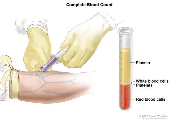
HEMATOLOGICAL INVESTIGATIONS

Also known as: CBC; Hemogram

Sample Required?

  • A blood sample drawn from a vein in your arm or a fingerstick or heelstick (newborns)

Test Preparation Needed?

  • None

Why get tested?

  • To determine your general health status; to screen for, diagnose or monitor any one of a variety of diseases and conditions that affect blood cells, such as anemia, infection, inflammation, bleeding disorder or cancer.

Also known as: Hgb; Hb; H and H (Hemoglobin and Hematocrit)

Sample Required?

  • A blood sample drawn from a vein in your arm or a fingerstick or heelstick (newborns)

Test Preparation Needed?

  • None

Why get tested?

  • To evaluate the hemoglobin content of your blood as part of a general health check-up; to screen for and help diagnose conditions that affect red blood cells (RBCs); If you have anemia (low hemoglobin) or polycythemia (high hemoglobin), to assess the severity of these conditions and to monitor response to treatment

• When to get tested?

  • With a hematocrit or as part of a complete blood count (CBC), which may be ordered as a component of a general health screen; when you have signs and symptoms of anemia (weakness, fatique) or polycythemia (dizziness, headache); at regular intervals to monitor these conditions or response to treatment

Also known as: Thrombocyte count; PLT; Platelet distribution width; PDW; Mean Platelet volume; MPV.

Sample Required?

  • A blood sample drawn from a vein in your arm or a fingerstick or heelstick (newborns)

Test Preparation Needed?

  • None

Why get tested?

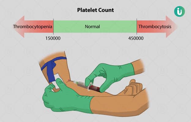
  • To determine the number of platelets in a sample of your blood as part of a health exam; to screen for, diagnose, or monitor conditions that affect the number of platelets, such as a bleeding disorder, a bone marrow disease, or other underlying condition.

• When to get tested?

  • As part of a routine complete blood count (CBC); when you have episodes of unexplained or prolonged bleeding or other symptoms that may be due to a platelet disorder

What is being tested?

  • Platelets, also called thrombocytes, are tiny fragments of cells that are essential for normal blood clotting. They are formed from very large cells called megakaryocytes in the bone marrow and are released into the blood to circulate. The platelet count is a test that determines the number of platelets in a person’s sample of blood. When there is an injury to a blood vessel or tissue and bleeding begins, platelets help stop bleeding.

Also known as: Leukocyte differential count; Peripheral differential; WBC count differential; Diff; blood differential; Differential Blood Count

Formal name: White blood cell differential

• Why get tested?

  • To help determine the cause of abnormal results on a WBC count; to help diagnose or monitor an illness affecting your immune system, such as an infection or inflammatory condition, or cancers that affect your white blood cells, such as leukemia.

• When to get tested?

  • As part of a CBC; when you have a routine health examination; when results of a CBC fall outside the reference range; when you have any number of signs and symptoms that may be related to a condition affecting white blood cells, such as infection, inflammation, or cancer, when you are receiving treatment that is known to affect WBCs, such as chemotherapy.

• What is being tested?

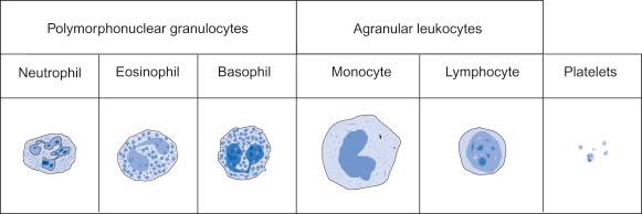
  • WBCs, also called leukocytes, are cells that circulate in the blood and the lymphatic system that help protect the body against infections. They are an important part of the body’s immune system and also have a role in inflammation, allergic responses, and protection against cancer. A WBC differential totals the number of each of the different types of WBCs in a person’s sample of blood.
  • There are five types of white blood cells, each with different functions.

  • Also known as: TLC; WBC count
  • Total WBC count is used as one of the index of presence of systemic infection and to rule out the possibility of leukemia & malignant neutropenia
  • Calculated with haemocytometer/ automated cell counts
  • RBCs are lysed by diluting the blood sample with dilute acetic acid leaving the WBCs intact.

  • Also known as: Red Blood Cell Count, RBC count
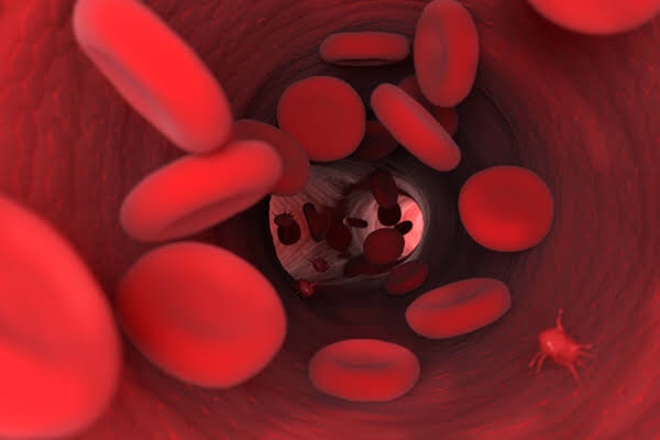
  • Red blood cells, also known as erythrocytes, make up the cellular part of blood, giving it its red color and also the ability to bind and carry oxygen to all parts of the body. Under a microscope, they appear to be circular and biconcave in shape.
  • Gives us the number of erythrocytes per cubic mm in circulating blood & Hb in blood.
  • Procedure done by office or chairside method and also automated procedure.
  • Hematological diseases of RBCs are anemia & polycythemia.

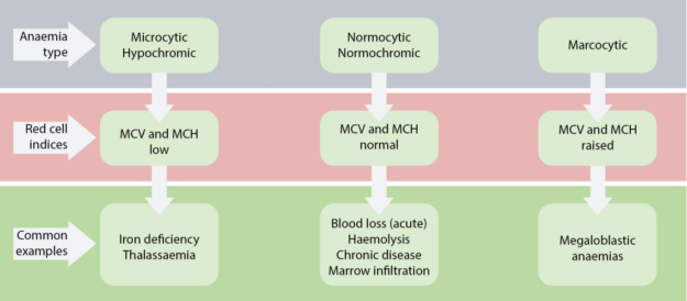
  • Categorized by mean corpuscular volume, anemia can be differentiated into microcytic, macrocytic and normocytic anemias. Normocytic anemia can be further divided into intrinsic and extrinsic RBC defect and blood loss.
  • MCV – Mean corpuscular volume is the average volume of red blood cells and is reflective of RBC size. When RBCs increase or decrease in size, the mean corpuscular volume changes; this helps physicians determine the type of anemia and its causes. Normal MCV is 80–96 µm³.
  • MCH stands for “mean corpuscular hemoglobin.” An MCH value refers to the average quantity of hemoglobin present in a single red blood cell.
  • MCHC is short for mean corpuscular hemoglobin concentration. MCHC refers to the average amount of hemoglobin inside a single red blood cell.
  • Hematocrit is the measure of the total volume % of red blood cells in the blood. The normal value for hematocrit is 45% for men and 40% for women. It is an important component of a patient’s complete blood profile.

Indications:

  • To prepare smears from paper points removed from root canals for evaluation of microcytic status of canal prior to filling.
  • A scraping or swab of an oral lesion is needed to confirm diagnosis of thrush
  • A scraping of gingival region or mucosal ulcer is sometimes used to confirm diagnosis of Acute Necrotising ulcerative stomatitis.
  • Identification of giant cells that accompany vesicular infections
  • Identification of Acantholysis

Dentowesome|@drmehnaz🖊


References: Google.com, lecturio.com, Study Notes✍🏻

Routes of metastasis

Spread of cancer to distant sites is by following routes:-

Lymphatic spread :-

•Generally, carcinomas spread by lymphatic route and sarcomas spread through hematogenous route.However few sarcomas may also spread by lymphatic route.

• Involvement of lymph nodes by malignant cells is by:-

– lymphatic permeation :- The walls of the lymphatics are readily invaded by cancer cells and may form continuous growth in lymphatic channels called lymphatic permeation.

– lymphatic emboli :- Malignant cells may detach to form tumour emboli that may be carried along to the next lymph node. Tumour emboli will enter the lymph node by convex surface and gets lodged in subcapsular sinus and start growing . Later the lymph node will be enlarged and will be replaced by malignant tumour.

Characteristics of lymphatic spread:-

• Regional lymph nodes are invariably involved producing regional nodal metastasis. From carcinoma of the breast to axillary lymph nodes ; from the carcinoma of thyroid to cervical lymph nodes and however all regional nodal enlargements are not because of nodal metastasis.

• And sometimes, Lymphatic metastasis do not develop in the 1st lymph node which is nearest to the tumour because of venous lymphatic anastomoses or due to radiation , obliteration of lymphatics by inflammation so called skip metastasis.

• Other times due to obstruction of lymphatics by the tumour cells the lymph flow is disturbed and the tumour cells spread against the flow of lymph causing retrograde metastasis at unusual sites. Metastasis of carcinoma prostate to supraclavicular lymph nodes.

Virchows lymph node is nodal metastasis preferentially to supraclavicular lymph node from cancers of abdominal organs like cancer of stomach , colon.

Hematogenous spread :-

• Sarcomas usually spread by hematogenous route. And few carcinomas spread through hematogenous route.

Few features of hematogenous route are :-

• systemic veins drain blood to inferior vena cava from limbs , head and neck and pelvis so tumour of these organs will mostly metastasize to lungs.

• portal veins drains blood from bowel, pancreas and spleen to liver so, tumour of these organs are secondarily metastasize to liver.

Gross appearance of affected organ:-

There are multiple ; rounded nodules of varying size and scattered throughout the organ.

Microscopically:- primary tumour upon metastasis at different sites show varying degree of differentiation due to influence of local environment surrounding the tumour for its growth.

Spread along the body cavities and natural passages:-

• It’s the uncommon route of spread of tumour.

• Transcoelomic spread :- certain cancers invade through the serosal wall of the coelomic cavity so that the tumour cells or its fragments breaks off and will be carried in the coelomic fluid and gets implanted else where in the body. Most often involved is peritoneal cavity and less occasionally pleural and pericardial cavity.

– examples :- carcinoma of the stomach seeding to both ovaries (krukenberg tumour)

• Spread along the epithelium lined surfaces :- it’s unusual because intact epithelium and mucus are resistant to be penetrated by tumour cells. Examples :-are through the bronchus into alveoli.

• Spread via CSF:- Malignant tumours of ependyma and leptomeninges may spread by release of tumour cells through CSF.

• Implantation :- rare cases of reports of spread of some tumours is by implantation.

Reference :- Harsh mohan

Thrombus

• Thrombus is a blood clot in the circulatory system. It attaches to the site at which it is formed and remains there, hindering the blood flow.

• Thrombus is most likely to occur in the people who are immobile and those with a genetic predisposition to blood clotting.

• When a thrombus forms in an artery such as heart or brain is called arterial thrombosis.

• When a thrombus occurs in a vein is called venous thrombosis and if it occurs in deep veins it’s called deep vein thrombosis(DVT).

• Section of blood clot that breaks free from the thrombus and circulates in the bloodstream it’s called as an Embolus . Embolus is a dangerous and fatal complication of thrombosis.

Causes of thrombus:-

• Use of tobacco

• High cholesterol level

• Atherosclerosis

• Obesity

•Diabetes

•Stress

• Inactive life style

Symptoms of arterial thrombosis:-

• Unstable angina

• Myocardial infarction

• Ischaemic stroke

Symptoms of venous thrombosis :-

• pain , swelling and tenderness , usually in the calf

• Aching and warmth of the skin in the affected area

• Red skin, particularly at the back of the leg below the knee

DVT can be life threatening conditions ; emboli can travel through the bloodstream before blocking arteries elsewhere in the body.

Diagnosis:-

• Duplex ultrasound:- This is the most common test for diagnosing DVT. Duplex ultrasound uses the sound waves to create the images of the blood flowing through arteries and veins.

• A D – dimer test :- This test measures the levels of a substance in the blood that results from the breakdown of blood clots. High levels of this substance may indicate the presence of DVT or another type of blood clot.

• Venography:- A dye will be injected into the affected leg. This dye makes the vein visible on X ray such as fluoroscopy. If the scan shows a slower than the usual blood flow through the vein a thrombus may be present.

• MRI and CT scans

• A VQ scan it uses radioactive substance called radiotracer to reveal, on the scan ,the flow of Air and Blood within the lungs.

Treatment :-

• Surgery may be done in the case of medical emergency.

• Inferior vena cava (IVC) filters :- They are small mesh devices that a surgeon can put in the inferior vena cava under a local anesthetic. Inferior vena cava filters traps the blood clot and prevents them from reaching the heart and lungs. An IVC filter can be permanent or may be combined with anticoagulant therapy.

• Anticoagulants reduce the risk of clot forming and which also reduces the size of the thrombus.

• Compression stockings:- The stockings help to prevent calf pain and swelling and also reduces the risk of complications.

• Excercise :-usually recommended to stimulate blood circulation.

Source:- Medicalnewstoday.com

Granuloma

• Granuloma is defined as a circumscribed tiny lesion and it’s about 1 mm in diameter. Granuloma is derived from word granule means granule like lesion and oma suffix generally refers to tumour but here it refers to collection of macrophages.

• Granuloma is type 4 hypersensitivity reaction. It’s protective but sometimes leads to tissue destruction because of poorly digestible antigen.

Pathogenesis :-

• Macrophages and monocyte engulf the antigen and tries to destroy it since the antigen is poorly degradable these cells fail to destroy antigen and macrophages undergoes morphological changes and transform into epitheloid cells.

• As macrophages have failed to destroy antigen they present it to CD 4 T lymphocytes. Lymphocytes get activated and releases interleukin 1 and interleukin 2 that stimulate more T cells, interferon gamma activates macrophages.

• And also TNF alpha that promotes proliferation of fibroblasts and activates the endothelium to secrete prostaglandin and growth factors which stimulate fibroblast growth.

Composition of granuloma

Epitheloid cells are nothing but modified macrophages these are elongated cells and have slipper shaped nucleus . Nuclear chromatin is vesicular and lightly stained and cytoplasm is abundant and pale staining with hazy outlines.

Multinucleate giant cells formed by the fusion of adjacent epitheloid cells containing more than 20 nuclei. The nuclei may be arranged like horseshoe or ring or may be clustered at 2 poles ( langhans gaint cell seen in tuberculosis ) or may be present centrally ( foreign body giant cell seen in foreign body tissue reaction).

lymphoid cells as response to cell mediated immunity to antigen lymphocytes are the integral composition of granuloma. In some granulomas presence of plasma cells is indicative of humoral immune response.

Necrosis is a feature of some granulomataous conditions like TB.

Fibrosis is a feature of healing formed by fibroblast at the periphery.

So, Granuloma is having epitheloid cells at centre with interspersed gaint cells , surrounded peripherally lymphocytes and healing by fibroblast. Granuloma is a microscopic finding and it indicates chronic inflammation.

Reference :- Harsh mohan

Aneurysm

Aneurysm is one of the most common words we see in our pathology books. Every year in India, around 76,000-200,000 cases of cerebral aneurysms are reported to have occurred. An estimated 6.5 million people in the United States have an unruptured brain aneurysm, or 1 in 50 people. The Centre for Disease Control and Prevention (CDC) states that aortic aneurysms contribute to over 25,000 deaths each year in United States. Let’s see what the word actually means and it’s related causes.

The word “aneurysm” comes from the Greek word “aneurysma” meaning “a widening”.

An aneurysm refers to a weakening of an artery wall that creates a bulge, or distension of the artery.

Types of aneurysms :

Aneurysms are classified by their location in the body. The arteries of the brain and the heart are the two most common sites of a serious aneurysm.

The bulge can take two main shapes:

  • Fusiform aneurysms bulge all sides of a blood vessel
  • Saccular aneurysms bulge only on one side

Aortic aneurysm : the aorta is the large artery that begins at the left ventricle of the heart and passes through the chest and abdominal cavities. The most common aneurysm of the aorta is an abdominal aortic aneurysm (AAA). Less commonly, a thoracic aortic aneurysm (TAA) can affect the part of aorta running through the chest.

Cerebral aneurysm : aneurysms of the arteries that supply the brain with blood are known as intracranial aneurysms. Ruptured cerebral aneurysms are the most common cause of a type of stroke known as “subarachnoid hemorrhage”.

Peripheral aneurysm : an aneurysm can also occur in a peripheral artery. Peripheral arteries are less likely to rupture than aortic aneurysms. Types of peripheral aneurysm include:

  • Popliteal aneurysm : this happens behind the knee. It is the most common peripheral aneurysm
  • Splenic artery aneurysm : this type of aneurysm occurs near the spleen
  • Mesenteric artery aneurysm : this affects the artery that transports blood to intestines
  • Femoral artery aneurysm : the femoral artery is in the groin
  • Carotid artery aneurysm : this occurs in the neck
  • Visceral aneurysm : this is the bulge of arteries that supply blood to the bowel or kidneys

Cause of aneurysm : aortic dissection is one identifiable cause of an aortic aneurysm. The arterial wall has three layers. Blood can burst through a tear in the weakened wall of the artery, splitting these layers. It can then fill the cavity surrounding the heart. Dissection leads to compression. Compression prevents blood from returning to the heart. This is also known as a pericardial tamponade.

Risk factors : smoking tobacco, hypertension or high blood pressure, poor diet, inactive lifestyle, obesity

Symptoms : most aneurysms are clinically silent. Symptoms donot usually occur unless an aneurysm ruptures. Rapidly growing abdominal aneurysms are sometimes associated with symptoms. Some people with abdominal aneurysms report abdominal pain, lower back pain, or a pulsating sensation in the abdomen. Similarly, thoracic aneurysms can affect nearby nerves and other blood vessels causing swallowing and breathing difficulties, and pain in the jaw, chest and upper back.

Complications : thromboembolism, severe chest or back pain, angina, a sudden extreme headache

Diagnosis : an MRI scan can identify an aneurysm that has not yet ruptured. CT scans are usually preferred for ruptured aneurysms

Treatment : a ruptured aneurysm needs emergency surgery. Without immediate repair, patients have a low chance of survival.

A large or growing aortic aneurysm is more likely to need surgery. There are two options for surgery

  • Open surgery to fit a synthetic or stent graft
  • Endovascular stent graft surgery

In the Endovascular surgery, the surgeon accesses the blood vessels through a small incision near the hip. Stent graft surgery inserts an endovascular graft through this incision through a catheter. The graft is then positioned in the aorta to seal off the aneurysm.

In the open AAA repair, a large incision is made in the abdomen to expose the aorta. A graft can then be applied to repair the aneurysm.

Source : medicalnewstoday.com

Apoptosis

• It is coordinated and internally programmed cell death it has significance in various physiological processes like involution of thymus in early age , sculpting of tissues during development of embryo and pathological process like diseases of heart like acute MI and CNS diseases like alzehmeirs disease and parkinson’s disease

In apoptosis single cells or small cluster of cells are involved. When compared to the normal cell the apoptotic cell has more eosinophilic cytoplasm and chromatin condensation.

Apoptotic bodies are membrane bound spherical bodies with cell shrinkage ,chromatin condensation and eosinophilic cytoplasm. Apoptotic bodies are also referred to as Mummified bodies.

DNA changes can be seen by gel electrophoresis by H and E stain to see the chromatin condensation and other stains. Flow cytometry for cell shrinkage.

Apoptosis is not accompanied by inflammation unlike necrosis

Mechanism of apoptosis :-

1. Initiators of apoptosis :- Every cell has built mechanism for the cell survival and to activate the signal of cell death. Apoptosis takes place when there is withdrawal of cell survival signals like growth factors , cytokines. And the agents of cell injury like heat and radiation leads to Apoptosis

2. Initial phase of apoptosis :- There are 2 pathways by which apoptosis takes place that is intrinsic and extrinsic pathway by activating the caspases. Intrinsic pathway is the major pathway of cell death. Caspases are nothing but the protein splitting enzymes that will act on nuclear proteins and other protein components.

Intrinsic pathway :-

This pathway of cell death is by release of protein cytochrome C from mitochondria to cytoplasm. This triggers the cell to Apoptosis. Protein C is the life line of intact mitochondria. This is regulated by pro and anti apoptotic genes. Anti apoptotic genes are bcl2 , Mcl 1 and bcl x. Pro apoptotic genes are bid , bad. The proapoptotic genes will trigger apoptosis that will release Bak and Bax that will damage the mitochondrial membrane and releases the cytochrome c from mitochondria to cytoplasm that will activate caspases

Extrinsic pathway :-

This pathway of cell death is by activation of cell death receptors present on the cell surface. Cell death receptor tumour necrosis factor and related transmembrane protein that is fas and it’s ligand fas l this binding is associated with Fas associated death domain. This will activate caspases

3. Final phase of apoptosis :- There is activation of caspases 8 by intrinsic pathway and caspases 9 and 10 by extrinsic pathway that will act on the nuclear proteins and this leads to the changes in the DNA and cell death.

4. Phagocytosis :- The surface of the cell will undergo changes in order to activate the adjacent macrophages for its phagocytosis. Phosphatidyl serine and thrombospondin molecules is present inside, during apoptosis it comes to the exterior that triggers the cell for its phagocytosis. Phagocytosis is rapid and it’s not accompanied by inflammatory cells.

Reference :- Harsh Mohan

Gastroesophageal reflux disease (GERD)

  • GERD is a digestive disorder that affects the ring of muscle between your Esophagus and stomach. This ring is called the lower esophageal sphincter.
  • Causes : in normal digestion, your LES opens to allow food into our stomach. Then it closes to stop food and acidic stomach juices from flowing back into your esophagus. GERD happens when the LES is weak or relaxes when it shouldn’t. This let’s the stomach’s contents flow up into the Esophagus.
  • Some doctors believe a hiatal hernia may weaken the LES and raise your chances of gastrophageal reflux. Hiatal hernia happens when the upper part of your stomach moves up into the chest through a small opening in the diaphragm.
  • Several other things can make it more likely that you’ll have GERD : being obese, pregnancy, delayed emptying of stomach (gastroparesis), diseases of connective tissue such as rheumatoid arthritis or lupus.
  • Symptoms : most common is heartburn, the burning or pain of heartburn can last as long as 2 hours. Besides pain, you may also have nausea, bad breath, trouble breathing, wearing away of tooth enamel, a lump in your throat.
  • Treatment – antacids : they neutralize acid in the Esophagus and stomach and stop heartburn.
  • H2 blockers – cimetidine, famotidine
  • Proton pump inhibitors – dexlansoprazole, esomoprazole, Omeprazole, pantoprazole
  • Prokinetics – they elp to empty the stomach faster – include domperidone, metoclopramide
  • GERD complications – esophageal ulcer, esophageal stricture, Barrett’s Esophagus, lung problems such as chest congestion or aspiration, asthma, bronchitis or even pneumonia.

Source : Robbins and Cotran’s book of pathology

Barrett’s Esophagus

  • Barrett’s Esophagus is a complication of GERD that is characterized by intestinal metaplasia within the esophageal squamous mucosa.
  • The greatest concern in Barrett Esophagus is that it confers an increased risk of esophageal adenocarcinoma.
  • Morphology – Barrett Esophagus can be recognised as one or several tongues or patches of red, velvety mucosa extending upward from the gastroesophageal junction
  • This metaplastic mucosa alternates with residual smooth, pale squamous mucosa and interferes with light brown columnar mucosa distally.
  • Goblet cells, which have distinct mucous vacoules that stain pale blue by H&E and impart the shape of a wine goblet to the remaining cytoplasm, define intestinal metaplasia and are necessary for diagnosis of Barrett Esophagus.
  • Clinical features – Barrett Esophagus can only be identified through endoscopy and biopsy, which are usually prompted by GERD symptoms

Treatment – endoscopic resection, which uses an endoscope to remove damaged cells to aid in the detection of dysplasia and cancer.

Radiofrequency ablation, which uses heat to remove abnormal Esophagus tissue.

Genetic disorders with mnemonics

Autosomal dominant disorders :

(Mnemonic : vo familial hypercholesterolemia autosomal dominant hai)

Vo – Von willebrand disease

Familial – Familial adenomatous polyposis

Hypercholesterolemia – hypercholesterolemia (familial)

Autosomal – Adult polycystic kidney

D – Dystrophia myotonica

O – Osteogenesis imperfecta

M – Marfan syndrome

I – Intermittent porphyria

N – Neurofibromatosis

A – Achondroplasia

T – Tuberous sclerosis

Hai – Huntington’s disease, Hereditady spherocytosis

Autosomal recessive disorders :

(Mnemonic : Fried Poori aur Garam chawal mast hai)

Fried – Friedrich’s ataxia

Poori aur – Phenylketonuria

Garam – Galactosemia

C Cystic fibrosis

H Hemochromatosis

A Alpha 1 antitrypsin deficiency

W Wilson’s disease

A Alkaptonuria

L Lysosomal and glycogen storage diseases

M Muscular atrophy

A Adrenal hyperplasia

S Sickle cell disease

TThalassemia

Hai – Homocystinuria

Atherosclerosis

  • Atherosclerosis is characterized by intimal lesions called atheromas that protrude into vessel lumens.
  • An atheromatous plaque consists of a raised lesion with a soft, yellow, grumous core of lipid (mainly cholesterol and cholesterol esterscocered by a white fibrous cap.

Major risk factors for atherosclerosis:

Non modifiable factors –

  1. Age : atherosclerosis is typically progressive, it does not usually manifest clinically until middle age or later
  2. Gender : premenopausal women are relatively protected aginst atherosclerosis compared to age-matched men. After menopause, however, the incidence of atherosclerosis related diseases increases and at older ages actually exceeds that of men
  3. Genetics : family history is theost significant independent risk factor for atherosclerosis. The well established familial predisposition to atherosclerosis is usually multifactorial, relating to inheritance of various genetic polymorphisms and hypertension or diabetes

Modifiable risk factors :

  1. Hyperlipidemia : and more specifically hypercholesterolemia is a major risk factor for atherosclerosis, hypercholesterolemia is sufficient to stimulate lesion development. LDL cholesterol is the form of cholesterol that is delivered to peripheral tissues. In contrast, HDL mobilizes cholesterol from tissue and transports it to the liver for excretion in the bile.
  2. Hypertension : it is the most important cause of left ventricular hypertrophy and hence the latter is also related to IHD
  3. Cigarette smoking : the increased risk and severity of atherosclerosis in smokers is due to reduced level of HDL, deranged coagulation system and accumulation of carbon monoxide in the blood that produces carboxyhemoglobin and eventually hypoxia in arterial wall favouring atherosclerosis.
  4. Diabetes mellitus : the incidence of atherosclerosis is twice as high in diabetics as in non diabetics. There is also an increased fold of strokes and a 100-fold increased risk of atherosclerosis induced gangrene of the lower extremities.

Pathogenesis of atherosclerosis :

  • Endothelial injury – endothelial loss due to any kind of injury results in intimal thickening.
  • The etiological culprits contributing to endothelial cell dysfunction in early atherosclerosis include hypertension, hyperlipidemia, toxins from cigarette smoke, homocysteine and even infectious agents
  • Hemodynamic disturbances – plaques tend to occur at Ostia of existing vessels, branch points, and along the posterior wall of the abdominal aorta, where there are disturbed flow patterns
  • Lipids – the dominant lipids in atheromatous plaques are cholesterol and cholesterol esters. Chronic hypercholesterolemia can directly impair endothelial cell function by increasing local oxygen free radical production; oxygen free radicals can injure tissues and accelerate nitric oxide decay, reducing its vasodulator activity
  • Inflammation – monocytes transform into macrophages and avidly engulf lipoproteins including oxidised LDL . Activated macrophages produce reactive oxygen species that aggrevate LDL oxidation and elaborate growth factors that drive smooth muscle cell proliferation
  • Infection – herpes virus, cytomegalovirus, Chlamydia pneumoniae have all been detected in Atherosclerotic plaques but not in normal arteries.
  • Smooth muscle cell proliferation – initial smooth muscle cell proliferation and ECM deposition convert a fatty streak, the earliest lesion into a mature atheroma and contribute to progressive growth of atherosclerotic lesions

Morphological features of atherosclerosis:

  1. Fatty streaks and dots : they may be the precursor lesions of atheromatous plaques and are prominent in aorta and major arteries
  2. Gelatinous lesions : they develop in the intima of the aorta and may also be the precursora of plaques
  3. Atheromatous plaques : a fully developed atheromatous lesion is called atheromatous plaque. Most often and severely affected is the abdominal aorta. Grossly they are white to yellowish white lesions varying in diameter from 1-2 cm. Microscopically, superficial luminal part of fibrous cap is covered by endothelium, and is composed of smooth muscle xells; cellular area under the fibrous cap is composed of mixture of macrophages, foam cells, lymphocytes; deeper central soft core consists of extracellular lipid material, cholesterol clefts,fibrin; in oldrr and more advanced lesions, the collagen in the fibrous cap may be dense and hyalinised
  4. Complicated plaques – various pathologic changes that occur in fully developed atheromatous plaques are called the complicated lesions. Calcification, ulceration, thrombosis, hemorrhage , aneurysm formation are the complications

Source : Robbins and Cotran’s book of pathology and Harsh Mohan’s textbook of pathology