Muhad Noorman p, final year, Team dentowesome

Reference – Internet
Muhad Noorman p, final year, Team dentowesome
Reference – Internet
Muhad Noorman p, Final year, Team dentowesome

Reference: Shafer textbook of Oral Pathology
Muhad Noorman p, Final year, Team dentowesome


Reference: Shafers, Textbook of oral pathology
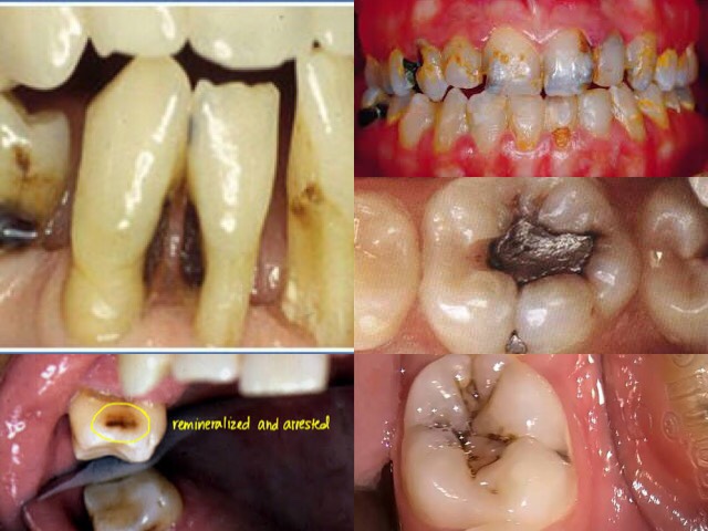
1️⃣ According to Anatomical Site –
2️⃣ According to rate of caries progression –
3️⃣ According to nature of attack-
4️⃣ Based on chronology –
https://dentowesome.wordpress.com/2020/05/11/pit-fissure-caries/
https://dentowesome.wordpress.com/2020/05/07/dental-caries/
Sudden, rapid & almost uncontrolled destruction of teeth affecting surface that are relatively caries free.
Affect deciduous teeth due to prolonged use of nursing bottle containing milk, sugar or honey.
💬 What is 👶 bottle decay? What causes it and how to prevent it? 👇🏻
Dentowesome|@drmehnaz🖊
Image Source: Google.com
Muhad Noorman P, Final year Team dentowesome.
CLASSIFICATIONS OF CLEFT LIP AND PALATE
Davis & Ritchie classification (1922)
Veau’s (1931)
Harkins and associates(1962)
Kernahan’s Classification (1971)
Spina (1974)
Tessiers’s Classification.
VEAU’S CLASSIFICATION
Group 1- Cleft of soft palate only
Group 2 – Cleft of hard and soft palate extending
no further extending than involving incisive foramen, (secondary palate only)
Group 3 – Complete unilateral cleft, extending from uvula
to incisive foramen in the midline, then deviating the one side and usually extending through the alevolus at the position of the future lateral incisor
Group 4- Complete bilateral cleft, extending forward through incisive foramen to alevolus. Premaxilla, suspended from the nasal septum .
Asian population have highest frequency often
CLINICAL FEATURES:-
Incidence- 1in 500,With african population the lowest at 1in 250. Cleft lip alone more common in males. Isolated Cleft palate more common in female. 50% are syndromic, and are born with other congenital abnormalities.Cleft lip appear as unilateral / bi lateral. Line of cleft start on lateral part of upper lip and continues through philtrum of alveolus between lateral incisor and canine. When cleft lip continues from incisive foramen through palatal suture middle in palate,cleft lip with palate (unilateral/bilateral) present . . Cleft palate appears with involving soft palate only, involving uvula (bifid uvula),isolated cleft palate also.
.Patient have significant physical and physiological effects like, difficulty in eating and drinking with regurgitation of food to nose.
. Speech problem
. Ear infection: Malposition of Eustachian tubes result in middle ear infection
.Cosmetic deformities.
MANAGEMENT
Management of Cleft lip and palate require, multidisciplinary coordinated approach by specialist including maxillofacial surgeon, pediatric surgeon, anesthetist, Prosthodontist, Orthodontist, Speech pathologist, otolaryngology ,audiologist etc..
Management is aimed at closure and correction of lip and palate, secondary correction of palatal fistulae, orthodontic management of malocclusion, Orthognathic surgery, Rhinoplasty,and providing prosthesis for patients.
Pre operative criteria selected by physicians for surgery is Millards Rule of 10’s
1) 10lb weight
2) 10mg/l of haemoglobin
3) 10 weeks of age
SURGICAL MANAGEMENT
1) Primary : Closure of lip & palate
2) Secondary : Closure of palatal fistula, Pharygoplasty ,Bone grafting, orthodontic management Rhinoplasty and Scar revision.
Reference: Oral and Maxillofacial surgery, Balaaji. Textbook of general surgery for dental students, SRB
Muhad Noorman P , team dentowesome, final year.
Cleft palate and Cleft lip comprises the complex of Orofacial clefts . Both comprises congenital malformations affecting oro-facial region.
Failure of fusion of nasal and maxillary process leads to cleft of primary palate which can lead to unilateral and bilateral. Cleft of secondary palate is medial, Varies from bifid uvula to complete cleft palate up to incisive foramen.
Etiology:-
Clinically, Isolated Cleft palate
and Cleft lip with or without Cleft palate has been established.
Factors playing role in Development of Orofacial cleft comprises
1)Heredity (40%of cleft lip and 20% cleft lip appear genetically- single gene/polygenic mutation, Monozygotic twins far likely to get)
2)Nutritional disturbances (experimentally proved in rat fed on abnormal dietary regimen caused cleft palate) mostlty- Riboflavin,Folic Acid Deficiency.
3) Physiologic, Emotional stress, Traumatic also thought to cause
4) Circulating Alcohol ,Drugs ,toxins.
5)Environmental Factors– Teratogens (phenytoin, methotrexate, Corticosteroid, Sodium Valproate)
6)Syndromic Clefting
Treacher collins syndrome
Pierre Robin Syndrome
Stickler syndrome
Oro Facial Digital syndrome
Trisomy of 13,18
Van Der Voude syndrome ( lip pit syndrome, autosomal dominant , deletion of 1q32 , clinically presenting with cleft lip and palate and medial pits on lower lips on vermillion border.Also include ankyloglossia,high arch palate ,maxillary hypodontia and sygnathia)
EMBRYOGENESIS AND CLEFTING
During sixth and seventh weeks of development upper lips forms when median nasal process merges each each other and fuses with maxillary process of 1st branchial arches. Mid portion of upper lip is derived from median nasal process, lateral derived from maxillary process. Lateral nasal process involved in ala of nose. Primary palate is formed from merging of median nasal process to form intermaxillary segment, which give rises to premaxilla
(bone including 4 incisor teeth). Secondary palate make up 90% of palate formed from maxillary process of first branchial arches.Defective fusion of median nasal process with maxillary process forms cleft lip. Failure of palatal shelves to fuse result in cleft palate
45% are Cleft lip with palate
30% Cleft palate alone
25% with isolated cleft lip


Reference:- Textbook of maxillofacial surgery- Balaji. Textbook of embryology- Inderbir Singh. Neville Oral pathology. Images credits : Image 1 – Internet medscape (https://emedicine.medscape.com/article/950823-overview
Image 2 :- Internet, Children Hospital of Philadelphia , ( https://www.chop.edu/conditions-diseases/van-der-woude-syndrome)
Muhad Noorman P, Team dentowesome, Final year
Anitschkow cell: Modified macrophages with nuclei having caterpillar apperance ( linear bar of chromatin with peripheral radiating chromatins . Found in Recurrent Apthous stomatitis, Iron deficiency Anemia, megaloblastic Anemia, children receiving chemotherapy.
Langerhans cells : Bone marrow derived antigen presenting cells found in epidermis positive for CD1a and Bierbeck Granules. Increased in Langerhans cell histocytosis and decreased in Psoriasis etc.. Named after Paul Langerhans
Tzank Cells: Enlarged , Balooned up degenerating keratinocytes with enlarged vesicular hyperchromatic nucleus, basophilic cytoplasm and diminished nucleoli and perinuclear halo appearance. Named after Arnault Tzank. Found in Herpes Simplex infection, Herpes Zoster, Pemphigus vulgaris, Varicella.
Langhans Giant cells: They are formed by the fusion of epithelioid cells and contain multiple nuclei arranged in a horseshoe-shaped pattern in the cell periphery or are arranged circumferentially. Named after Theodor Langhans. Found in granulomatous lesions like Tuberculosis, tuberculous Leprosy.
Downey cells : Atypical lymphocytes, abundant pale blue cytoplasm and irregular chromatin . Found in infectious mononucleosis. Named after Hal Downey.
Warthin Finkeldy Giant cells : Giant cells with upto 100 nuclei, cytoplasmic and nuclear inclusions. Pathognomic of measels infection. Named after Warthin and Finkeldy.
Reed–Sternberg cells: They are named after Dorothy Reed Mendenhall and Carl Sternberg. They are large cells that either are multinucleated or have a bilobed nucleus (having an “owl’s eye” appearance) with prominent eosinophilic nucleoli.Reed–Sternberg cells are giant cells found in Hodgkin’s lymphoma (HL).
Gaucher Cells : Glucocerebroside laden macrophages containing tubular cytoplasmic inclusions. (Crumbled tissue paper apperance) Hallmark of gaucher disease. Staining positive for Wright and PAS stain.
Rushton bodies : Peculiar linear , curved- hyaline homogeneous structure found in wall of ODONTOGENIC cysts like periapical cyst, dentigerous cyst etc..
Ghost cells: Well defined, eosinophilc, elliptoid fused epithelial cells with blurred apperance. Found in Ghost cell odontogenic tumor, Calcifying epithelial odontogenic cyst, odontoma, craniopharyngioma etc..
Rusells bodies : Large eosinophilc immunoglobulin containing inclusion bodies found in plasma cells. Distented endoplasmic reticulum staining positive for PAS, CD38 etc. Found in chronic inflammations. Aggregate Is called Mott bodies.
Reference : Internet, Shafers oral pathology, Neville Oral pathology
Muhad Noorman, Team dentowesome , Final year
Apple jelly nodules in nasal septum: It is the nodular form of the tuberculosis in nasal mucosa. It begins in the vestibule and extends to adjoining skin and mucosa.
Arnold head: In Cleidocranial dysplasia, the fontanelles may remain open until adulthood, but the sutures often close with interposition of wormian bones
Blue Sclera: Osteogenesis imperfecta , EHLER danlos syndrome, Fetal Rickets, MARFANS Syndome etc.. Asymptomatic bluish discoloration of sclera due to thinning of sclera and exposing underlying vascular choroid.
Ash-leaf spots: Hypomelanic macules in Tuberous sclerosis.
Buffalo hump: Cushing’s syndrome, the fat relocalization in nape of the neck resembling the buffalo’s hump
Bull neck: Diptheria, Cherubism
Cerebriform tongue: Pemphigus vegetans. Also known as Furrowed tongue with numerous sulci and gyri
Chipmunk facies: Thalassemia and Sickle cell anemia. The bones of the head and face become enlarged and deformed causing an abnormal appearance resulting in a typical “chipmunk/ rodent facies” appearance. This occurs because the bone marrow, the site of red blood cell production, becomes hyperactive in an attempt to produce sufficient red cells to over profound anemia.
Cobble stone appearance: Lymphangioma, Inflammatory papillary hyperplasia, Heck’s disease
Forschemmier spots : Forscheimer spots are enanthem seen as small, red spots (petechiae) on the soft palate in patients with rubella. Also found in palatal mucosa of Scarlet fever.
Fournier’s molars: congenital syphilis, Mulberry molars
Hamman’s crunch: Cervicofacial emphysema. It’s a crunching, rasping sound, synchronous with the heartbeat, heard over the precordium .
Hebra nose: Rhinoscleroma. Epistaxis, nasal deformity, and destruction of the nasal cartilage are also noted.
Iris pearl’s: Leprosy. Miliary lepromas or iris pearls near the pupillary margins, which are spherical yellowish opaque micronodules
Koplik’s spots: Measles. Koplik spots are a prodromic viral enanthem of measles manifesting two days before the measles erythmatous cutaneous rash itself. They are characterized as clustered, white lesions on the buccal mucosa ( table salt appearance) near each Stensen’s duct (on the buccal mucosa opposite the maxillary 2nd molars) and are pathognomonic for measles.
Lisch nodules: Neurofibromatosis. A Lisch nodule is a pigmented hamartomatous nodule found in iris which is an aggregate of melanocytes.
Pastia’s lines: Scarlet fever. Pastia’s sign, Pastia lines or Thompson’s sign is a clinical sign in which pink or red lines formed of confluent petechiae are found in skin creases, particularly the crease in the antecubital fossa. Caused by erythrogenic toxins staphylococcus.
References : Shafers Oral pathology. Burkets Oral Medicine
Dentowesome- Muhad Noorman P
Final year
Asboe-Hansen sign (also known as indirect Nikolsky sign) refers to the extension of a blister to adjacent unblistered skin when pressure is put on the top of the intact bulla.
Auspitz’s sign is the appearance of punctate bleeding spots when psoriasis scales are scraped off, named after Heinrich Auspitz.This happens because there is thinning of the epidermal layer overlying the tips of the dermal papillae and blood vessels within the papillae are dilated and tortuous, which bleed readily when the scale is removed.
Button-hole sign: Neurofibromatosis. invagination of a nodule when pressed with a finger, a characteristic of neurofibromatosis
Carpet tack lesions: Discoid lupus erythematosus. There are follicular hyperkeratotic plugs causing a carpet tack appearance as they project from the undersurface of the scale when it is removed from advanced lesions.
Charcot’s triad: Seen in multiple sclerosis, characterized by intention tremor, nystagmus, dysarthria
Crowe’s sign: Found in Neurofibromatosis. Presence of axillary freckling in people with neurofibromatosis type I.
Gorlin sign: Ehler -Danlos Syndrome. Ability to touch the tip of the nose with tongue and touch the elbow with the tongue
Higomenakis’s sign: Congenital syphilis. A unilateral enlargement of the sternoclavicular portion of the clavicle, seen in congenital syphilis
Millian sign: Erysipelas. Involvement of the ear (Milian’s ear sign) is a distinguishing feature for erysipelas since this region does not contain deeper dermis tissue.
Nikolsky’s sign: Epidermolysis bullosa, pemphigus, Severe Steven –Johnson syndrome etc. Application of lateral oblique pressure to skin yeild formation of new blisters.
Oil drop sign: Psoriasis. A translucent discolouration in the nail bed that resembles a drop of oil beneath the nail plate.
Reference: Neville Textbook of oral Pathology. Cawsons essential oral pathology
Regezi and Batsakis Oral pathology

It is purpulish discoloration of skin and mucous membrane due to sub-cutaneous and sub-mucous extravasation of blood.

Image :- Google
Writing :- notes made from mastering bds and Sanjay Kumar Purkait books
