IMPORTANT SIGNS-I

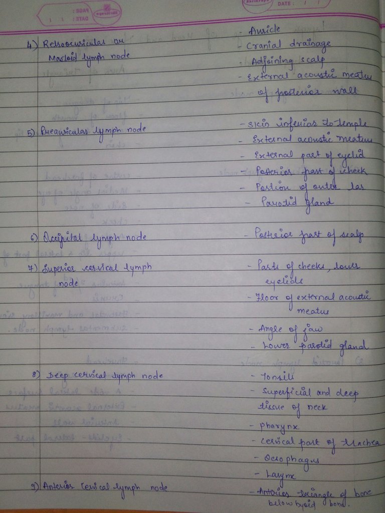
  • Allis sign- seen in developmental dysplasia
  • Asboe sign- seen in pemphigus (Bulla spread sign)
  • Auspitz sign- seen in psoriasis -peeling away scales result in pin point bleeding spots
  • Albright sign -dimple at the metacarpelopharyngeal joint, due to short metacarpal.Seen in pseudohypoparathyroidism,Turner’s syndrome,basal cell carcinoma
  • Babinski’s sign- by stroking the lateral aspect of the dorsum of foot
  • Barber’s chair sign- seen in multiple sclerosis
  • Battle’s sign-in basal skull fracture(sphenoid bone)blood pigment stain behind the ear over the mastoid
  • Benda’s sign- in tuberculous meningitis.Spasm of the trapezius muscle that the shoulder on affected side is lifted and at times brought forward
  • Blue berry muffin sign-raised purple lesions seen in dermal metastases of neuroblastoma
  • Brim sign- seen in Paget’s disease
  • Brudzinski’s sign- seen in meningitis
  • Button hole sign- seen in neurofibromatosis
  • Carpet track sign- seen in discoid lupus erythematosis
  • Cerebriform tongue sign- seen in pemphigus veterans
  • Chandelier’s sign- seen in gonorrhea in women
  • Chvostek sign- elevation of corner of mouth
  • Coleman’s sign- in fracture of body of mandible ( hematoma seen in the floor of the mouth)
  • Corner’s sign- seen in scurvy
  • Crowe’s sign- seen in neurofibromatosis
  • Darier sign- seen in urticaria pigmentosa
  • Dubois sign- seen in congenital syphilis
  • Erb’s sign- seen in latent tetany
  • Paget’s sign- seen in yellow fever
  • Falling fragment sign- seen in solitary bone cyst
  • Fitzpatrick sign- seen in dermatofibroma
  • Flag sign- seen in kwashiorkor
  • Floating membrane sign-seen in hydatid cyst
  • Floating teeth sign- seen in eosinophilic granuloma
  • Forscheimer sign- seen in rubella
  • Head drop sign- seen in poliomyelitis
  • Heel pad sign- seen in acromegaly
  • Higoumanaki’s sign- seen in late syphilis
  • Hutchinson sign-seen in heroes zoster ophthalmicus
  • Jellinek sign-seen in hyperthyroidism
  • Joffroy’s sign-seen in thyrotoxicosis

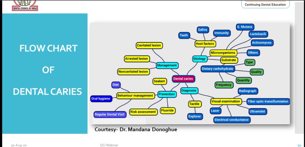
Source: http://www.slideshare.net ( signs in medicine),www.clinicalskillz.com

Hand Schuller Christian Disease (Multifocal Eosinophilic Granuloma)

  • Characterized by widespread skeletal & extra – skeletal lesions & chronic clinical course.
  • Occurs primarily in early life, before the age of 5; more common in boys & girls.
  • Most cases reported from Western literature, authentic cases from India are few.

Clinical features:-

  • Classic triad of :-
  1. Single or multiple areas of ‘punched out’ bone destruction in skull
  2. Unilateral or bilateral exophthalmos
  3. Diabetes insipidus with or without other manifestations of dyspituitarism
  • Involvement of facial bones frequently associated with soft tissue swelling & tenderness; causes facial symmetry.
  • Classically involves the flat bones of the skull, ribs, pelvis, and scapula
  • Diabetes insipidus affects 5 to 50% of patients.
  • Skin involvement in the form of erythematous scaly rash or papular or nodular lesions which is seen in only 30% of patients.
  • Poor sexual development and retarted growth is another feature.
  • Vision loss or strabismus caused by optic nerve or orbital muscle involvement occurs rarely.
  • Chronic otitis media and otitis externa due to involvement of the mastoid and petrous portions of the temporal bone with partial obstruction of the auditory canal are fairly common

Oral manifestations:-

  • Earliest signs of the disease; may be present in 5 – 75% of cases.
  • Often non specific; sore mouth, halitosis, gingivitis, ulcerations, loose teeth, suppuration, failure of healing of extraction sockets
  • Loss of supporting alveolar bone mimicking advanced periodontal disease is characteristic.

Radiological features:-

  • Skull lesions sharply outlined; jaw lesions may be diffuse.
  • Jaw lesions manifested as bone destruction with tooth displacement.
  • Panoramic may show remarkable atrophy of the alveolar ridge and severe parodontitis.
  • Remaining teeth abnormally sited in an extra alveolar position


Histological features:-

  • Manifesting in four stages during progression of characteristic lesion
  • Proliferative histiocytic phase with accumulation of collections of eosinophilic leukocytes scattered throughout sheets of histiocytes.
  • Vascular – granulomatous phase with persistence of histiocytes & eosinophils, sometimes with aggregation of lipid laden (cholesterol) macrophages.
  • Diffuse xanthomatous phase with abundance of ‘foam cells’
  • Fibrous healing phase.
  • Infiltration with CD1 positive histiocytes disclosing intracytoplasmic Birbek granules at the electron microscopic examination .
  • Histological diagnosis is based on the presence of a histiocytic infiltrate in the upper and middle dermis.
  • The optical microscope examination reveals in the papillary dermis an important oedema, large cells with an indented nucleus and abundant eosinophilic cytoplasm
  • Histochemical colouring, showing positiveness for S-100 protein, the presence of CD1, CD4 and HLA-DR surface antigens confirm the diagnosis

Treatment & prognosis:-

  • Therapy of the Hand-Schuller-Christian disease varies according to the age of the patient, the severity and extent of the clinical picture.
  • Approximately half of the patients undergo spontaneous remission over a period of years.
  • Treatment of choice is curettage or excision of lesions.
  • Inaccessible lesions may be irradiated.
  • Some patients may benefit from chemo – therapeutic drugs like Prednisone, Vinblastine & Cyclophosphamide.

Reference:

1.Faculty notes

2.Google

PIGMENTS & SPOTS

Pigmentation is the process of deposition of pigments in the tissue.Mostly found in the mouth and could be attributed to oral manifestations of systemic diseases or malignancies.This could be due to :

  • Increased number of melanocytes
  • Augmentation of melanin production
  • Deposition of accidentally introduced exogenous materials

The various oral pigmentations can be in the form of:

1)Blue /Purple vascular lesions-

  • Hemangioma
  • Varix
  • Angiosarcoma
  • Kaposi’s sarcoma
  • Hereditary hemorrhagic telangiectasia

2)Brown melanotic lesions-

  • Ephelis & oral melanotic macule
  • Nevus
  • Malignant melanoma
  • Drug induced melanosis
  • Physiologic pigmentation
  • Cafè au lait pigmentation
  • Smoker’s melanosis
  • Endocrinopathic pigmentation
  • Peutz-jeghers syndrome
  • HIV oral melanosis

3)Brown heme associated lesions-

  • Ecchymosis
  • Petechia
  • Hemochromatosis
  • Hemorrhagic mucocele
  • Thrombosed varix

4)Gray/black pigmentations-

  • Silver amalgam tattoo
  • Graphite tattoo
  • Heavy metal ingestion-lead,mercury,bismuth
  • Hairy tongue

SPOTS

Appearance of peculiar spots in specific sites on the body are often diagnostic for major systemic disease or condition or an infection.Some of them are discussed here;

  • Koplik spots -measles (rubeola)
  • Pink spots on teeth – internal resorption
  • Roth spots – subacute endocarditis, typhoid fever
  • Bitot’s spots white plaque on conjunctiva of vitamin A deficient children
  • Herald spots -primary lesion seen in pitryiasis rosea
  • Sore spots -traumatic ulcers from denture irritation mostly
  • Cafè au lait spot-neurofibromatosis, Macune-Albright syndrome,Peutz-jeghers syndrome

Sources: Shafers textbook of oral pathology ,http://www.ncbi.nlm.nih.gov (oral pigmentation : A review),http://www.gibbleguts.com