Amelogenesis Imperfecta

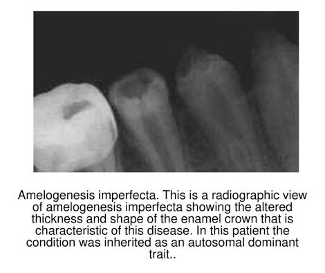
➡️ Represents a group of hereditary defects of enamel unassociated with any other generalized defects. It is entirely an ectodermal disturbance, since the mesodermal components of the teeth are basically normal.

➡️ Otherwise known as…

  • AI
  • Hereditary enamel dysplasia
  • Hereditary brown enamel
  • Hereditary brown opalescent teeth
AI can be inherited as an X-linked Autosomal Recessive or Autosomal Dominant condition

Prevalence: 1 in 700 to 1 in 15,000

Etiology:

  • Dental enamel is a highly mineralized tissue with over 95% of the volume occupied by unusually large, organized structures called the hydroxyapatite crystals.
  • The formation of these is controlled in Ameloblasts through the interaction of a no. of organic matrix molecules that include –
MMP20 (Matrix Metallopeptidase 20)
DSPP (Dentin sialophosphoprotein)

Develoment of Enamel:

➡️ 3 stages:

  1. Formative – deposition of organic matrix
  2. Calcification – Matrix mineralization
  3. Maturation – Crystallites enlarge & mature

Types of AI classification (Witkop and Sauk)

Based on clinical, histological & genetic criteria:

🔹 TYPE I HYPOPLASTIC

  • Pitted Autosomal dominant
  • Local Autosomal dominant
  • Local Autosomal Recessive
  • Smooth Autosomal dominant
  • Smooth, X-linked dominant
  • Rough Autosomal dominant
  • Enamel agenesis, Autosomal Recessive

🔹 TYPE II HYPOMATURATION

  • Diffuse Pigmented, Autosomal Recessive
  • Hypomaturation
  • Snow-capped teeth, X-linked
  • Autosomal Dominant

🔹 TYPE III HYPOCALCIFICATION

  • Diffuse Autosomal dominant
  • Diffuse Autosomal Recessive

🔹 TYPE IV COMBINATION TYPE

  • Hypomaturation – Hypoplastic with taurodontism
  • Hypomaturation – Hypoplastic with taurodontism, Autosomal Dominant
  • Hypoplastic – Hypomaturation with taurodontism, Autosomal Dominant

Clinical Features:

1) Hypoplastic – Enamel not formed to full normal thickness.

2) Hypomaturation –

  • The enamel can be pierced by an explorer point under firm pressure.
  • Can be lost by chipping away from the underlying normal appearing dentin.
  • Teeth normal in shape but exhibit a mottled, opaque white, brown-yellow discoloration.
  • Snow capped pattern – Zone of white opaque enamel on the incisal or occlusal third of crown.

3) Hypocalcified

  • The enamel is so soft that it can be removed by a prophylaxis instrument.
  • Yellow, brown or orange on eruption. Stained brown to black with time.
  • Rapid calculus apposition.
  • Coronal enamel lost with function except for the cervical portion which is mineralized better.
  • Autosomal Recessive – More severe.

Other Features:

  • Both dentition affected
  • Crown – Yellow to dark brown
  • Enamel might have numerous parallel vertical wrinkles or grooves.
  • Open Contact points.
  • Occlusal & incisal edges frequently abraded.

Radiographic Features:

Source: SlidePlayer
  • The enamel may appear totally absent.
  • When present may appear as a thin layer, chiefly over tip of cusps & on inter-proximal surfaces.
  • In some cases, calcification is so much affected that enamel and dentin seem to have the same radio density, making differentiation b/w the two difficult.

Histological Features:

  1. Hypoplastic: Disturbance in the differentiation/viability of Ameloblasts. Defect in matrix formation.
  2. Hypomaturation: Alteration in enamel rod & rod sheath structures.
  3. Hypocalcified: Defects of matrix structure & of mineral deposition.

Management:

  • Sealants/bonding
  • Prosthetic reconstruction

References: Shafer’sTextbook Of Oral Pathology; Internet

SIDE EFFECTS OF ANTI HYPERTENSIVE DRUGS

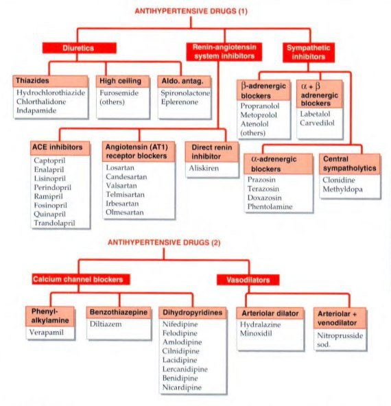
The objective of antihypertensive therapy is to reduce the incidence of adverse cardiovascular events, particularly CAD stroke and heart failures.

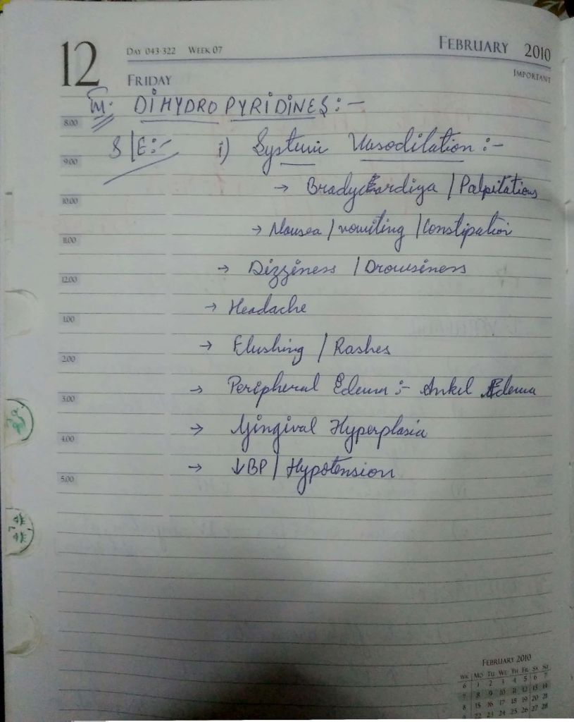
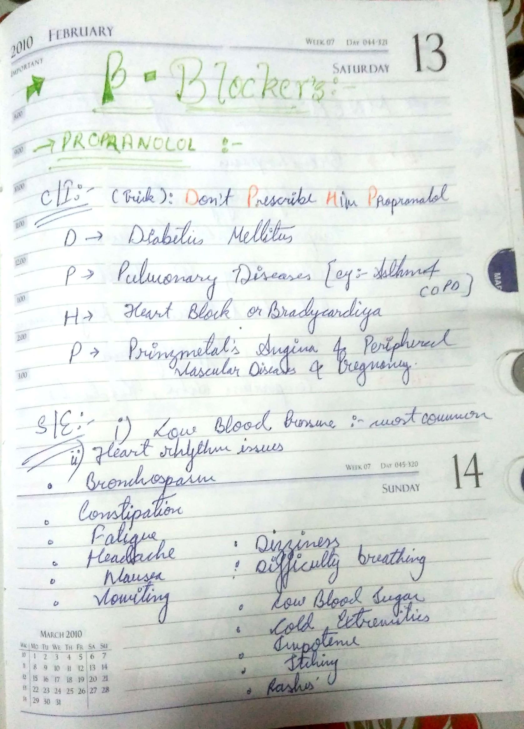
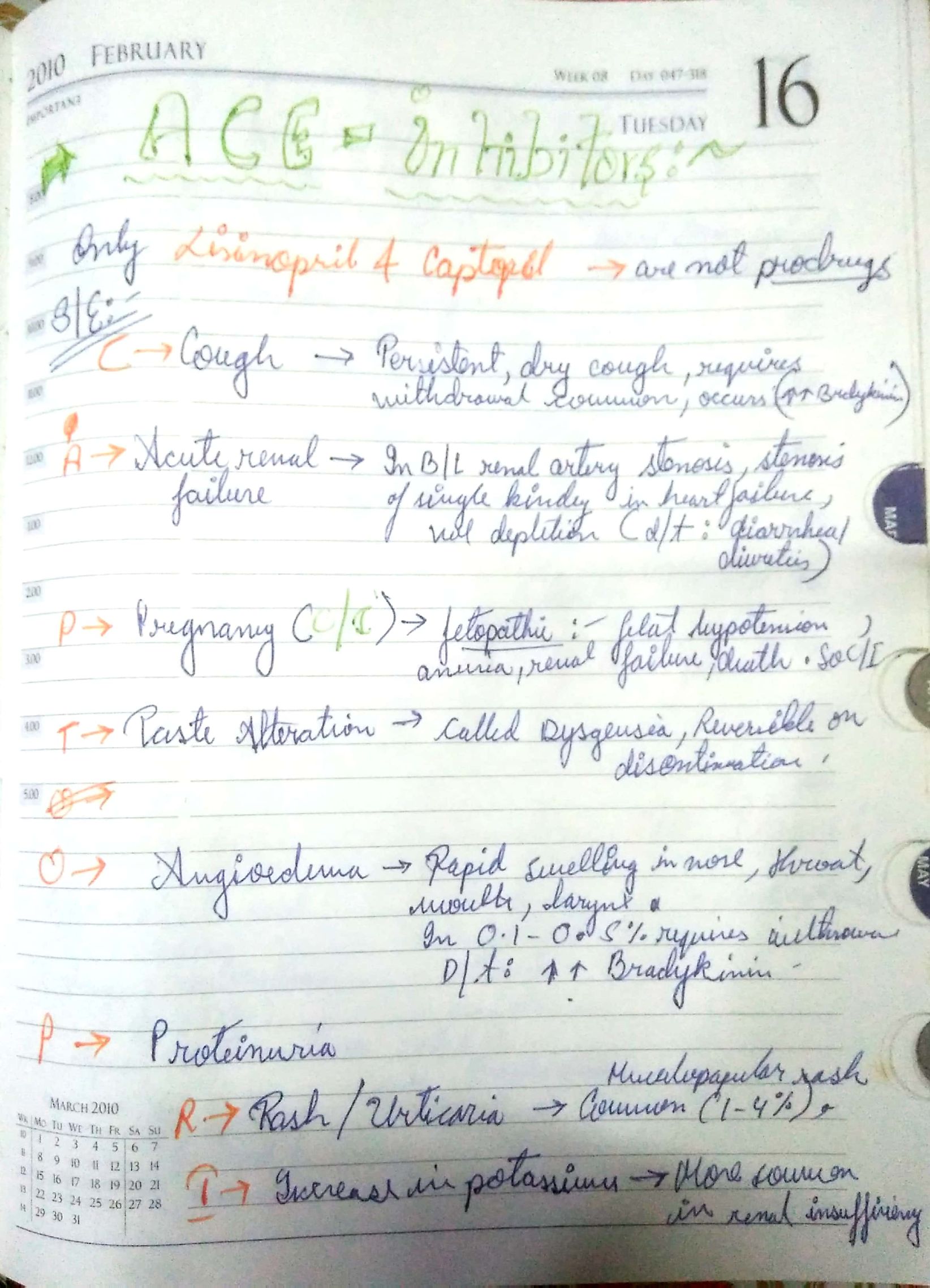
Here , we discuss some side effects of the Major drugs which is mainly prescribed for the treatment:-

The choice of antihypertensive therapy is initially indicated by the patient’s age and ethnic background.

Comorbid conditions also have an influence on initial drug selection,for example:-

•beta blocker might be the most appropriate treatment for a pateint with angina.

•Thiazides diuretics and dihydropyridines calcium channel blockers antagonists are the most suitable drugs for treatment in older people.

Thank you,

Regards,

KRITI Naja Jain 🙂

REference:-

  1. KDT 8th e
  2. Davidson 23rd e
  3. YouTube video

Pit & Fissure Caries

➡️ Morphology of Fissures:

Based on morphological alphabetical description of shape – 4 types:

  1. V + U (Self-cleansing)
  2. U
  3. K

Note: Pit & fissure with high steep walls & narrow bases are more prone to caries.(Developmental faults)

Occlusal fissures: Deep invagination of enamel, described as broad/narrow funnels, constricted hour glasses, multiple invaginations with inverted Y-shaped divisions & irregularly shaped.

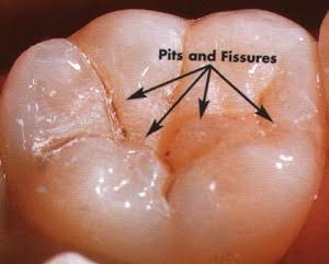
Deep and narrow Pit & Fissure

⬇️

Retention of food debris & microbes

⬇️

Fermentation of food by microbes

⬇️

Formation of Acid

⬇️

Caries

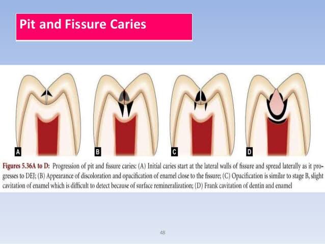
➡️ The lesions develops from attack on their walls.
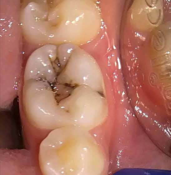
  • Cross section: Inverted “V” (A narrow entrance & wider involvement closer to DEJ)
  • Therefore, Greater no. of Dentinal Tubules are involved.
  • Early dentin involvement – When enamel at bottom of Pit & fissure is thin.

Caries when occur at Pit & Fissure follow direction of ENAMEL RODS

ENAMEL LAMELLAE – Initiation of Caries

The Initial Carious lesion of Enamel:-

• Clinical View:

  • Visual Changes – Chalkiness, yellow/brown/black discoloration.
  • Soft & ‘catch’ a fine explorer point.
  • Enamel bordering them is opaque bluish white & undermined ➡️ Lateral spread of caries at DEJ
  • Sign on stained tooth (Brown P/F)
  • Newly erupted teeth – underlying decay; Older: Arrested lesion

References: Wheeler’s Textbook, Google images