Viral Hepatitis


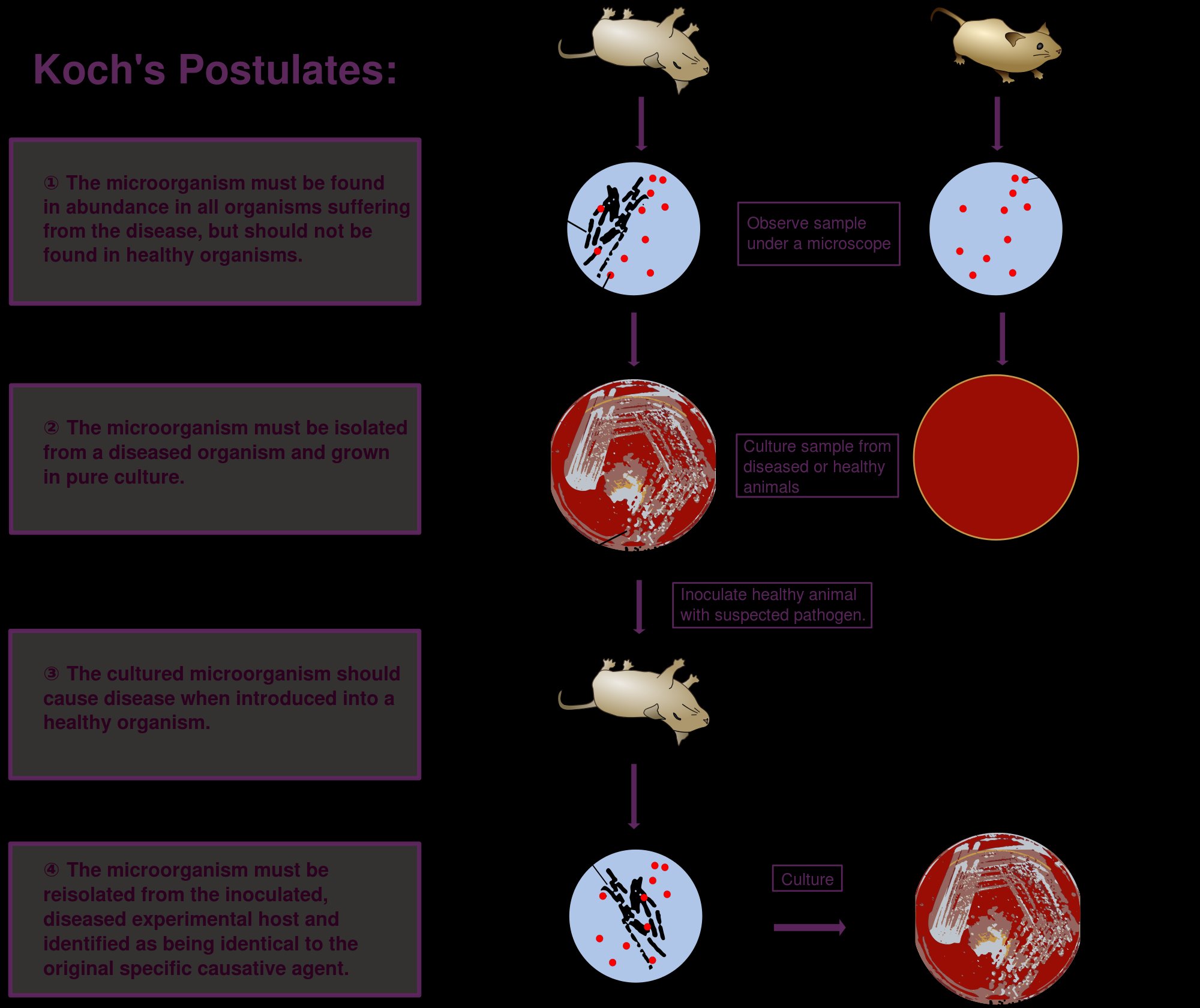
A wide range of viruses may cause liver inflammation (hepatitis) and several
are relevant to dentistry. Here, I describe the recognized family of hepatitis viruses A to G.


Hepatitis A virus (HAV)
The hepatitis A virus is:

  • A member of the Enteroviridae.
  • It is a spherical, non-enveloped virus.
  • It has a single-stranded RNA genome.
  • HAV initially replicates in the gut, followed by a viremic phase, during which the virus enters the liver.

Transmission
• Fecal-oral transmission.
• Endemic worldwide.

Diagnosis
• Demonstration of HAV antigen in feces.
• Serology: detection oflgM anti-HAV.


Clinical features
• The incubation period is 2-7 weeks.

Many infections are asymptomatic. Clinical disease is mild with few complications. There is no carrier state.

Prevention and Control

Good hygienic measures and sanitary disposal of excreta.
Passive immunization gives immediate protection for 3-6 months.
Active vaccination: the formalin-inactivated vaccine provides protection for up to 10 years.
HAV is not a major cross-infection hazard in dentistry but is a hazard if traveling, especially to the tropics.

Hepatitis B virus (HBV)

This highly infectious blood-borne virus poses a major cross-infection hazard
in surgery and dentistry:
• It is a member of the hepadnavirus family.
• The intact viral particle (Dane particle) has a double-shelled structure, with the outer hepatitis B surface antigen (HBsAg) coat surrounding the central hepatitis B core antigen (HBcAg), DNA, and DNA polymerase.
• Peripheral blood of infected patients also contains non-infective spherical and filamentous particles of HBV

Transmission

• HBV can be present in blood, saliva, cervical secretions, and semen.

• Spread is via the parenteral route, especially by intravenous drug use, but transmission by intimate contact and sexual activity also occur.

• Perinatal infection is important in certain parts of the world, for example east and southeast Asia.

• There is a large reservoir of unidentified carriers within the population.

• Infected patients may have up to 1010 Dane particles per ml of blood; as little as 0.0001 ml of blood may transmit the infection.

• HBV has been transmitted in dentistry, to patients and dental staff. Some have died from infection.

Diagnosis

• Serological.

• Initial screening is for HBsAg; if present, it indicates infection with HBV.

• Screen then for HBeAg. If present, the person is at high risk for transmission.

• A minority who are HBeAg negative can also transmit infection. Hepatitis B carriers produce HBsAg and, in high-risk carriers, HBeAg for many years.

• Development of anti-HBs, anti-Hbe, and anti-HBc antibodies is associated with recovery. The incubation period is 2-3 months duration. There are a number of possible outcomes of exposure to HBV:

• Subclinical infection (65%).

• Acute hepatitis B with full recovery (30%).

• Chronic carriage (up to 9% of adults): this gives a long-term risk of cirrhosis, liver failure, and hepatocellular carcinoma. Carriers remain infectious to others.

• Fatal fulminant hepatitis (1 %).

Prevention and Control

• Modifications to behavior.

• Adequate infection control procedures in clinical practice.

• Passive immunization: hyperimmune hepatitis B immunoglobulin is used following a single acute exposure in an unprotected individual.

• Active immunization. Hepatitis B vaccine consisys of20 mg ofHBsAg given intramuscularly at 0, l, and 6 months. Boosters have been recommended at 5-year intervals. All vaccinees should have their serum antibody level assessed after vaccination. High-risk carriage of HBV should be excluded in non-responders who are health care workers.

• Interferon may be effective in the treatment of chronic HBV infection.

Hepatitis C virus (HCV)

This blood-borne virus, discovered in 1989, is responsible for most cases of what was previously known as parenterally transmitted non-A, non-B hepatitis(NANBH). Is an enveloped RNA virus.
• Is related to animal pestiviruses and human flaviviruses.
• Has multiple genotypes.
• Cannot be grown in tissue culture.

Diagnosis
• Serological.
• Initial detection of HCV antibodies.
• Confirmation by PCR for HCV RNA.

Transmission
• The prevalence of HCV antibodies among UK blood donors is 0.1-0.3%.
• In recipients of blood products and among intravenous drug users the seroprevalence is high(> 80%).
• Parenteral transmission is the major route, especially in intravenous drug use.
• Sexual transmission is inefficient.
• Occupational transmission may be through needlestick injuries, though it is less infectious than HBV.
• Undefined routes: in a significant number ofHCV-infected individuals, the route of infection is unknown.

Clinical features
• The mean incubation period is 6-12 weeks.
• Acute disease is mild and often subclinical.
• Chronic disease is common (> 60%). These patients may develop longterm liver disease, including hepatocellular carcinoma. Some patients may develop oral disorders similar to Sjogren’s syndrome or lichen planus.

Prevention and Control
• Changes in behavior, e.g. needle exchange schemes for intravenous drug users.
• Screening of donated blood.
• Effective universal infection control in health care settings.
• No vaccine is available.
• Treatment of chronic carriers with interferon and ribavirin is effective in
about 40% of cases.

Hepatitis D virus (HDV)


• A defective, independently transmissible agent which requires hepatitis B virus for replication.
• In developed countries it is mainly a problem among intravenous drug users.
• The genome is single-stranded RNA.
• Transmission is primarily parenteral, either at the time of first infection with HBV (co-infection) or during a subsequent exposure in a patient already infected with HBV (superinfection).
• HDV increases the severity of HBV infection and fulminant hepatitis is common.
• HDV has been transmitted in dentistry, to patients and dental staff. Dental patients have died from infection.

Hepatitis B vaccination is protective.

Hepatitis E virus


This recently discovered virus causes the disease described previously as enterically transmitted non-A, non-B hepatitis:
• It is a spherical, non-enveloped, RNA virus.
• Transmission is via fecal!y contaminated drinking water.
• The incubation period is 2-9 weeks.
• It mainly affects young adults.
• Infection is usually self-limiting.
• There are no chronic carriers.
• Infection carries a high mortality (up to 20%) in pregnancy.
• It is not a major cross-infection risk in surgery.

Hepatitis G virus


• Hepatitis G is a flavivirus, first isolated in 1995 from a patient with chronic hepatitis.
• Seroprevalence studies show evidence of infection in 3% of blood donors in the United Kingdom, 18% of hemophiliacs, and 33% of intravenous drug users.
• Hepatic damage appears mild or absent and the virus is not considered an important pathogen.
• It is not a major cross-infection risk in surgery.

Dr Iswarya V

General Practitioner,

Trivandrum.

Reference : Oxford Clinical Dentistry

Koch’s postulates

Robert Koch was a German practicioner. He is also known as the father of microbiology.

Koch’s postulates:

According to Koch’s postulates, a microorganism can be accepted as the causative agent of an infectious disease only if the following conditions are fulfilled.

(i) The organism should be constantly associated with the lesions of the disease.

(ii) It should be possible to isolate the organism in pure culture from the lesions of the disease.

(iii) The isolated organism (in pure culture) when inoculated in suitable laboratory animals should produce a similar disease.

(iv) It should be possible to re-isolate the organism in pure culture from the lesions produced in the experimental animals.

Source- textbook of microbiology C P Baveja and Google images

IgD and IgE

1. Immunoglobulin D( IgD)

(i) IgD resembles IgG structurally.

(ii) IgD is present in a concentration of 3 mg per 100 ml in serum. It is mostly intravascular in distribution.

(iii) Molecular weight is 180000 (7S monomer).

(iv) Half life is about three days.

2. Immunoglobulin E (IgE)

(i) IgE is mainly produced in the linings of respiratory and intestinal tracts. It is mostly distributed extravascularly.

(ii) It is also referred to as reagins.

(iii) Molecular weight is 190000 (8 S molecule).

(iv) Half life is 2-3 days.

(v) It resembles IgG in structure.

(vi) It is heat labile whereas other immuno-globulins are heat stable.

(vii) It has affinity for surface of tissue cells, particularly mast cells of the same species (homocytotropism).

(viii) IgE mediates type I hypersensitivity (atopic) reaction. This is responsible for asthma, hay fever and eczema.

(ix) It cannot cross the placental barrier.

(x) IgE is responsible for anaphylactic type of reaction.

Source- textbook of microbiology for dental students c p baveja and Google images

Pathogenesis of Staph. aureus

Staph aureus | Neuros- Social Networking For Medical Students ...

What is Staphylococcous ?

A gram positive cocci and that is arranged in grape like clusters. They mainly cause suppuration

The important species of Staphylococcus are –

  • Staph.aureus
  • Staph.epidermidis
  • Staph.saprophyticus
Staphylococcus epidermidis - Wikipedia
staph.epidermidis
Staphylococcus Saprophyticus - Stock Image - B234/0182 - Science ...
staph . saprophyticus
Difference Between Staphylococcus Epidermidis and Staphylococcus ...

Staphylococcus aureus

  • They are gram positive cocci
  • Arranged in grape like clusters
  • They are non motile,non sporing
  • Diameter – 1 micrometer approx.
  • Grow in culture medium between 10-42 degree celcius but approximately at 37 degrees celcius
  • Grow at a pH of 7.4-7.6
  • They are aerobes and facultative anaerobes (which can also grow without oxygen)
Staphylococcus aureus (S .aureus) | SnackSafely.com
staph. aureus

Pathogenesis

  • It is a very important pyogenic organism and lesions are localized in nature.
  • They produce thick cream colored colonies

They are classified as –

  • cutaneous and deep infections
  • nosocomial infections
  • food poisoning
  • skin exfoliative diseases
  • toxic shock syndrome

1.Cutaneous infecctions – superficial infecctions

  • pustules
Pustule: Causes, Symptoms, Diagnosis, Treatment and Prevention
  • boils
Boils: Pictures on Skin, Causes, and Treatment
  • carbuncles
Carbuncles: Treatment, pictures, causes and symptoms - TODAY
  • impetigo
Impetigo - Wikipedia
  • pemphigus neonatrum
Pemphigus - Symptoms and causes - Mayo Clinic
  • styes
Stye Symptoms and Treatment in Children
  • abscesses
Abscess - NHS
  • wound and burn infections
Treatment of Infection in Burn Patients - ScienceDirect

2. Deep infections

  • osteomyelitis
  • tonsillitis
  • pharyngitis
  • pneumonitis
  • endocarditis
  • empyema
  • bacteremia
  • septicemia
  • meningitis
  • sinusitis
The role of nasal carriage in Staphylococcus aureus infections ...

3. Food poisoning –

Staphylococcal food poisoning may follow 2-6 hrs after injestion of contaminated food that has preformed entertoxin.

Food Poisoning: Tummy Trouble | BusinessMirror

4.Nosocomial infections –

They are important cause of hospital acquired infections

5.Skin exfoliative Diseases

  • These are produced by strains of Staph.aureus .
  • There is stripping of the superficial layers of skin from the underlying tissue .
  • They are Ritters disease ,pemphigous neonatrum,bullous impetigo.
  • Staphylococcocal scaled skin syndrome (SSSS) is the example. In this the toxin is spread systemically.
Nepal STAPHYLOCOCCUS n Staphylococcus causes diseases ranging from ...

6. Toxic shock syndrome –

  • It is caused by TSST-1 ( toxic shock syndrome toxin ). This is characterised by high fever,hypotension, diarrhoea, scarletiniform rash,vomiting.
emDOCs.net – Emergency Medicine EducationStaphylococcal toxic ...
  • It is also associated with use of tampons by mentruating women which is rare.
Toxic shock syndrome is rare. Be vigilant but not alarmed

source – C P Baveja textbook of microbiology for dental students and google images

DERMATOPHYTES

(1) Dermatophytes are a group of fungi that infect only superficial keratinised tissue (skin, hair and nails) without involving the living tissue.

(ii) They break down and utilise keratin.

(iii) They are incapable of penetrating subcutaneous tissue.

(iv) They cause dermatophytosis, also known as tinea or ringworm

Ring worm

Classification-

Dermatophytes are classified into three genera as follows:

Genus. Infection of

  1. Trichophyton- hair,skin,nail
  2. Microsporum- hair ,skin
  3. Epidermophyton- skin, nail

Clinical types

Clinically, ringworm can be classified depending on the site involved. These include

1.Tinea capitis (scalp)

2.Tinea corporis (non-hairy skin of the body)

3. Tinea cruris (groin)

4.Tinea pedis (foot) or athlete’s foot

5. Tinea barbae or barber’s itch (bearded areas of the face and neck).

🔶Favus- is a chronic type of ringworm involving the hair follicles. It leads to alopecia and scarring.

• In favus, there is sparse hyphal growth and formation of air spaces within the hair shaft.

Two types of hair infection may be present, ectothrix and endothrix.

🛑Ectothrix

In ectothrix, a sheath of arthrospores is present on the surface of hair shaft.

🛑Endothrix

In endothrix the arthrospore formation occurs entirely within the hair shaft.

Source – slide share

Lab diagnosis-

🔻Specimens

• Skin scrapings

• Hair clippings

• Nail

🔻Direct microscopy

• Direct 10% KOH mount may show fungal hyphae.

🔻Culture

SDA and SDA with antibiotics are used.

• Culture media are incubated at 25-30°C for three weeks.

• Identification of dermatophytes is based on

🔶colony morphology

🔶pigment production

🔶microconidia and macroconidia

🔻Colony characters

🔶Reverse of media is red in T. rubrum.

🔶White to creamy, cottony growth

Dermatophytes On SDA media

🔻Microscopy

🔶 Lactophenol cotton blue preparation from colony reveals microconidia, macroconidia or both. The following are the characteristics of three genera:

🔶Genus TrichophytonMore microconidia, very few macroconidia

🔶Genus Microsporum- Predominant macroconidia

🔶Genus Epidermophyton-macroconidia

🔻Treatment of dermatophytoses

🔶Topical antifungal agents are generally used for treatment.

🔶Oral griseofulvin is the drug of choice.

Source – textbook of microbiology for dental students c p baveja

Major Histocompatibility Complex ( MHC)

Transplants from one individual to another member of the same species (‘allografts’) are recognised as foreign and rejected.

Gorer (1930) identified the antigens responsible for allograft rejection in inbred mice that led to the discovery of the major histocompatibility complex (MHC).

The MHC in humans is known as the human leukocyte antigen (HLA) complex.

HLA complex

Histocompatibility antigens mean cell surface antigens that evoke immune response to an incompatible host resulting in allograft rejection.

These alloantigens are present on surface of leucocytes in man and are called human leucocyte antigens (HLA) and the set of genes coding for them is named the HLA Complex.

The HLA complex of genes is located on short arm of chromosome 6 and is grouped in three classes

Class I

HLA-A, HLA-B and HLA-C

Class II

HLA-DR, HLA-DQ and HLA-DP (All of these are present within HLA-D region of HLA complex.)

Class III ( Complement loci encode for C2, C4 and Factor B of complement system and tumour necrosis factors (TNF) alpha and beta)

A locus is the position where a particular gene is located on the chromosome.

HLA loci are multiallelic i.e. the gene present on the locus can be any one of several alternative forms (alleles).

Each allele determines a distinct antigen. There are 24 alleles at HLA-A locus and 50 at HLA-B. HLA system is very pleomorphic. Every individual inherits one set of HLA-genes from each parent.

1.Class 1 MHC Antigens (A, B, C)

The MHC class I antigens are present on the surface of all nucleated cells. They are involved in graft rejection and cell mediated cytolysis. The cytotoxic T cells (CD8) recognise MHC class I antigens for their action.

2 Class II MHC Antigens (DR, DQ and DP)

They have a very limited distribution and are principally found on the surface of macrophages, monocytes, activated T-lymphocytes (CD4) and B-lymphocytes. They are primarily responsible for the graft-versus-host response and the mixed leukocyte reaction (MLR).

3.Class III MHC Antigens

Class III genes encode C2, C4 complement components of the classical pathway and properdin factor B of the alternative pathway.

HLA complex

Source – textbook of microbiology for dental students – harsh mohan

Endotoxins and Exotoxins

Endotoxins –

🔻They are Lipopolysaccharide in nature and former integral part of the gram negative bacteria cell wall.

🔻 They are Heat stable.

🔻Form integral part of the cell wall; released only on disruption of bacterial cell.

🔻Weakly antigenic; antitoxin is not formed but antibodies against polysaccharide are raised.

🔻Cannot be toxoided.

🔻No enzymatic action.

🔻Non-specific action of all endotoxins.

🔻Low potency

🔻Non-specific in action.

🔻Usually produce fever.

🔻Produced by Gram-negative bacteria.

🔻 Massive gram negative septicemia may cause a syndrome of endotoxic shock characterized by fever, leukopenia, thrombocytopenia ,profound fall of blood pressure and circulatory collapse  to death.

Exotoxins

🔻Protein (polypeptides) M.W. 10,000 to 900,000.

🔻 Heat labile (more than 60°)

🔻Actively secreted by living cells into medium.

🔻 Highly antigenic, stimulates formation of antitoxin which neutralises toxin.

🔻Converted into toxoid by formaldehyde.

🔻Enzymatic in action.

🔻Specific pharmacological effect for each exotoxin.

🔻Very high potency.

🔻Highly specific for particular tissue eg. tetanus toxin for CNS.

🔻Don’t produce fever in host.

🔻Produced mainly by Gram-positive bacteria and also by some Gram-negative bacteria.

Source- textbook of microbiology for dental students c p baveja

Hot air oven

🔻It is the most widely used method of sterilisation by dry heat.

🔻 The oven is electrically heated and is fitted with a fan to ensure adequate and even distribution of hot air in the chamber

🔻It is also fitted with a thermostat that maintains the chamber air at a chosen temperature.

Temperature and time

160°C for two hours (holding time) is required for sterilisation.

Uses

It is used for sterilisation of

(i) Glassware like glass syringes, petri dishes, flasks, pipettes and test tubes.

(ii) Surgical instruments like scalpels, scissors, forceps etc.

(iii) Chemicals such as liquid paraffin, fats, sulphonamide powders etc.

Precautions-

(i) It should not be overloaded.

(ii) The material should be arranged in a manner which allows free circulation of air.

(iii) Material to be sterilised should be perfectly dry.

(iv) Test tubes, flasks etc. should be fitted with cotton plugs.

(v) Petri Dishes and pipettes should be wrapped in craft paper.

(vi) Rubber materials (except silicone rubber) or any inflammable material should not be kept inside the oven.

(vii) The oven must be allowed to cool for two hours before opening the doors, since the glasswares may crack by sudden cooling.

Sterilisation control

(i) The spores of nontoxigenic strain of Clostridium tetani are kept inside the oven. These spores be destroyed if the sterilisation is proper

(ii) Thermocouples may also be used.

(iii) Browne’s tube with green spot is available .

After proper sterilisation a green colour is produced (after two hour at 160°C).

Source – textbook of microbiology for dental students c p baveja

IgM

Immunoglobulin M (IgM)

(i) IgM is a pentamer consisting of 5 immuno globulin subunits and one molecule of J chain, which joins the Fc region of the basic subunits.

(ii) It constitutes about 5-8 percent of total serum immunoglobulins. The normal level in serum is 0.5-2 mg/ml.

(iii) Half life is about five days.

(iv) It is heavy molecule (19S) with a molecular weight 900,000 to 1000000 hence also called the ‘millionaire molecule‘.

(v) IgM is mainly distributed intravascularly (80%).

(vi) It is the earliest synthesised immunoglobulin by foetus.

(vii) It appears early in response to infection before IgG.

IgM antibodies are short lived, and disappear earlier than IgG. Hence, its presence in serum indicates recent infection

(viii) It can not cross the placenta, presence of IgM antibody in serum of newborn indicates congenital infection.

(ix) It is very effective antibody in agglutination and complement fixation. It is more efficient than IgG in these reactions.

(x) IgM provides protection against blood invasion by microorganisms.

Source – textbook of microbiology for dental students c p baveja

IgG

  • Immunoglobulin G ( IgG)

(i) IgG is the major serum immunoglobulin (about 80% of the total amount). The normal serum concentration is about 8-16 mg/ml.

(ii) Molecular weight is 150,000 (7S)

(iii) Half life is about 23 days (longest amongst all the immunoglobulins).

(iv) It is the only immunoglobulin that is transport through placenta and provides natural passive immunity to newborn.

(v) It is distributed equally between extravascular and intravascular compartments.

(vi) IgG appears late but persists for longer period. It appears after the initial immune response which is IgM in nature.

(vii) It participates in precipitation, complement fixation and neutralization of toxin and viruses.

(viii) It is protective against those microorganisms which are active in the blood and tissues.

Source – textbook of microbiology C P Baveja