

Source- textbook of microbiology for dental students c p baveja
Source- textbook of microbiology for dental students c p baveja
Reference : Paniker’s textbook of microbiology
Reference : Paniker’s textbook of microbiology

Reference : Paniker’s textbook of microbiology

Reference : Paniker’s textbook of microbiology

Reference : Paniker’s textbook of microbiology
Reference : Paniker’s textbook of microbiology

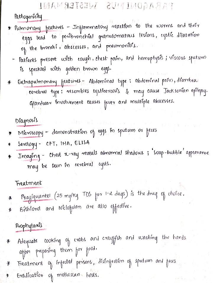
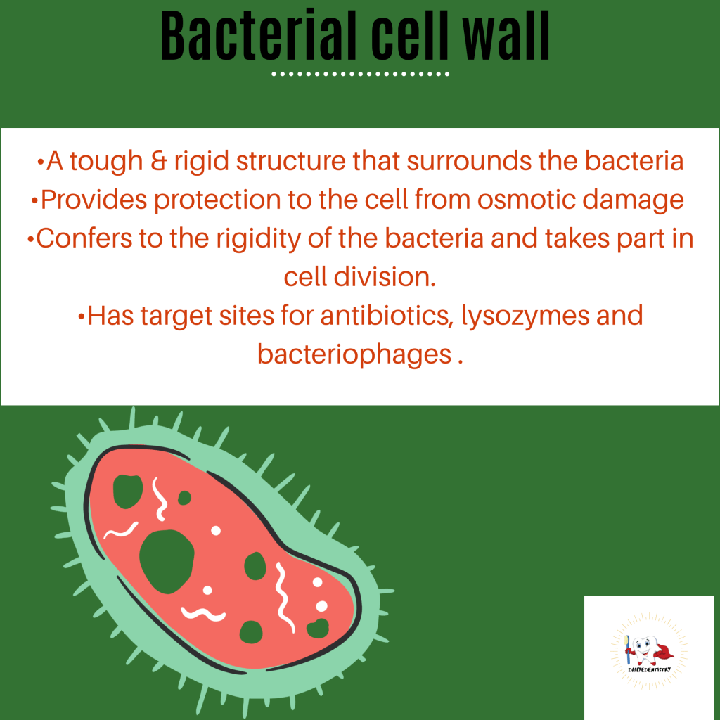
The cell wall is a tough and rigid structure surrounding the bacterium like a shell. It weighs about 20-25% of the dry weight of the cell.
FUNCTIONS:
1.Shape
2. Protection against osmotic damage
3.Rigidity
4.Cell division
5.Possesses target site for antibiotics, lysozymes and bacteriophages. Carries bacterial antigens that are important in virulence and immunity.
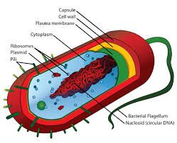
The rigid part of the cell wall is a peptidoglycan which is a mucopeptide (murein) composed of N-acetyl muramic acid and N-acetyl glucosamine molecules alternating in chains, cross-linked by peptide sub-units.
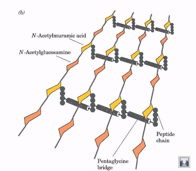
DIFFERENCES BETWEEN GRAM POSITIVE AND GRAM NEGATIVE CELL WALL
| CHARACTER | GRAM POSITIVE | GRAM NEGATIVE |
| Thickness | 20-80nm | 10nm |
| Periplasmic space | Absent | Present |
| Lipids and Glycoproteins | Few(0.3%) | Many(58%) |
| Teichoic acid | Present | Absent |
| Peptidoglycan | <15% | 10-20% |
| Gram’s reaction | Violet | Pink |
| Outer membrane | Absent | Present |
| Lipopolysaccharides | Absent | Present |
DEMONSTRATION OF CELL WALL
1.Plasmolysis
2,Microdissection
3.Differential staining
4.Reaction with specific antibody
5.Electron microscopy
BACTERIA WITH DEFECTIVE CELL WALL
PLEOMORPHISM AND INVOLUTION FORMS
Certain species of bacteria exhibit great variation in shape and size of individual cells are called pleomorphic bacteria.
Some bacteria show swollen and aberrant forms in ageing laboratory cultures and are known as involution forms.
Defective cell wall synthesis is responsible for development of these two forms 🙂
SOURCE: MICROBIOLOGY – C.P BAVEJA
Reference : Paniker’s textbook of microbiology
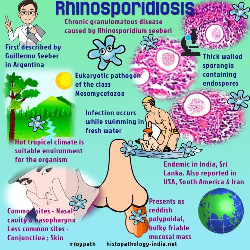
(i) Rhinosporidiosis is a chronic granulomatous disease characterised by formation of friable polyps, usually confined to the nose, mouth or eye.

(i) Causative agent is Rhinosporidium seeberi.
(iii) More than 80% cases are reported in India and Sri Lanka.
(iv) The mode of infection is not known but most infections occur in males who have frequent contact with stagnant water or aquatic life.
ORAL MANIFESTATIONS
•Oronasopharyngeal lesions appear as soft red polypoid growth which spread to pharynx and larynx.
• These lesions often contain mucoid discharge and are vascular.
LAB DIAGNOSIS
• The fungus has not been cultivated.
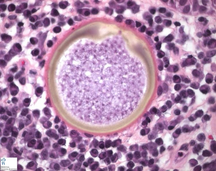
• Diagnosis depends on the demonstration of sporangia
•Tissue sections stained with H & E stain show large number of endospores within the sporangia embedded in a stroma of connective tissue and capillaries.
The sporangium (10-200 um) contains thousands of endospores (6-7 µm in diameter)

Source – textbook of microbiology for dental students c p baveja and Google images
