HISTOPATHOLOGY OF SKIN LESIONS

BY: Dr.Kriti Naja Jain :-
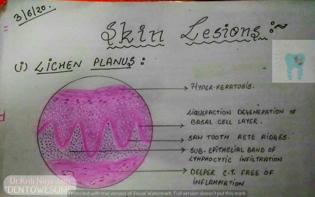
1.LICHEN PLANUS:-

*Lichen planus is a chronic mucocutaneous disorder manifested in a various forms in the oral cavity.

*The most characteristic pattern is” RETICULAR TYPE” with the interlacing white stripe called “WICKHAM’S STRIAE”.

*HISTOPATHOLOGY:-

  •   Histopathology FIRST DESCRIBED BY DUBRENILL 1906
  • later revised by Shklar in 1972
  • Hyper orthokeratinisation or hyper parakeratinisation
  • ◦Thickening of granular layer
  • ◦Acanthosis of spinous layer
  • ◦Intercellular oedema in spinous layer
  • ◦“ Saw-tooth” rete pegs
  • ◦Liquefaction necrosis of basal layer- Max Joseph spaces
  • Civatte ( hyaline or cytoid) bodies
  • ◦Juxta epithelial band of inflammatory cells
  • ◦An eosinophilic band may be seen just beneath the basement membrane and represent fibrin covering lamina propria.

2.PEMPHIGUS :-

Pmphigus is a tissue specific autoimmune disease affecting the skin and mucosa. Clinical manifestations is in the from of “vesiculobullous lesions”  that rupture to form ulcer and erosions .

*Vesiculobullous lesions develop due to immune mediated acantholysis causing intraepithelial vesicle formation.

*HISTOPATHOLOGY :-

  • Formation of the vesicle or bullae within the epithelium that often results in a supra-basilar spilt or separation.
    • Following this suprabasilar spilt in the epithelium, the basal cell layer remains attached to the lamina propria, and it often appears as a row-of-tomb stones.
    • Loss of intercellular bridges and collection of edema fluid result in acantolysis within the spinus cell layer, which causes disruption of the prickle cells.
    • As a result of acantholysis, clumps of large hyperchromatic epithelial cells desquamate that are often seen lying free within the vesicular fluid, these desquamated cells are often rounded and smooth in appearance and are known as “Tzanck cells”.
  • Small number of polymorphonuclear neutrophil (PMN) and lymphocytes may be found within the vesicular fluid, but there is minimum inflammatory cell infiltration in the underlying connective tissue (unlike any other vesiculobullous lesion).

3.PEMPHIGOID :-

Pemphigoid is a vesiculobullous lesions that develop due to an autoimmune reaction directed against some components of basement membrane.

*This results in seperation of epithelium from the connective tissue with sub epithelial vesicles formation .

*Bullous pemphigoid and cicatricial pemphigoid are two different types of pemphigoid lesions.

*HISTOPATHOLOGY:-

  • The inflammatory infiltrate is typically polymorphous, with an eosinophilic predominance.
  • Mast cells and basophils may be prominent early in the disease course.
  • Electron microscope shows basement membrane attached to the connective tissue rather than overlying separated epithelium.
  • Tzanck smear shows only inflammatory cells.
  • Sub epithelial vehicle formation.
  • Intact epithelium without acantholysis.

REFERENCE:-

  1. Pic – Maji Josh 2nd edition

Primary Immunodeficiency Disorders (Overview)

OVERVIEW

  • Primary immunodeficiencies (aka, PIDs) are inherited, as opposed to the acquired secondary disorders.
  • PIDs can arise as defects in the innate and/or adaptive immune systems.
  • Defects in the innate system include: dysfunctional leukocytescomplement proteins, and toll-like receptors.
  • Defects in the adaptive immune system include dysfunctional or absent T cells, natural killer cells, B cells, or antibodies (aka, immunoglobulins).
  • Primary immunodeficiencies increase an individual’s susceptibility to infection, allergy, and autoimmune disorders; they are often diagnosed in infancy or early childhood.

INNATE IMMUNITY DEFECTS

  • Adhesion deficiencies impair leukocyte trafficking:
    – Leukocyte adhesion deficiency 1 is due to defects in the CD 11/CD18 integrins, which adhere neutrophils to the vessel endothelium during recruitment.
    – Leukocyte adhesion deficiency 2 is due to defects in the selectin receptor, which neutrophils use to roll along the vessel wall to the site of diapedesis.
    – See acute inflammation for a reminder of leukocyte trafficking.
  • Chediak-Higashi syndrome: defective phagosome-lysosome fusion prevents neutrophilic antimicrobial products from reaching their pathogen targets.
  • Chronic granulomatous disease is caused by defects in NADPH oxidase; reactive oxygen species production and neutrophil respiratory burst are inhibited.
  • Myeloperoxidase deficiency also impairs antimicrobial effects, though this deficiency tends to have less severe consequences.
  • Dysfunctional complement proteins interrupt the complement cascade, and, in many cases, leave individuals more susceptible to invasive meningococcal infections.
    – C2 and/or C4 deficiencies interrupt the classical pathway, and are associated with the symptoms of lupus due to persistent immune complexes.
    – Defects in C3 interrupt both classical and alternative pathways.
    – Properdine &/or factor D deficiencies inhibit the alternative pathway.
    – Defects in late-acting complement proteins inhibit the formation of membrane attack complexes (MACs).
    – C1 inhibitor deficiency leads to angioedema caused by Bradykinin production; swellings in the gastrointestinal and respiratory tracts can have dire consequences.
  • Dysfunctional toll-like receptors impair cytokine production; defective TLR also contribute some adaptive immune disorders.

ADAPTIVE IMMUNITY DISORDERS

Lymphocyte Cell Lineage

  • Common lymphoid progenitor gives rise to Pro-T and Pro-B cells.
  • Pro-T cells give rise to immature T cells; these cells ultimately give rise to CD4+ Helper and CD8+ Cytotoxic cells.
  • Pro-B cells give rise to Pre-B cells.
  • Upon stimulation by Bruton’s Tyrosine Kinase, Pre-B cells become immature B cells with IgM antibodies on their cell surfaces.
  • Further maturation and class switching produces the full range of antibodies, including IgM, IgG, IgA, and IgE antibodies (we omit IgD, here, for simplicity).

Maturation Defects:

  • Adenosine deaminase deficiency – severe combined immunodeficiency (ADA SCID) impairs T lymphocyte maturation.
    – This is an autosomal recessive form of SCID.
    – Individuals have low T cells and natural killer cells, and, because T cells are required for most B cell activation, reduced B cells and antibodies.
    – Clinically, ADA SCID manifests as thrush, rash, diarrhea and susceptibility to infections; neurologic abnormalities, pulmonary proteinosis, and liver dysfunction are also possible.
  • X-linked SCID impairs progression from the Pro-T cell to immature T cell stage.
    – Serum values include low T cells, natural killer cells, and antibodies.
    – Individuals have increased susceptibility to thrush, rash, diarrhea, slow growth, and infection, particularly pneumonia.
  • DiGeorge Syndrome impairs progression from the immature T cell to mature T cell stage.
    – Low T cell counts, and normal to low antibody levels.
    – Also known as thymic hypoplasia, DiGeorge syndrome presents with hypoparathyroidism, hypoplastic thymus, conotruncal heart defects, and facial abnormalities.
  • X-linked ammaglobulinemia, aka, Bruton’s agammaglobulinemia, interrupts the development of Pre-B cells to the immature B cell stage.
    – Low B cells and antibodies, and an absence of plasma cells.
    – Individuals present with recurrent bacterial infections, especially in the respiratory tract, and viral infections in the gastrointestinal tract. Antibody therapy is required.

Activation defects:

  • Common variable immunodeficiency (CVID) is a group of disorders that blocks activation of B cells.
    – This is the most common symptomatic primary adaptive immunity disorder.
    – Reduced antibody production.
    – Susceptible to pyogenic (pus-forming) and sinopulmonary infections, herpesvirus, enterovirus, and autoimmune disorders.
    – Unlike most of the other primary immunodeficiency disorders, CVID is commonly diagnosed in adults.
  • Hyper-IgM disorder occurs when class switching fails to occur, for example, when the gene for the CD40 ligand is mutated.
    – As its name suggests, IgM levels are high, but all other antibody levels are reduced.
    – Thus, individuals are susceptible to pyogenic and sinopulmonary infections, autoimmune disorders, particularly hemolytic anemia, and diseases of the liver.
  • Selective IgA deficiency is the most common asymptomatic disorder.
    – Occurs when class-switching to IgA is inhibited.
    – Though usually benign, individuals may have increased susceptibility to sinopulmonary and gastrointestinal infections, autoimmune disorders, and allergy.
  • X-linked lymphoproliferative syndrome results in abnormal, usually low, antibody production and reduced levels of natural killer cells.
    – In many cases, this disorder is triggered by Epstein-Barr virus, and is associated with increased risk of lymphoma and fulminant infectious mononucleosis.

Systemic disorders that have immunological components:

  • Wiskott-Aldrich syndrome is characterized by low levels of IgM, but elevated levels of IgE and IgA; individuals tend to present with thrombocytopenia, eczema, recurrent infections, autoimmune disorders, and B-cell lymphoma.
  • Ataxia telangiectasia is characterized by low levels of A, G, and E antibodies; as the name suggests, individuals present with ataxia and telangiectasia, and have increased susceptibility to respiratory tract bacterial infections and cancers. They are sensitive to radiation exposure (including X-rays).

Primary immunodeficiencies of the adaptive system are often treatable with hematopoietic stem cell transplants or antibody therapy.

Type IV Hypersensitivity

Overview

  • Type IV reactions are T cell mediated.
  • Because of the time it takes to recruit and activate T cells and their products, these reactions are delayed – they occur 1-3 days after antigen exposure; in contrast, recall that the other types of hypersensitivity reactions occur within minutes to hours after exposure.
  • CD4+/Helper T cells induce hypersensitivity reactions via cytokine recruitment of inflammatory cells.
  • CD8+/Cytotoxic T cells directly destroy tissues.

CD4+ T Cell Mechanism

  • CD4+ T cells are activated when they recognize and interact with cells displaying the antigen-MHC II complex.
  • As a result, CD4+ cells proliferate and differentiate:
    — Under direction from interferon-gamma and IL-12, Helper T cells of the Th1 subset are produced.
    — Under direction from IL-1, IL-6, and IL-23, cells of subset Th17 are produced.
  • In turn, these Helper T cells release cytokines that recruit and activate inflammatory cells:
    — Th1 Helper T cells release interferon-gamma, which recruits macrophages; show that, upon activation, macrophages induce tissue damage and fibrosis.
    — Alternatively, show that Th17 Helper T cells release IL-17 and IL-22, which recruit and activate neutrophils; show that neutrophils cause inflammation.

Examples:

  • Tuberculosis is characterized by the formation of granulomas, aka, tubercles, which comprise special populations of epithelial cells and macrophages that gather around the M. tuberculosis bacteria. In a more magnified view, label a giant cell, which formed from macrophages that merged together.
  • The tuberculin reaction test uses TB antigens, called Purified Protein Derivatives, to determine whether an individual has been previously exposed to the M. tuberculosis bacteria.
    — In our image, we can see that a positive test result shows induration, which is caused by macrophage activities, and, erythema, which is caused by neutrophil-induced inflammation.

Cytotoxic T Cell

  • Destruction of host tissues is more direct:
    — When the cytotoxic T cell recognizes the antigen-MHC I complex, it releases granzymes and other harmful molecules into the tissues.

Example

  • Type I diabetes mellitus can be caused by insulitis:
    — Cytotoxic T cells target beta cells of the Islet of Langerhans; recall that beta cells are responsible for insulin secretion.
    — Indicate that, in an affected islet, we would see infiltration of destructive lymphocytes.

Additional Disorders

Helper T cell-mediated damage

  • Psoriasis involves macrophage release of Tumor Necrosis Factor (TNF) and subsequent destruction of the epidermis; thus, dead skin cells build up to form scaly, flaky plaques, often on the hands and feet, scalp, and places where the skin folds, such as elbows and knees.
    — The nails can also be affected; show that the fingernails become thick and broken, with a yellowish tint.
  • Multiple sclerosis is a demyelinating disorder caused by inflammatory cell destruction of myelin sheaths.
    — We can see areas of periventricular white matter lesions in a radiograph.
    — In a histological sample, we can see perivascular cuffing, which is characterized by aggregation of lymphocytes and macrophages around the blood vessels.
  • Rheumatoid arthritis is caused by inflammation and tissue erosion.
    — Indicate neutrophil and macrophage destruction of the cartilage and bone of a synovial joint of the hand; rheumatoid arthritis tends to affect the bones of the hands and feet, first.
    — In an x-ray, we can see how tissue erosion has led to deformation of the hands.

Cytotoxic T cell-mediated damage

  • Contact dermatitis is characterized by itching, redness, and blisters.
    Common causes of contact dermatitis include:
    — Urusiol oil, found in poison ivy.
    — Heavy metal; many people are allergic to nickel, which is a common component of jewelry and clothing. For example, we show that the nickel buttons on denim pants causes a characteristic umbilical rash in susceptible individuals.
  • Stevens-Johnson Syndrome (SJS), and the related, more severe Toxic Epidermal Necrolysis (TEN), are potentially life-threatening disorders that can be triggered by drugs, especially some antibiotics.
    — Cytotoxic release of granzymes causes severe blistering; SJS is characterized by blistering of less than 10% of the skin, and TEN is characterized by blistering of more than 30% of the skin.
    — Early recognition is key for effective treatment; causative agents, such as drugs, must be removed immediately.

Type II Hypersensitivity

Overview

  • Type II Hypersensitivity reactions occur 1-3 hours after antigen exposure.
  • Mediated by IgG antibodies and have cytotoxic and complement-activating effects. Recall that the complementcascade produces various proteins that promote inflammation, phagocytosis, and cell lysis.
  • Three mechanisms of antibody-mediated hypersensitivity:
    — Opsonization, inflammation, and, cellular dysfunction.

Opsonization

  • Coats cells in antibodies, leads to phagocytosis and/or complement activation.
    — IgG binding of cell-bound antigens initiates the complement cascade, which generates active proteins.
    — Some of these proteins, including C3b, are deposited on the cell surface.
  • Thus, neutrophils can recognize the opsonized cell via two mechanisms:
    — Its high-affinity Fc receptor can bind with the Fc region of the IgG antibody.
    — Its C3b receptor can bind with the deposited complement on the cell’s surface.
  • In both cases, binding promotes phagocytosis of the host cell.

Examples:

  • Transfusion reactions can occur when donor cell antigens are bound by host IgG antibodies.
    — If complement protein C1 binds also those antibodies, the complement cascade ultimately produces a Membrane Attack Complex (MAC); the MAC allows massive water influx into the cell, causing its lysis.
  • Hemolytic disease of the fetus and newborn: maternal anti-Rh+ antibodies attack fetal Rh+ red blood cells.
    — The maternal antibodies were produced in response to prior pregnancy with a Rh+ fetus; upon subsequent exposure to a another Rh+ fetus, the maternal antibodies readily react.
  • Autoimmune blood cell destruction occurs when one’s own blood cells are targeted:
    — Anemia can result when red blood cells are destroyed.
    — Agranulocytosis, when granulocytes, such as neutrophils, are targeted.
    — Thrombocytopenia, when platelets are removed.
  • Drug hapten reactions occur when haptens adhere directly to the cell surface.
    — When IgG antibodies bind the hapten, the complement system is activated and phagocytosis occurs. Penicillin is an example of a drug that can cause type II hypersensitivity reactions.

IgG-induced inflammation

  • Inflammation occurs when antibodies are deposited in the tissues.
    — Neutrophil Fc receptor binding and complement activation leads to recruitment of additional leukocytes, including macrophages and additional neutrophils.
    — The released inflammatory products, including enzymes and reactive oxygen species, cause local tissue injury.
    IgG antibodies specific to host tissues can cause severe destruction.

Examples

  • Vasculitis: Anti-neutrophil cytoplasmic antibodies (ANCAs) can induce vasculitis, which is inflammation of the blood vessels. This may show up as small reddish purple lesions in the skin, or larger bruise-like patterns of decay.
    — Treatments include methotrexate, prednisone, and cyclophosphamide, which suppress the immune response.
  • Goodpasture’s syndrome is caused by antibodies that attack the basement membranes of renal glomeruli and/or respiratory alveoli; specifically, the antibodies attack type IV collagen in the tissues.
    — In a histological sample of an affected glomerulus, we can see the characteristic crescent-shaped area formed by excessive fibrin and cellular material.
    — In a sample of lung tissue, we highlight hemorrhaging in the inflamed alveoli.
    — Goodpasture’s syndrome may be treated with corticosteroids and cyclophosphamide, or plasmapheresis, which removes the attacking antibodies.

Cellular Dysfunction

Antibodies bind cellular receptors and cause dysfunction; two classic examples are myasthenia gravis and Graves’ disease.

  • In Myasthenia gravis, IgG binds the acetylcholine receptors on the muscle tissue; thus, acetylcholine released from the nerve cell cannot bind and stimulate muscle contraction.
    — For example, eyelid drooping, called ptosis, is common in individuals with myasthenia gravis. Drooping occurs because antibodies block or destroy the acetylcholine receptors, thus inhibiting eyelid muscle contraction.
    — Immuno-suppressive steroids may reduce circulating IgG antibodies to treat muscle weakness.
  • Graves Disease
    — Caused by binding of thyroid-stimulating antibody to the TSH receptor; as its name suggests, this has a stimulatory effect, and induces excessive thyroid hormone production.
    — In the histological sample, we can see the colloid-filled lumen, and, in a more magnified view, we highlight the hyperactive thyroid epithelium.
    — Indicate that excessive thyroid hormone production in periorbital tissues can produce Graves’ opthalmopathy, which is characterized by proptosis caused by swelling and adipose deposition.
    — Show that enlargement of the thyroid gland, called goiter, is caused by chronic thyroid stimulation. However, be aware that goiter can be indicative of hyper- or hypo-thyroidism; both states can involve overstimulation of the thyroid gland.
  • TSH-stimulating blocking antibody can inhibit thyroid hormone production.

Type III Hypersensitivity

Overview

  • Type III hypersensitivity reactions are mediated by IgG (or IgM) antibody-antigen complexes; recall that these responses occur 1-3 hours after antigen exposure.
  • Normally, antibody-antigen complexes, aka, immune complexes, are removed by phagocytes of the spleen and lymph nodes.
  • However, persistent complexes can concentrate and cause tissue damage via complement and immune cell activation.
  • This most commonly occurs where blood or plasma is filtered through fenestrated capillaries; for example, in the synovial joints and renal glomeruli.

Pathogenesis of immune complex-mediated hypersensitivity:

  • Immune complexes become deposited on the vessel wall (or, in some cases, in the tissues).
  • As a result, complement and neutrophil activation occurs, leading to the release of pro-inflammatory cytokines, enzymes, and reactive oxygen species.
  • Increased vessel permeability allows the inflammatory molecules to cause additional tissue damage outside of the vessel.

Examples

  • Vasculitis, which is inflammation of the vessel walls, is a common manifestation of type III hypersensitivity. In the image, we can see small purple lesions in the skin.
    — In the histological sample showing fibrinoid necrosis of a vessel, highlight where immune complex deposits promote cell death and excessive fibrin synthesis.
  • Arthritis occurs when immune complexes deposit in the synovial joints.
  • Post-infectious glomerulonephritis, which involves inflammation of the renal glomeruli, is associated with Staphylococcus bacterial infection (and, increasingly, Streptococcus infections).
    — In the histological sample of a glomerulus, show that antibody-antigen complexes aggregate in the basement membrane, and, that neutrophils infiltrate the glomerulus and damage the renal filtration system.

Local Arthus Reaction

  • Produces localized vasculitis and tissue necrosis; rarely, it is associated with tetanus or diphtheria-containing vaccines.
  • It can be produced by re-injecting antigens too soon after initial exposure:
    — The antigens form immune complexes with circulating IgG antibodies.
    — Then, the immune complexes activate complement and recruit inflammatory cells, including neutrophils and mast cells.
    — Inflammatory cell degranulation releases cytokines, enzymes, and other molecules that produce local inflammation at the injection site, which is marked by pain, swelling, and redness that typically subsides after a few days.
  • As an additional clinical correlation, write that hypersensitivity pneumonitis, aka, allergic alveolitis, is a type III hypersensitivity reaction that takes place in the lungs (we discuss this in more detail in the hypersensitivity essentials tutorial).

Systemic Serum Sickness

  • Manifests throughout the body.
  • Serum sickness can be induced by older vaccines that used antibodies from other species, such as horses and rabbits; though rare, it can also occur after transfusions. Anti-venom reactions can occur when, for example, anti-snake venom treatment is administered to a previously sensitized individual. Newer anti-venom treatments avoid this reaction.
  • Graphically:
    — X-axis = “Time in Days”; Y-axis = “Plasma levels”
    — At time 0, show that plasma levels of foreign proteins spike immediately after administration, then slowly decline for a period; Initially, plasma antibody concentration is low.
    — Then, show that as production of plasma antibodies increases, immune complexes are formed; thus, the concentration of foreign protein is rapidly reduced.
    — Furthermore, show that the appearance of serum sickness symptoms coincides with immune complex formation, and typically resolve on their own.
  • Common manifestations of serum sickness include: blotchy skin rashes, joint pain, peripheral edema, and fever.

Systemic Lupus Erythematosus (SLE)

  • Genetically-influenced disorder caused by autoantibodies that target self-antigens.
  • SLE is associated with a wide variety of autoantibodies, some of which are specific to the disorder; furthermore, some of the autoantibodies are correlated with specific outcomes.
    — Some key autoantibodies include:
    Antinuclear antibodies, which target components of host nuclei; these are non-specific to SLE.
    Anti-double stranded DNA antibodies, which are specific for SLE and may be associated increased renal damage;
    Anti-sm antibodies, also specific to SLE, may be associated with increased pulmonary arterial hypertension;
    Antiphospholipid antibodies, which are non-specific and are associated with thrombosis, hemolytic anemia, and, possibly, problematic pregnancies.
    Anti-Ro and anti-La antibodies are commonly found in SLE patients, but are not-specific; importantly, anti-La antibodies may cross the placenta and interfere with fetal heart development.
  • Clinical manifestations of autoantibody actions include:
    — Lupus nephritis, of which there are 6 patterns of glomerular damage;
    Here, we indicate that the most common includes widespread destruction characterized by thickened “wire loops” and necrosis; often, crescent-shaped damage in Bowman’s capsule is also visible.
    — SLE produces specific rash patterns; the malar rash pattern, also known as the “butterfly rash,” and, “discoid rash,” which is disc-shaped and tends to be scalier.
    — The cardiovascular system may also be affected. Here, show Libman-Sacks endocarditis, in which sterile vegetations form along the margins of the valve leaflets; recall that sterile vegetations are especially prone to breaking free and embolizing.
    — Pericarditis and/or peritonitis are also common, though not shown here.
  • Common environmental triggers for SLE flares include: ultraviolet light, some medications, cigarette smoking, and certain viral infections.
  • Some of the immune-complex mediated symptoms of systemic lupus erythematosus can be treated with hydroxychloroquine (originally used to treat malaria) and corticosteroids, such as Prednisone, which dampen the immune response.

Type I Hypersensitivity

Overview

  • Type I hypersensitivity reactions, which are characterized by immediate allergic responses and mediated by IgE and Mast cell activation.
  • “Atopy” is the susceptibility to develop immediate hypersensitivity reactions, and is characterized by elevated levels of IgE antibodies.
    — Atopy is influenced by genetic and environmental factors.

Primary humoral response to antigens

  • When Helper T cells recognize the antigen-MHC complex, intercellular interactions activate the B cell and induce production of large quantities of antigen-specific IgE antibodies.
  • Mast cell (aka, mastocyte) display high affinity IgE Fc receptors on their surfaces; basophils (which we haven’t shown) also have high-affinity IgE receptors.
  • Circulating IgE antibodies readily bind these receptors, “sensitizing” the mast cell.
  • The next time the antigen is present tissues, it binds with the IgE antibodies and cross-links the mast cell receptors.
    This triggers release of:
    — Cytoplasmic granules, which include histamines, proteases, and heparin
    — Membrane phospholipids, which comprise platelet activating factor and arachidonic acidProstaglandin D2 and Leukotrines B4, C4, and D4. Recall that prostaglandin causes vasodilation and increases vascular permeability; leukotrienes recruit pro-inflammatory leukocytes and promote bronchoconstriction.
    — Pro-inflammatory cytokines, including Tumor Necrosis Factor (TNF), IL-1, IL-4, and chemokines.
    These degranulation products mediate early and late phases of the hypersensitivity reaction.

Early Phase

  • Vasodilation, vascular leakage, and increased glandular secretion; notice that are the early events of the acute inflammatory response.
  • Anti-histamines, which prevent histamine binding, are often effective at suppressing the early phase.

Late Phase

  • Eosinophil activity, particularly their degranulation and release of Major Basic Protein and Eosinophil Cationic Protein, among other proteins, and IL-4 and other cytokines.
  • Broad-spectrum anti-inflammatory drugs, such as corticosteroids, can reduce eosinophilia; thus, they are more effective than anti-histamines at the late phase of type I reactions.

Skin Prick Test

  • Clinically, allergy diagnosis can be achieved with skin prick tests, in which small quantities of common allergens are introduced via small pricks or scratches to the skin.
    — In the image, we can see that this patient had several positive reactions, which are characterized by wheal-like swelling and flare-like erythema, in response to antigens from spiders, moths, caterpillars, scorpion, tick, and, to a lesser extent, worker honey bees. In contrast, there were negative responses to spider web, caterpillar web, and house dust.

Common clinical manifestations of hypersensitivity type 1 reactions

  • Asthma is characterized by bronchioles with thickened smooth muscle, inflamed mucosal linings with hyperactive goblet cells – in our sample, we can see that the lumen is filled with mucus. Upon antigen exposure, the bronchi constrict and the goblet cells are activated to secrete mucus, all of which reduces airflow.
    — Treatments include corticosteroids, leukotriene antagonists, and phosphodiesterase inhibitors, which suppress the inflammatory actions of the immune system.
  • Allergic hypersensitivity can produce various skin reactions; eczema and hives are common.
    — Eczema (aka, atopic dermatitis) is characterized by red, scaly, flaky areas that may become “weepy;”
    flares can be treated with corticosteroids or topical calcineurin inhibitors, which block cytokine expression; thick emollients that maintain the skin barrier may prevent flares.
    — Hives are characterized by red areas with raised, swollen plaques; anti-histamines or corticosteroids are used to suppress edema and erythema.
  • Allergic rhinitis involves inflammation and stimulation of the mucosa of the nasal passages and is often associated with allergic conjunctivitis.
    — Anti-histamines, corticosteroids, or immunotherapy are used to suppress inflammation, and avoidance of the responsible allergens is advised.
  • Gastrointestinal allergic reactions are characterized by increased smooth muscle contraction and, therefore, peristalsis and increased gastric secretions; thus, vomiting and diarrhea occur.
    — Aside from allergen avoidance, antihistamines to suppress smooth muscle contraction may be helpful. If the allergy is severe, anaphylaxis, which we address next, can be treated with epinephrine.
  • Systemic anaphylaxis, which can be fatal, has multifold effects:
    Bronchoconstriction, which limits airflow
    Edema, which, if in the larynx, further obstructs airflow
    Vasodilation, which can lead to hypotension and shock
    Cardiac arrhythmia or even cardiac arrest
    — Immediate treatment with epinephrine, which reverses the cardiopulmonary effects, is necessary to prevent serious organ damage or death.

Common Allergens

  • Common environmental allergens include insect venom (i.e., bee stings), pollen, dust (specifically, the feces of dust mites), animal dander, and air pollution.
  • Certain foods, specifically the proteins in some foods, can trigger allergic responses; nuts, fish, milk, eggs, soy, wheat, and gluten are the most common culprits.
  • Some medications are responsible for type I reactions; specifically, penicillin, aspirin, and other non-steroidal anti-inflammatory drugs. Paradoxically, topical corticosteroids are also known to cause allergic reactions.

Long-term desensitization

Can be achieved by regular administration of increasing quantities of the antigen, which suppresses IgE activity.

Hypersensitivity Overview

  • Hypersensitivity reactions are mediated by antibodies and T cells (if you are unfamiliar with antibodies and T cells, we recommend you review our adaptive immunity tutorials).
    — Types I, II, and II are antibody-mediated.
    — Type IV is T cell-mediated.

Type I Hypersensitivity

  • Type I immediate hypersensitivity is typified by allergy.
    — Mediated by IgE antibodies and mast cell and basophil activation.
    — This reaction occurs within minutes of antigen presentation.

Mechanism

  • Initial exposure to antigen
    — IgE antibodies are released from mature B cells (plasma cells).
    — These antibodies bind to mast cells (and basophils, not shown); the mast cells are now “sensitized.”
  • Subsequent exposure to the same antigen
    — Antigen binding cross-links the antibodies bound to mast cells, leading to degranulation.
    — As a result, cytokines, membrane phospholipids, and granules are released form the mast cell.
  • Some key mediators of type I hypersensitivity that are released upon degranulation include histamine, leukotrienes, prostaglandins, and platelet activating factor.
    — Recall that these are the mediators of acute inflammation, which, when excessive, cause damage to the tissues.

Manifestations

  • Early edema and erythema are often characterized by “wheal and flare” – the “wheal” is caused by vascular leakage and swelling; the “flare” is caused by vasodilation and reddening of the skin.
  • The later stage of type I hypersensitivity is characterized by eosinophilia in the effected tissues; chemokines released during degranulation and leukocyte activation attracts eosinophils, which release proteins that cause more tissue damage.
  • Anaphylaxis is a systemic and potentially fatal form of type I hypersensitivity; epinephrine is administered to reverse respiratory and cardiovascular effects.

Type II Hypersensitivity

  • Type II antibody-mediated hypersensitivity is characterized by cytotoxic IgG or IgM complement activation.
  • Reactions occur within 1-3 hours after antigen exposure.

Mechanisms

  • Example: transfusion reaction
    — Donor red blood cell with surface antigens;
    — Host IgG antibodies bind those antigens, which initiates the complement cascade.
    — As a result, a Membrane Attack Complex (MAC) forms and allows water influx into the red blood cell, causing lysis.

Manifestations

  • Blood cell destruction
  • Goodpasture’s syndrome: damage to renal and lung tissue by anti-basement membrane antibodies.
  • Cellular dysfunction: antibodies that bind to cellular receptors, altering their activity.
    — For example, IgG antibodies can bind TSH receptors, with inhibitory OR stimulatory consequences, depending on their configuration.

Type III Hypersensitivity

  • Characterized by deposition of IgG or IgM antibody-antigen complexes (also called “immune complexes”) in vessels walls and/or tissues.
  • Type III reactions occur 1-3 hours after antigen exposure.

Mechanism

  • When IgG-bound antigen deposits in tissues, complement and neutrophils are activated, with tissue destruction as a result.

Manifestations

  • Hypersensitivity pneumonitis, aka allergic alveolitis, is a localtype III response to inhaled antigens; historically common in farmers who inhaled mold, fungi, and other environmental pathogens, it is increasingly common in office workers exposed to microorganisms in humidifiers and air conditioning systems.
    — Immune complexes activate complement in the alveoli, the site of gas exchange in the lungs. Then, local Arthus reactions thicken the lung interstitium, making gas exchange more difficult.
  • Serum sickness is a systemic example of a type III reaction;
    — It is induced by antibodies from other species. It is characterized by widespread effects, including rash, edema, joint pain, and fever.

Type IV Hypersensitivity

  • Also called delayed hypersensitivity, it’s mediated by helper and cytotoxic T cells.
  • These responses appear 1-3 days after exposure, because it takes additional time to recruit and activate T cells and their products.

Mechanisms

  • In response to antigens, helper T cells release cytokines that recruit macrophages and neutrophils, which damage host tissues.
  • Cytotoxic cells directly damage host tissues via granzymes.

Manifestations

  • An example of helper T cell-mediated hypersensitivity is the tuberculin reaction test: a positive result, characterized by induration and erythema, indicates that an individual has been previously infected by M. tuberculosis.
  • An example of cytotoxic T cell-mediated hypersensitivity is contact dermatitis; in response to certain organic or metallic substances, such as poison ivy, cytotoxic T cells destroy host tissues and cause an itchy, red, vesicular rash.

Autoimmune Disorders (Systemic Overview)

SELF-TOLERANCE

  • Failure of self-tolerance results in autoimmune disorders, in which abnormal immune cells provoke a damaging immune response to otherwise benign molecules.
  • These disorders have genetic components, often related to HLA alleles, and are often triggered by environmental or infectious stimuli. Furthermore, women are more often affected than are males, though the reasons for this are undetermined.
  • Tolerance is achieved by selective removal of T and B cells that respond to self-antigen via central and peripheral mechanisms

Central Tolerance & Peripheral Tolerance

  • Hematopoietic bone marrow houses the B and T cell precursors.
  • B cells
    — During central tolerance, show that B cells remain in the bone marrow: here, they are exposed to self-antigen.
    — B cells that respond to self-antigen can undergo receptor editing, in which they develop new receptors that are do not recognize self-antigen, or, they can undergo apoptosis.
    — When self-reactive B cells in the periphery are exposed to self-antigen, they enter an unresponsive state of anergy. It is thought the absence of co-stimulation by T cells renders B cells anergic.
  • T cells
  • T cells undergo central tolerance in the thymus, where they are exposed to self-antigen by antigen-presenting epithelial cells.
  • In response, they can differentiate to become T regulatory cells, or, undergo apoptosis.
  • Peripherally, T cells reactive to self-antigens may become anergic and unresponsive (particularly in the absence of co-stimulation); or, they may be suppressed by T regulatory cells, which inactivates them; or, they may undergo apoptosis.

Unfortunately, some self-reactive B and T cells end up in the circulation, where they act as autoantibodies that cause chronic, often progressive, damage.

SYSTEMIC AUTOIMMUNE DISORDERS

Bear in mind that autoimmunity can also be organ-specific, as in multiple sclerosis or Type I diabetes mellitus.

Rheumatoid arthritis

  • Tends to affect the synovial joints of the hands and feet early in the disorder.

Key mediators and their effects:

  • Helper T cells release interferon gamma and IL-17.
    — Interferon gamma activates macrophages
    — IL-17 recruits inflammatory neutrophils and monocytes.
  • Macrophages release tumor necrosis factor and IL-1.
    — Trigger the release of degradative proteases.
  • Activated T cells
    — Express RANKL, which promotes bone resorption.

Characteristic damage of rheumatoid arthritis in a zoomed in view:

  • Inflammation of the synovium
    — Results in pannus formation, which comprises thickened synovial fluid with infiltrating inflammatory cells, fibroblasts, and synovial cells that erode the cartilage.
    — Over time, the pannus on opposing bones forms a fibrous ankylosis, which may then become bony ankylosis.
  • These changes cause stiffness, swelling, and joint deformities.
    — In a photograph and x-ray, we can see swan neck deformity, a common consequence of rheumatoid arthritis in which the proximal interphalangeal joint is hyperextended and the distal interphalangeal joint is flexed (thus, it looks like the neck of a swan).

Serum Markers:

  • Antibodies against citrullinated protein antigens (ACPAs), such as citrullinated fibrinogen, which is produced during inflammation.
  • Rheumatoid factor is also found in patients with rheumatoid arthritis; however, despite its name, rheumatoid factor is non-specific to rheumatoid arthritis and is present in other autoimmune disorders.

Systemic Sclerosis

  • Aka, scleroderma

Key mediators and their effects:

  • Caused by a combination of autoimmunity, vascular damage, and fibrosis.
    — Tumor growth factor Beta is a key mediator of this disorder.

Serum Markers:

  • Anti-centromere antibody (ACA)
  • Anti-Scl-70

Limited Systemic Sclerosis

  • Affects the skin of the fingers, forearms, and face; visceral involvement, if any, occurs late.
  • Limited systemic sclerosis is characterized by the presence of anti-centromere antibodies.
    CREST
    Calcinosis, in which calcified nodules form under the skin; they may progress to lesions.
    Raynaud’s phenomena, in which the small vessels of the fingers and toes severely constrict in response to stress and/or cold.
    — Initially, reduced blood flow turns the fingers white; as oxygen levels fall, the fingers appear bluish; and, finally, after blood flow is re-established, they become red.
    Esophageal dysmotility is caused by collagen replacement of the muscularis layer of the esophagus; dysphagia and/or heart burn can result.
    Sclerodactyly occurs when hands take on a claw-like appearance because fibrosis of the dermis pulls the skin taught; dermal tightening can also occur in the face.
    Telangiectasia is characterized by dilation of superficial micro-vessels; it commonly looks like a rash on the face or chest.

Diffuse Systemic Sclerosis

  • Manifests as widespread cutaneous fibrosis with quick progression to the viscera.
  • It is associated with Scl-70.
  • Examples of Organ Effects:
    — Pulmonary fibrosis, in which the interstitial tissue becomes thick and fibrotic, can occur. Pulmonary fibrosis and hypertension are the leading causes of death from systemic sclerosis.
    — Renal failure, due to systemic vascular damage and hypertension.
    — Pericarditis and pericardial effusion.
    — Damage to the villous structures in the small intestine, which leads to malabsorption.

Sjögren’s syndrome

  • Characterized by damage to the lacrimal and salivary glands, and is largely mediated by interferons (types 1 & 2).
  • Key autoantibodies are anti-SSA/Ro, anti-SSB/La, which are anti-nuclear autoantibodies (which are also implicated in the pathogenesis of systemic lupus erythematosus).

Xerostsomia

  • Aka, dry mouth
  • Can lead to ulcers and increased dental caries
  • In a histological sample of the minor salivary glands of the lip, we indicate the leukocyte infiltration that has damaged the tissues.

Keratoconjuctivitis sicca

  • Aka, dry eye.
  • Occurs when the lacrimal gland, which is located laterally above the eye, does not produce tears.
  • Thus, the eyes become dry, irritated, and more susceptible damage; vision may be impaired.
  • Furthermore, Sjörgen’s syndrome may have non-glandular effects, particularly in the joints, lungs, kidneys, and cardiovascular system.

Be aware that systemic lupus erythematosus is another autoimmune disorder with multi-organ effects; we discuss it in more detail, elsewhere.

Chronic Inflammation

Chronic inflammation occurs when tissue injury and repair attempts overlap.

  • It is characterized by infiltration of mononuclear cells, particularly macrophages and lymphocytes, which interact dynamically over the course of inflammation (recall that acute inflammation is characterized by neutrophil infiltration).
  • As a result of infectious agents and/or prolonged inflammatory response, tissue damage occurs.
  • Tissue repair attempts comprise angiogenesis (formation of blood vessels, which ultimately regress) and fibrosis, aka, scarring.
  • Because macrophages constitute the primary leukocyte in chronic inflammation, we’ll focus on their origins and activation; however, be aware that other leukocytes, including neutrophils, can be found at sites of chronic inflammation.

MACROPHAGE ORIGINS & ACTIVATION

  • Macrophages arise from hematopoietic cells that reside in the bone marrow of adults (in the fetus, hematopoietic cells reside in the yolk sac and liver).
    — These cells give rise to various blood cell lines, including the monocytes, which circulate in the vasculature.
  • Outside of the circulation, monocytes differentiate to become macrophages, and reside scattered throughout the connective tissues of the body.
    – Additionally, tissue resident macrophages reside in the liver (Kupffer cells), spleen and lymph nodes (sinus histocytes), central nervous system (microglial cells) and in the alveoli of the lungs.
    – Collectively, we can refer to these macrophages as the mononuclear phagocyte system (formerly called the “reticuloendothelial system”; some authors even reject the notion of a “mononuclear phagocyte system”).
  • Upon activation, macrophages engage in various activities:
    – Presentation of antigens to T lymphocytes.
    – Production of cytokines, which mediate inflammatory responses.
    – Production of growth factors and enzymes that promote tissue repair and inhibit inflammation.
  • To accomplish these goals, macrophages have dynamic phenotypes reflective of their microenvironments.

Macrophage Activation

Be aware that some texts describe two distinct types of macrophage activation, classical and alternative, and the resulting macrophages as either Type 1 (aka, classical type) or Type 2 (aka, alternative type); however, research now suggests that these should not be thought of as dichotomous types, but, rather, as potential phenotypes that macrophages express in response to environmental stimuli.

Pro-inflammatory effects:

  • Stimulation by specific cytokines, such as interferon gamma (aka, type II interferon) and/or by microbes activate macrophages to produce inducible nitric oxide, reactive oxygen species, and lysosomal enzymes, which are antimicrobial.
  • These activated macrophages also produce inflammatory cytokines and chemokines:
    – Tumor necrosis factor and chemokines activate lymphocytes.
  • Activated T lymphocytes produce interferon gamma, which, as we’ve indicated, triggers pro-inflammatory macrophage activation. Thus, T lymphocytes can act as part of a positive feedback loop with pro-inflammatory activated macrophages.

Anti-inflammatory effects:

  • Anti-inflammatory stimuli include interleukins 13 and 4 (IL-13, IL-4).
    – In response to these stimuli, activated macrophages produce cytokines, such as interleukin-10 (IL-10), that inhibit inflammatory activity of T lymphocytes, natural killer cells, and macrophages; thus, interleukin 10 prevents excessive inflammation and damage to host cells.
    – These “alternatively activated” macrophages also produce growth factors that promote tissue repair, including vascular endothelial growth factor (VEGF), which facilitates angiogenesis, and transforming growth factor beta (TGF-ß), which facilitates deposition of extracellular matrix proteins for fibrosis.
  • Interleukins 13 and 4 (IL-13 and IL-4) are produced by T lymphocytes; thus, T lymphocytes can also promote anti-inflammatory macrophage activation.

Fibrosis

Unfortunately, the pro-inflammatory feedback loop can be maladaptive when prolonged macrophage-lymphocyte interactions promote chronic inflammation, as seen in fibrosis of the lung.
– In the example, chronic inflammation led to the deposition of extracellular proteins at the cost of healthy lung alveolar tissues.

  • Fibrosis is characterized by excessive collagen deposition in response to persistent stimulation; it results in tissue loss and organ failure.
    – Some common pathological conditions caused by organ fibrosis include liver cirrhosis, constrictive pericarditis, scleroderma, and lung fibrosis disorders.

Acute Inflammatory Response

  • The acute inflammatory response is activated in the presence of infectious agents and/or damaged tissues.
  • Acute inflammation triggers vascular and cellular responses that deliver cells and proteins to the site of cell injury.
    Key steps of this process include:
    – Recognition of inflammatory agents.
    – Leukocyte and plasma protein recruitment from the blood to the tissues.
    – Leukocyte activation.
    – Control and termination of inflammatory reactions, which are otherwise harmful to healthy cells.

DETAILS

Recognition of offending agents

  • Cellular receptors for microbes exist in the plasma membranes, endosomes, and cytosol of host cells.
    – For example: Toll-like receptors (TLR) enable dendritic and other “sentinel cells” to recognize invading microbes.

Other sensors are specialized to recognize signs of host cell damage:

  • Cytosolic sensors recognize various molecules, such as uric acid, ATP, DNA, and reduction of intracellular potassium concentrations, that indicate cellular damage.
    – For example: Multi-protein cytosolic complexes called inflammasomes respond to the cytosolic sensors and trigger the release of cytokines, which, as we’ll see, are key mediators of the inflammatory response.
  • Circulating proteins act as pattern recognition molecules that recognize invaders by their display of abnormal, non-self patterns.
    – For example: mannose-binding lectin protein binds to mannose, which is a characteristic microbial sugar; after binding, MBL facilitates microbe ingestion and activates the immune system.

Recruitment of plasma proteins and leukocytes from the blood

  1. Vasodilation and increased permeability of the vessel wall:
  • Occur in response to inflammatory mediators, importantly: histamine, prostaglandins, platelet activating factor (PAF), thromboxane A2 (generated from prostaglandins), bradykinin, and leukotrienes.
  1. Plasma proteins and fluid exit the vessel (aka, exudation); this process can lead to excess fluids in the interstitial tissues, a condition called “edema.”
    – Clinical correlation: fibrinous pericarditis is a form of acute inflammation; as a result of the inflammatory response, fibrin and leukocytes infiltrate the pericardium (specifically the visceral pericardium, aka, the epicardium) and can cause “friction rub.”
  2. Neutrophil recruitment from the blood involves: Capture, Rolling, Adhesion, Diapedesis, and Chemotaxic Migration.
  • Capture:
    – Capture occurs via E-selectins, which are a type of cell adhesion molecule.
    – Cytokines, specifically Tumor Necrosis Factor (TNF) and Interleukin-1, upregulate the expression of E-selectins on the endothelial lining of the vessel.
    – Correspondingly, neutrophils express PSGL-1 (P-selectin glycoprotein ligand -1), which binds with selectins.
  • Rolling:
    – Achieved via binding with P-selectins; their expression is upregulated by cytokines, thrombin, and histamine.
  • Adhesion:
    – Firmer adhesion occurs when endothelial ICAM-1 (Intracellular Adhesion Molecule) binds with neutrophil LFA-1 ligands (Lymphocyte Function-Associated).
  • Diapedesis:
    – The process of movement across the vessel wall typically occurs via the paracellular route, and is assisted by PECAM-1 (Platelet Endothelial Cell Adhesion Molecule).
    – Once outside of the vessel, neutrophils generate more cytokines, which further promotes the inflammatory response.
  • Chemotaxic Migration:
    – Chemokines guide neutrophils to the site of inflammation along chemotactic gradients.

Phagocytosis and destruction of inflammatory agents

We’ll use neutrophil destruction of microbes as an example.

  1. Neutrophil recognition of the microbe via sensors.
  2. The neutrophil engulfs the microbe and moves it into a phagosome.
  3. Lysosomes merge with the phagosome, which exposes the microbe to lysosomal degradative enzymes in a phagolysosome.
  4. Lysosomal enzymes, reactive oxygen species (ROS, aka, reactive oxygen intermediates), and inducible nitric oxide (iNO) destroy the microbe.
    – Inflammatory cytokines, such as interferon gamma, trigger the production of ROS and iNO within the lysosomes and phagolysosomes.
  • NETs
    In addition to phagocytosis, neutrophils can produce extracellular traps (aka, NETs) to destroy infective pathogens.
    – In this process, the neutrophil exudes its nuclear materials to envelop the microbes in chromatin and concentrated antimicrobial peptides and enzymes.
  • Macrophages
    Although neutrophils are the primary leukocytes active in acute inflammation, other cell types, particularly macrophages, have important roles.
    – Macrophages release both pro- and anti-inflammatory cytokines that mediate the inflammatory response; they also release growth factors and enzymes that promote tissue repair. We learn more about the complex actions of macrophages, elsewhere.

Control and Termination

Control and, ultimately, termination of the acute inflammatory response is necessary to avoid destruction of healthy host cells. Thus, it is not surprising that the mechanisms for control are built into the process:

  • Neutrophils have short half-lives outside of the blood stream, so their destructive capabilities in the tissues are limited.
  • Lipoxins, which are secreted by neutrophils and macrophages, prohibit continued recruitment of new neutrophils.
  • Also, as we mentioned earlier, macrophages release various anti-inflammatory molecules.

Possible outcomes of acute inflammation:

  • In many cases, full resolution occurs, in which return to normal tissue functioning is possible.
  • In others, scarring or fibrosis occurs, in which the damaged tissues are replaced by connective tissues.
  • Chronic inflammation results when inflammatory agents persist; we’ll learn more about chronic inflammation, elsewhere.

Pharmacological correlation

  • NSAIDS – non-steroid anti-inflammatory drugs – inhibit cyclooxygenase (COX), which is the enzyme responsible for prostaglandin synthesis.
  • By inhibiting COX production, these medications, which include aspirin and ibuprofen, limit inflammation and pain. Elsewhere, we’ll learn their affects on blood coagulation.