BECHET’S SYNDROME

  • Behçets disease (BD) was initially described by the Turkish dermatologist Hulusi Behçet as a triad of symptoms including 

                        1. Recurring oral ulcers, 

                         2.Recurring genital ulcers, and 

                         3.Eye involvement.

ETIOLOGY

  • The cause of BD is unknown, 
    • but immune dysregulation, 
    • including circulating immune complexes, 
    • autoimmunity, cytokines, and 
    • heat shock proteins, 
    • major factor in the pathogenesis of BD.

CLINICAL MANIFESTATIONS

  • Highest incidence is in young adults between the ages of 25 and 40.
  • The most common site of involvement is oral mucosa. 
  • The genital area is the second most common site of involvement and presents as ulcers
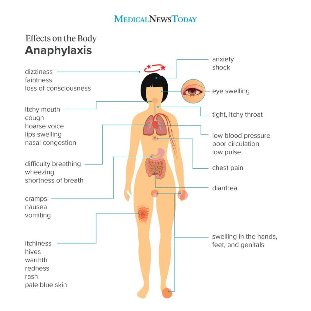
  • The eye lesions consist of uveitis, retinal vasculitis, vascular occlusion, optic atrophy, and conjunctivitis. 
  • Blindness is a common complication of the disease, and periodic evaluation by an ophthalmologist is necessary.

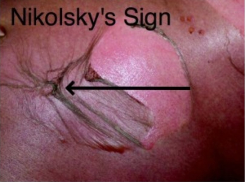
  • Positive pathergy is defined as  an inflammatory reaction  forming within 24 hours of a needle puncture scratch, or saline injection.
  • A positive pathergy test, which is performed by placing a 20 gauge needle 5 mm into the skin of the forearm. The test is positive if an indurated papule or pustule greater than 2 mm in diameter forms within 48 hours.
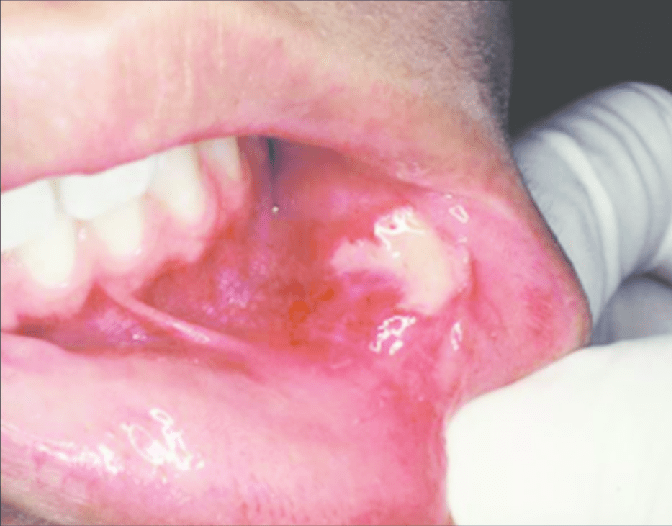
ORAL MANIFESTATIONS

  • The most common single site of involvement is the oral mucosa. 
  • Recurring oral ulcers appear in over 90% of patients; these lesions cannot be distinguished from RAS .
  • Some patients experience mild recurring oral lesions; others have the deep large scarring lesions characteristic of major RAS.
  • These lesions may appear anywhere on the oral or pharyngeal mucosa.

Histologic Features

The microscopic findings are not diagnostic. They consist of parakeratosis, acanthosis, and polymorphonuclear leukocyte infiltration of epithelium, sometimes with mi- croabscess formation similar to psoriasis. The connective tissue shows a lymphocyte and plasma cell infiltrate.

Laboratory Findings

The patients usually have a mild leukocytosis, an elevated erythrocyte sedimentation rate, and pyuria.

DIFFERENTIAL DIAGNOSIS

  • Sweet Syndrome : oral ulcers, conjunctivitis, episcleritis, Inflamed tender skin papules or nodules.
  • Erythema Multiforme: erosions, target(iris) lesions.
  • Pemphigoid: bullae, erosions.
  • Pemphigus : erosions, flaccid skin bullae.
  • Reiter syndrome: ulcers, conjunctivitis,
  • Herpes simplex virus
  • Lupus erythematosus

TREATMENT

The management of Behçet’s syndrome depends on the severity and the sites of involvement.

  • Azathioprine combined with prednisone has been shown to reduce ocular disease as well as oral and genital involvement.
  • Pentoxifylline, which has fewer side effects than do immunosuppressive drugs or systemic steroids, has also been reperted to be effective in decreasing disease activity, particularly of oral and genital lesions.
  • Dapsone, colchicines and thalidomide have also been reported to be effective to treat mucosal lesions of Behcet’s disease.

REFERENCE- BURKET TEXTBOOK OF ORAL MEDICINE AND SHAFER’S TEXTBOOK OF ORAL PATHOLOGY {8TH EDITION}

Systemic lupus erythematosus{SLE}

  • SLE is a multisystem autoimmune inflammatory disorder of unknown etiology.
  • Main feature is the formation of antibodies to DNA, which may initiate immune complex reactions, in particular a vasculitis. 
  • Female to male ratio of 9:1
  • More common in persons of non-European descent.
  • Etiology
  • Geneticpredisposition—relativeofpatientshavehigher incidences of auto-antibodies, immune deficiency and connective tissue disease. This tendency is greatest among identical twins.
    • Immunological abnormality possibly mediated by viral infection—immune complex consisting chiefly of nucleic acid and antibody account for majority of the tissue changes.
    • Autoimmune disease—as these patients develop antibodies to many of their own cells.
    • Endocrine—thereishighincidenceinfemalesinpreg- nancy. This finding suggestive of increased estrogen level.
    • Biochemicalincreaseinexcretionofmetabolicproducts, particularly tyrosine and phenylalanine, in certain SLEpatient.
  • CLINICAL MANIFESTATIONS
  • Lupus is known as “the great mimic.”
  •  Skin lesions of lupus can be classified 
    • lupus-specific (having diagnostic clinical or histopathologic features) 
    • nonspecific lesions.
  • Three subtypes of lupus-specific 
    • Acute
    • subacute 
    • chronic. 
  • Acute cutaneous lupus occurs in 30 to 50% of patients and is classically represented by the butterfly rash-mask-shaped erythematous eruption involving the malar areas and bridge of the nose
  • Chronic cutaneous lupus occurs in 15 to 20% of cases and affects the skin of the face or scalp in about 80% of cases.
  • The least common subtype, subacute cutaneous lupus, occurs in 10 to 15% of patients and includes papulo­squamous (psoriasiform) and annular-polycystic eruptions, usually on the trunk and arms.
  • Nonspecific but suggestive skin manifestations of lupus are common and include 
    • alopecia (both scarring following discoid lesions and non-scarring)
    • Photosensitivity
    • Raynaud’s phenomenon
    • Urticaria
    • Erythema
    • Telangiectases
    • cutaneous vasculitis.

  • ORAL MANIFESTATIONS
  • Two predominant types of oral lesions are
    •  discoid lesions 
    • ulcerations.
  • Oral ulcerations associated with SLE  they occur with increased frequency on the palate and in the oropharynx and are characteristically painless.
  • Histologically, they are characterized by lymphocytic infiltrate at the base of the ulcer and in the perivascular distribution, which is similar to that observed in discoid lesions.
  • Discoid oral lesions, appear as whitish striae frequently radiating from the central erythematous area, giving a so-called “brush border.”
  • Buccal mucosa, gingiva, and labial mucosa are the most commonly affected intraoral sites.
  • Direct immunofluorescent staining for immunoglobulins and complement C3 factor is a useful aid to diagnosis. Granular deposition of IgM, IgG, and C3 along the basement membrane is characteristic

Diagnosis

• Clinical diagnosis—skin lesion with lesion present on oral mucosa which is atrophic and erythematous will suspect lupus erythematous. Oral and nasopharyngeal ulceration is major diagnostic criteria for SLE.

Laboratory diagnosis—L.E. cell inclusion phenomenon with surrounding pale nuclear mass apparently devoid of lymphocytes. Anemia, leukopenia and thrombocyto- penia, with sedimentation rate increased. Serum gamma globulin increased and Coomb’s test is positive.

Positive lupus band test—it shows deposition of IgG,IgM or complement component in skin.

  1. Differential Diagnosis
    • Lichenplanus—homogenouspicture,nodarkerythema and no telangiectasia. Mucosal changes are usually extensive and symmetrical.
    • Lichenoidreaction—historyofdrugisalwaysthere.
    • Ectopic geographic tongue—systemic manifestation present is lupus erythematous, which is absent in ectopicgeographic tongue.
    • Psoriasis—Auspitz’s’signispositive.
    • Electrogalvanic lesion—dissimilar restorations are seenin oral cavity.
    • Leukoplakiaanderythroplakia—lesionstendtomaintainsame appearance and there are no skin changes.
    • Geographic stomatitis—no skin changes, mucosal lesionschange location rapidly.
    • Benign mucous membrane pemphigoid—no systemiccomplain and serology test to be done.
  • TREATMENT
  • Corticosteriods are the cornerstone of therapy
  • A pulse i.v cyclophosphamide regimen for remission induction followed by quarterly infusions
  • Recently, mycophenolate mofetil and azathioprine
  • NSAIDs for arthritis relief
  • Antimalarial like hydroxychloroquinine – effective in cutaneous lupus 
  • DENTAL MANAGEMENT
  • Recommended prophylactic antibiotics if ANC count falls below 500 – 1000 cells/mm3
  • Adrenal supression –
  • Adenocorticotropic hormone supression test is used to evalute
  • Current guidelines – Replacement therapy with hydrocortisone is unnecessary

REFERENCE- ANIL GHOM TEXTBOOK OF ORAL MEDICINE; BURKIT TEXTBOOK OF ORAL MEDICINE AND GOOGLE[SLIDE SHARE]

IMPORTANT SIGNS-II

  • Kocher’s sign-thyrotoxicosis
  • Macewen’s sign- alcohol poisoning
  • Murphy’s sign-acute cholecystitis
  • Nicolsky’s sign-application of pressure on normal skin produces a new lesion.Seen in pemphigus,TEN,Stevens Johnson syndrome ,Staphylococcla scalded syndrome
  • Osler’s sign- alkaptonuria
  • Pastia sign-scarlet fever
  • Pelken’s sign- scurvy
  • Prayer sign- diabetes
  • Raccoon eye sign-fracture of base of skull
  • Schamroth’s sign-clubbing
  • Signet ring sign-xray of patient with scurvy
  • Spatula sign-tetanus
  • Steinberg sign-Marfan’s syndrome
  • Tear drop sign- orbital blowout fracture
  • Trail sign- tracheal displacement
  • Tram track sign- Struge Weber syndrome
  • Trousseaus sign-seen in tetany
  • Trumbling bullet sign- post traumatic bone cyst
  • Water lily sign-hydatid cyst
  • Wimberger’s sign-congenital syphilis,scurvy
  • Wrist sign-Marfan’s syndrome

Sources:www.slideshare.net.com(signs in medicine),http://www.emdocs.net

IMPORTANT SIGNS-I

  • Allis sign- seen in developmental dysplasia
  • Asboe sign- seen in pemphigus (Bulla spread sign)
  • Auspitz sign- seen in psoriasis -peeling away scales result in pin point bleeding spots
  • Albright sign -dimple at the metacarpelopharyngeal joint, due to short metacarpal.Seen in pseudohypoparathyroidism,Turner’s syndrome,basal cell carcinoma
  • Babinski’s sign- by stroking the lateral aspect of the dorsum of foot
  • Barber’s chair sign- seen in multiple sclerosis
  • Battle’s sign-in basal skull fracture(sphenoid bone)blood pigment stain behind the ear over the mastoid
  • Benda’s sign- in tuberculous meningitis.Spasm of the trapezius muscle that the shoulder on affected side is lifted and at times brought forward
  • Blue berry muffin sign-raised purple lesions seen in dermal metastases of neuroblastoma
  • Brim sign- seen in Paget’s disease
  • Brudzinski’s sign- seen in meningitis
  • Button hole sign- seen in neurofibromatosis
  • Carpet track sign- seen in discoid lupus erythematosis
  • Cerebriform tongue sign- seen in pemphigus veterans
  • Chandelier’s sign- seen in gonorrhea in women
  • Chvostek sign- elevation of corner of mouth
  • Coleman’s sign- in fracture of body of mandible ( hematoma seen in the floor of the mouth)
  • Corner’s sign- seen in scurvy
  • Crowe’s sign- seen in neurofibromatosis
  • Darier sign- seen in urticaria pigmentosa
  • Dubois sign- seen in congenital syphilis
  • Erb’s sign- seen in latent tetany
  • Paget’s sign- seen in yellow fever
  • Falling fragment sign- seen in solitary bone cyst
  • Fitzpatrick sign- seen in dermatofibroma
  • Flag sign- seen in kwashiorkor
  • Floating membrane sign-seen in hydatid cyst
  • Floating teeth sign- seen in eosinophilic granuloma
  • Forscheimer sign- seen in rubella
  • Head drop sign- seen in poliomyelitis
  • Heel pad sign- seen in acromegaly
  • Higoumanaki’s sign- seen in late syphilis
  • Hutchinson sign-seen in heroes zoster ophthalmicus
  • Jellinek sign-seen in hyperthyroidism
  • Joffroy’s sign-seen in thyrotoxicosis

Source: http://www.slideshare.net ( signs in medicine),www.clinicalskillz.com

Syncope

Syncope refers to generalised weakness of muscles, loss of postural tone , inability to maintain erect posture and loss of consciousness.

If u find it difficult to remember the causes here is a MNEMONIC

Source- textbook of general medicine for dental students SN Chugh and pinterest images .

Oral manifestations of systemic diseases-II

Hematologic Diseases :-
• Hematologic diseases are disorders which
primarily affect the blood.
Anemia is usually defined as a decrease
in the amount of red blood cells (RBCs) or
hemoglobin in the blood.

Oral Manifestations:
– folate and vit. B12 deficiency
– iron deficiency
– glossitis
• red colour
• athrophic papilae
• recurrent aphthae
– candidal infection
– angular stomatitis
– oral pain

Leukemia is a group of cancers that
usually begins in the bone marrow and
results in high numbers of abnormal white
blood cells.

*Oral Manifestations:-

– gingival hypertrophy
– petechiae
– mucosal ulcers
– hemorrhage
Treatment of leukemia
– reactivation of herpes simplex virus – oral mucosistis.

References:-

1.Google- slideshare.

Oral manifestations of systemic diseases – I

Careful examination of the oral cavity may
reveal findings indicative of an underlying
systemic condition, and allow for early diagnosis
and treatment. Examination should include
evaluation for mucosal changes, periodontal
inflammation and bleeding, and general
condition of the teeth.

I.GIT Diseases
• Gastrointestinal diseases refer to diseases involving
the gastrointestinal tract, namely the esophagus,
stomach, small intestine, large intestine and rectum,
and the accessory organs of digestions, the liver,
gallbladder, and pancreas.

Crohn’s disease, also known as Crohn
syndrome and regional enteritis, is a type of
inflammatory bowel disease (IBD).
Ulcerative colitis is a form of inflammatory
bowel disease (IBD) that causes inflammation
and ulcers in the colon.
Gastroesophageal reflux is a chronic symptom
of mucosal damage caused by stomach acid
coming up from the stomach into the
esophagus.
Chronic liver disease in the clinical context is a
disease process of the liver that involves a
process of progressive destruction and
regeneration of the liver parenchyma leading to
fibrosis and cirrhosis.

1. Crohn disease:
– diffuse labial, gingival or mucosal swelling
– „cobblestoning“ of buccal mucosa and
gingiva
– aphtous ulcers
– mucosal tags
– angular cheilitis
– oral granulomas

2.Ulcerative colitis:-
– oral signs are present in periods of
exacerbation of disease
– aphtous ulceration or superficial
hemorrhagic ulcers
– angular stomatitis
– pyostomatitis vegetans, pyostomatitis
gangrenosum.

3.Gastroesophageal reflux:-
– reduction of the pH of the oral cavity below
5,5

– enamel damage
– damage of the dentin – higher sensitivity (to
temperature..), caries

4. Chronic liver diseases:-
– jaundice
– petechiae or gingival bleeding (hemostasis
disorder)

RREFERENCES:-

1.Google -slideshare

2.Davidson-22nd edition

COVID-19 (PART -2)

Written by : Dr. Urusa I Inamdar

Using mask and sanitizer

  • Person not showing any symptoms :

A medical mask should not be used as it creates a false sense of security that can lead to neglecting other essential measures . Resort to other measures like – washing hands , cover your nose and mouth while sneezing , monitor your body temperature and oxygen levels .

  • So when should you use medical mask ( apart from medical health care workers ) :

When a person develops symptoms like cough or fever , while visiting a health care facility .

Taking care of an ill person .

If your family member is a suspected or confirmed case undergoing home care .

ENSURE PROPER FIT OF MASK
  • Washing your hands :

Washing your hands frequently is essential . Use soap and water for atleast 20 seconds for most effective results .

  • Sanitizing your hands :

Hand sanitizers are to be used , when hand wash was not possible . An alcohol based sanitizer with 70% alcohol content must be used for 20 seconds .

  • REFERENCES:
  1. Self notes
  2. World health organization
  3. Google.com
  4. ayush.gov.in
  5. IGOT ( Integrated Government Online Training )
  6. AIMS , New Delhi