APICOECTOMY


• It is the surgical resection of the apex of the root.
• It is the procedure done in case of root canal treatment failure.
• If an infection does not subside even after root canal treatment,it may concern enquire a surgical procedure.
• In this procedure, the apical region of root is visualised by reflecting a flap and performing an osteotomy.


INDICATIONS :
• Aberrant Anatomy: Dilaceration of root apex do not allow endodontic restoration of apex.
• Obliteration of apex by secondary dentin.
• Iatrogenic repair: A broken endodontic file which cannot be retrieved by conventional means.
• Apex perforation.
• Improper apical seal which cannot be removed.
• Increased drainage of pus from root canal will not allow adequate apical seal.
• Open apex.
• Non healing periapical granuloma.
• Fracture of apical third of root.
• Periapical cyst/granuloma.

CONTRAINDICATIONS:

  1. Local Contraindications:
    • Poor periodontal status of tooth.
    • Grossly decayed tooth.
    • Inadequate tooth length.
    • Acute infection.
    • Traumatic occlusion.
    • Uncooperative patients.
    • Close proximity of root apex to vital anatomic structures such as Maxillary antrum & Nasal floor.
  2. Systemic Contraindications:
    • Poor medical status of diabetes,Bleeding disorders.etc

STEPS IN ENDODONTIC SURGERY:

  1. Cleaning of the area involved with antiseptic solutions.
  2. Local anaesthesia.
  3. Design of mucoperiosteal flap & reflection of flap.
  4. Bone removal for access to root tip.
  5. Root tip resection & curettage.
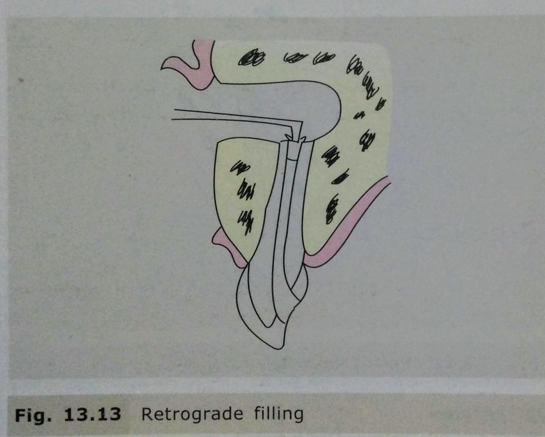
  6. Retro preparation & retrograde filling.
  7. Suturing & Follow up.

COMPLICATIONS:
• Mobility of tooth/adjacent tooth.
• Haemorrhage.
• Nasal perforation.
• Oroantral Fistula.
• Mental Nerve Damage.
• Inferior Alveolar Nerve damage.

REFERENCES:
• Textbook of Oral & Maxillofacial Surgery, Chitra Chakravarthy (2nd Edition)
• DentaGama.com

Root Canals

Written by : Dr. Urusa I Inamdar

It is the portion of the pulp cavity from the canal orifice to the apical foramen.

It is divided into 3 sections :

  • Coronal
  • Middle
  • Apical
  1. Accessory canals or lateral canals : lateral branching of the main root canal generally occurring in the apical third or furcation area of a root.
  2. Lateral canal : accessory canal that branches to the lateral surface of the root and may be visible on a radiograph.
  3. Apical foramen : aperture at or near the apex of a root through which the blood vessels and nerves of the pulp enter or leave the pulp cavity.
  4. Accessory foramina : openings of the accessory and lateral canals in the root surface.

A straight root canal extending the entire length of the root is uncommon. Either a constriction is present before the apex is reached or , as is often the case , a curvature is present.

The curvature may be :

  • A straight canal extending with minimal apical curvature.
  • A gradual curvature of the canal with a straight apical ending.
  • A gradual curvature of the entire canal.
  • A sharp curvature of the canal near the apex.

A curvature of about 20° in a narrow root canal may be difficult or even impossible to negotiate with endodontic instruments, whereas a curvature of even 30° may be negotiated if the root canal is wide.

Success in negotiating a narrow , curved canal depends on following :

  • Degree of curvature.
  • Size and constriction of the root canal.
  • Size and flexibility of the endodontic instrument blade.
  • Skill of the operator.

The various classification proposed are as follows :

  • Vertucci’s Classification:
  1. Type I : Single canal extends from the pulp chamber to the apex (1)
  2. Type II : Two separate canals leave the pulp chamber and join short of the apex to form one canal (2-1)
  3. Type III : One canal leaves the pulp chamber and divides into two in the root , the two then merge to exit as one canal (1-2-1)
  4. Type IV : Two separate distinct canals extend from the pulp chamber to the apex (2)
  5. Type V : One canal leaves the pulp chamber and divides short of the apex into two separate distinct canals with separate apical foramina (1-2)
  6. Type VI : Two separate canals leaves the pulp chamber , merge in the body of the root , and redivide short of the apex to exit as two distinct canals (2-1-2)
  7. Type VII : One canal leaves the pulp chamber, divides and then rejoins in the body of the root , and finally redivides into two distinct canals short of the apex (1-2-1-2)
  8. Type VIII : Three separate distinct canals extend from the pulp chamber to the apex (3)
  • Weine’s classification :
  1. Type I : Single canal from pulp chamber to apex
  2. Type II : Two canals leaving from the chamber and merging to form a single canal short of the apex
  3. Type III : Two separate and distinct canals from chamber to apex
  4. Type IV : One canal leaving the chamber and dividing into two separate and distinct canals
  • Classification based on canal cross – section:
  1. Round (circular)
  2. Oval
  3. Long oval
  4. Flattened (flat/ribbon)
  5. Irregular

References:

  • Dental notes
  • Grossman’s Endodontic Practice (13th edition)

ROOT CANAL SEALERS 🦷

These are used in conjugation with semi solid or solid obturating materials to establish an adequate seal of root canal system.This is a brief note on the various root canal sealers ,composition ,salient features and brand names of some sealers which would prove helpful in clinical setup.

Classification :(according to Grossman)

1)ZnOE based sealers:

i. Grossman’s Formula

ii. Roth’s 801

iii. Tubliseal

iv. Wachs Sealer

2)CaOH based sealers:

i. Sealapex

ii. Apexit

3)Glass ionomer based sealers:

4)Resin based sealers:

i. AH Plus

ii. AH 26

iii. Epiphany

iv. Diaket

ZnOE based sealer-

Composition:

Base ingredient of powder -ZnOE,

Liquid usually consists -Eugenol alone or in combination with other liquids such as Canada balsam.

Features:

.Hardens in 2 hours approx at 37 degree celsius & under 100% relative humidity.

.Setting time in canal is less.(10-30 mins).

. Tissue tolerance is satisfactory with little inflammation & no inhibition of repair.

CaOH based sealers-

Composition:

Base consists -Zinc oxide, CaOH, butyl benzene, sulfonamide & Zinc stearate

Catalyst consists – resin,isobutyl salicylate, barium sulfate,titanium dioxide & aerosol

Features:

.antimicrobial

.osteogenic-cementogenic

.better healing capability

Glass ionomer based sealer-

Feature : dentin bonding ability but are unpopular due to difficulty in removal from root canal walls after treatment.

Resin based sealer-

Composition:

Paste A -Epoxy resin ,Calcium tungstate,zirconium oxide,silica,iron oxide

Paste B – Admantaneamine-N,calcium tungstate,zirconium oxide,silica-silicone oil

Features:

. Dentinal adhesion (strong adhesive properties).

.Contracts slightly while hardening.

.Good sealing ability.

.Bio compatibility to periapical tissues.

.Moderate antimicrobial activity.

.Long working time and easy for manipulation.

List of various root canal sealers and brand names

Sources: Grossman’s endodontic practice (13 th edition ),Instagram – @__dentistars__