Systemic Implications in Dental practice

Cyclosporine therapy may cause gingival hyperplasia

Gingival growth occurs in patients taking phenytoin.

Patients with cardiac disease should receive dental treatment in minimal stressful environment. Anxiety,exertion and pain should be minimized.

Irregular pulse, engorged jugular veins and tachypnea may indicate the presence of cardiac disease.A history of hypertension, ischemic heart disease or any other cardiac problem particularly congenital heart disease and drug intake (anticoagulant, aspirin) should be sought.Angina may present as pain in the mandible, teeth and other oral Tissues Epinephrine in the local anesthesia may raise the blood pressure and precipitate dysarrhythmias.In patients with IHD, facilities for medical help, oxygen and nitroglycerine should be Available General anesthesia should be avoided for at least three months in patients with recent onset angina

Patient’s with Cushing’s syndrome more prone to get infections.(candidiasis)

Elective dental surgery should be deferred for 6 months following acute MI.Prophylaxis for infective endocarditis is mandatory in cases where there is a risk.Cardiac patients on anticoagulant drugs or aspirin are at increased risk of bleeding following dental procedures.Hence, these drugs should preferably be stopped a week before the procedure.Calcium channel blockers may cause gingival swelling and lichenoid lesions in the oral cavity. ACE inhibitorscan cause loss of taste, burning sensation in oral cavity,and angioedema. Dry mouth can result due toantihypertensive drugs such as d

Rifampicin can cause red saliva.

Elective dental care is avoided in patients with acuterenal failure

Elective dental procedures are better tolerated on non-dialysis days

Blood pressure measurement is advised at every visit.

Brown to black macular pigmentation in oral mucosa can be suspected for Addison disease.

Gonorrhea may present uncommonly with oral manifestations like tonsillitis, lymphadenitis, and painful oral and pharyngeal ulcers.

Oral manifestations in peptic ulcer disease are rare.However erosive dental lesions could be appreciated on lingual surface of lower incisors or palatal surface of upper maxillary teeth.

Overview of the Respiratory System

Key Function:

  • Provides mechanisms for the exchange of oxygen and carbon dioxide between the blood and the outside environment.

*Anatomically, the respiratory system divides into upper and lower respiratory tracts

The upper respiratory tract:

  • Nose
  • Nasal cavity
  • Paranasal sinuses
  • Pharynx

The lower respiratory tract:

  • Larynx
  • Trachea
  • Bronchi
  • Bronchioles
  • Respiratory bronchioles
  • Alveolar ducts
  • Alveolar sacs
  • Alveoli

Functionally, the respiratory system divides into conducting and respiratory portions

The conducting portion

Moves (aka, conducts) air between the lungs and the outside environment.

  • Nose
  • Nasal cavity
  • Pharynx
  • Larynx
  • Trachea
  • Bronchi
  • Bronchioles

The respiratory portion

Participates in gas exchange

  • Respiratory bronchioles
  • Alveolar ducts
  • Alveolar sacs
  • Alveoli

Anatomical Details:

Upper Respiratory Tract

  • The nose opens the respiratory system to the outside environment.
  • The nasal cavity has a mucosal lining, which “conditions” the air; specifically, it moistens, warms, and cleans it. The posterior-superior wall of the nasal cavity houses special sensory cells (olfactory cells) that detect smells.
  • The paranasal sinuses:
    – Frontal sinus of the frontal bone
    – Ethmoid sinus (aka, ethmoidal air cells) of ethmoid bone, which lies posterior to the frontal sinus.
    – Sphenoidal sinus, which is located posterior to the nasal cavity.
    – Maxillary sinus in the maxilla on either side of the nasal cavity.

Because the paranasal sinuses are open to the nasal cavity, infection can spread to the paranasal sinuses, which results in sinus infection (aka, sinusitis).

  • The pharynx is the muscular tube that lies behind the nasal cavity, oral cavity, and larynx; it is a conduit for air and food/liquid.

Lower Respiratory Tract

  • The larynx is the cartilaginous start of the lower respiratory tract; it prevents food/liquid from entering the lower respiratory tract and is often referred to as the “voice box” because it produces and modifies sounds.
  • Tracheobronchial tree = trachea and its branches.
    • The trachea (the “windpipe”) descends through the neck to the thorax (it terminates posterior to the sternal angle, at the level of T2); it comprises C-shaped (vertically-stacked) cartilaginous rings.
    • Right and left primary (aka, main) bronchi arise from trachea; they enter the lungs.
    • Secondary bronchi (aka, lobar bronchi) serve specific lobes of the lungs.
    • Tertiary bronchi (aka, segmental bronchi) serve specific bronchopulmonary segments.
    • Bronchioles are histologically different.
    • Terminal bronchioles are the final branches of the conducting portion of the respiratory system.
    • Respiratory bronchioles are the start of the respiratory portion, as they can participate gas exchange.
    • Respiratory bronchioles open to alveolar ducts.
    • Alveolar ducts open to alveolar sacs.
    • Alveolar sacs comprise outpockets called alveoli.
  • Aveloli:
    – Very thin walls, and are the primary sites of gas exchange.
    – The hundreds of millions of alveoli within give the lungs a light, spongy texture.
  • Lungs
    – Separated into lobes:
    – Right lung = 3 lobes
    – Left lung = 2 lobes
    – The left lung is slightly smaller to accommodate the heart

Clinical Correlations

  • Tuberculosis
  • Viral respiratory infections
  • Pneumonia

Anti-arrhythmic Drugs: Classes II through IV (+ Others)

Class II: Beta blockers

  • Treat both supraventricular and ventricular arrhythmias.
  • Slow channel blockers, so they work at the sinoatrial and atrioventricular nodes; more generally, as we’ve learned elsewhere, beta blockers reduce sympathetic stimulation.
  • We draw the normal curve of an action potential through the nodes.
  • Then, we show that beta blockers inhibit phase 4 depolarization and slow conduction through the atrioventricular node.
  • Suppress ectopic pacemakers.
  • Slow heart rate and reduce contractility.
  • Prolong the PR interval on ECG.
  • Examples
    – Drugs that are often used to prevent recurrent myocardial infarction:
    Propranolol, which also stabilizes membranes (a class I action)
    Metoprolol
    Nadolol
    Timolol
    – Esmolol is a cardioselective beta-1 receptor blocker and is short-acting.
    – Thus, it is used to treat acute arrhythmias.
  • Common beta blocker side effects: bradycardia, hypotension, bronchospasm (with non-selective beta blockers, specifically), and, myocardial depression.

Class III: Potassium Channel Blockers

  • Treat tachyarrhythmias, including re-entry arrhythmias.
  • We Draw the normal curve of an action potential in the ventricles.
  • Then, we indicate that potassium channel blockers delay repolarization, prolong the action potential, and lengthen the effective refractory period.
  • The drugs we’ll focus on the most, amiodarone and its derivative, slow the heart rate and atrioventricular node conduction; these additional actions are due to their abilities to block beta adrenoreceptors, sodium, potassium, and calcium channels.
  • Class III drugs prolong the QT interval on the ECG.
  • Examples
    – Amiodarone, which, due to the various channels and receptors it blocks, is a mix of classes I-IV. Its wide spectrum of action makes it a popular drug of choice in a variety of arrhythmias.
    Amiodarone is lipophilic, and is widely distributed through the body tissues.
    Widespread potential side effects:
    Liver toxicity and cirrhosis
    Interstitial pneumonitis, which can lead to pulmonary fibrosis
    Corneal microdeposits, which are caused by amiodarone-induced lipidosis in the eye
    (we show their “whorl-like” pattern(
    Photosensitivity/phototoxicity can produce a rash or even give the skin a gray-blue tint
    And, because amiodarone is iodine-rich, it can cause hypo- or hyperthyroidism.
    Amiodarone can also cause sinus bradycardia
    – Dronedarone is a synthetic, non-iondinated derivative of amiodarone (it doesn’t have iodine).
    It is used to treat atrial fibrillation, and is generally considered less toxic, but less powerful, than amiodarone.
    It is, however, associated with liver toxicity.
    – Sotalol, which also has class I beta blocking actions, is associated with torsades de pointes.
  • As a warning, write that most class III drugs are significantly pro-arrhythmic; amiodarone and dronedarone are important exceptions, as they less likely to precipitate arrhythmias.

Class IV: Calcium Channel Blockers

  • Treat supraventricular arrhythmias and to reduce ventricular rate in atrial flutter and atrial fibrillation.
  • They block calcium channels, so they are most effective at the sinoatrial and atrioventricular nodes, which rely on calcium ions for rate control.
  • We draw the curve of a normal nodal action potential, and show that calcium channel blockers prolong nodal conduction and the effective refractory period.
  • They prolong the PR interval on ECG (like the Class II drugs).
  • We use non-dihydropyridine calcium channel blockers
  • Examples
    – Verapamil and Diltiazem; both target myocardial cells, and diltiazem has some vasodilator effects.
    Both drugs are associated with constipation, and, due to their negative inotropic effects, can cause bradycardia and lower cardiac output.
    Thus, use caution when combining these drugs with beta blockers, which have similar effects, and, avoid using in patients with heart failure with reduced ejection fraction.

Class V: Others

Finally, let’s consider the “others” category, sometimes collectively called “Class V”.

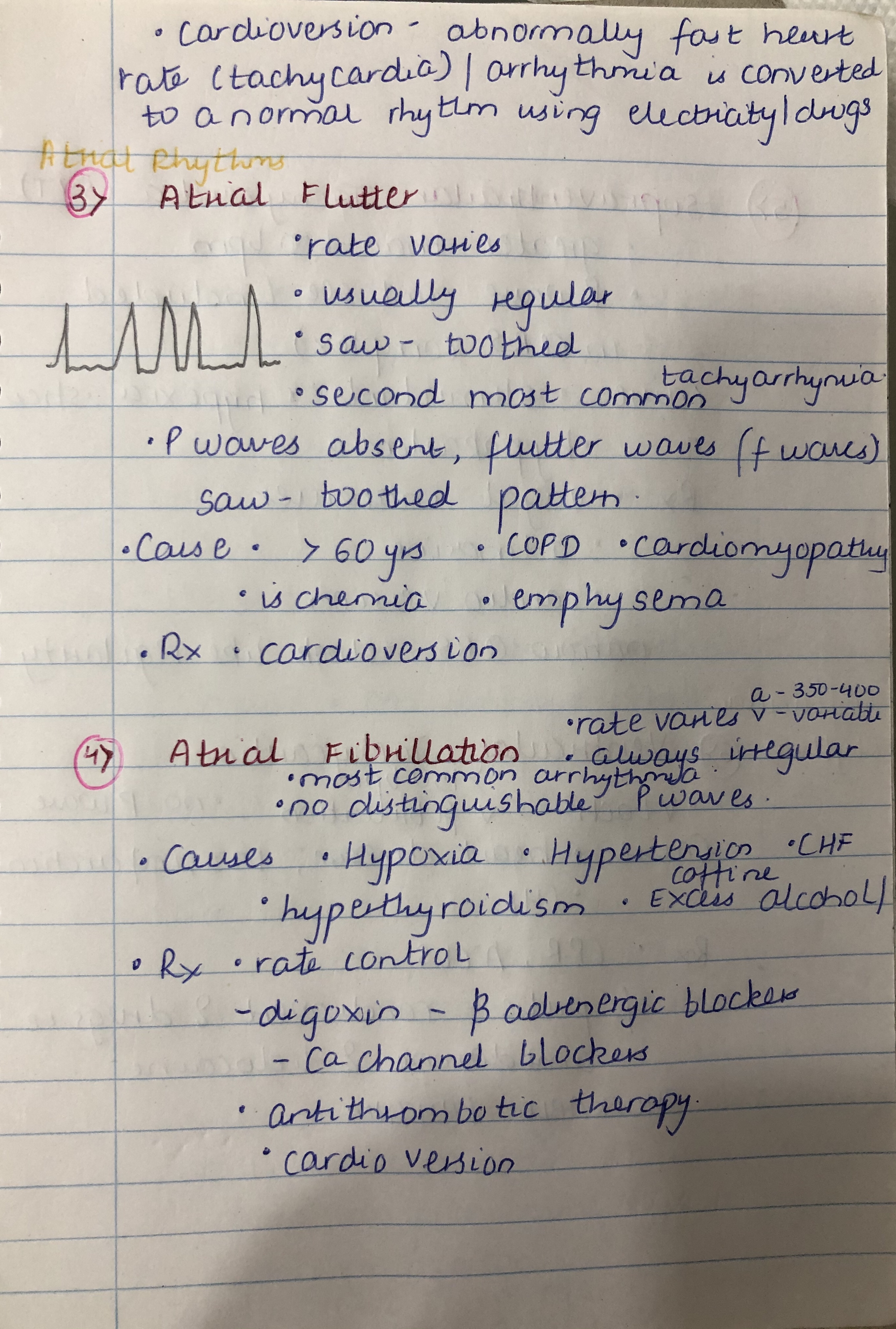
Adenosine is used to treat supraventricular tachycardias; it slows or blocks conduction in the atrioventricular node.
– It’s important to know that theophylline (a common asthma medication) and caffeine reduce adenosine’s efficacy by blocking its receptors.
– Adenosine may trigger bronchospasm.
Magnesium ions are sometimes used to treat torsades de pointes and digoxin toxicity.
Potassium ions may be used in some patients to slow conduction and can suppress ectopic pacemakers.
Digoxin can be used in some patients to treat atrial flutter or atrial fibrillation; it slows or blocks conduction in the atrioventricular node by inhibiting sodium-potassium ATPase.
– However, digoxin can cause ectopic arrhythmias, gastrointestinal and visual side effects, and breast enlargement (gynecomastia, in males), and has been associated with increased risk of breast and uterine cancer, likely due to its phyto-estrogen effects.

Anti-Arrhythmic Drugs: Class I

Anti-Arrhythmic Drugs Overview

  • Arrhythmias are characterized by abnormal generation and/or conduction of electrical impulses, which produce abnormal rate and/or activation sequences in the cardiac tissue. Ultimately, this can reduce cardiac output.
    Supraventricular Arrhythmias
    Ventricular Arrhythmias
    AV Node block
  • Class 1 drugs are sodium channel blockers; we’ll focus on this group in this tutorial.
  • Class 2 drugs comprise the beta blockers.
  • Class 3 drugs include the potassium channel blockers.
  • Class 4 drugs comprise the calcium channel blockers.
  • Magnesium and adenosine are also used to treat arrhythmias.
  • Paradoxically, anti-arrhythmic drugs can actually precipitate fatal arrhythmias; thus, the potential harm of these drugs may outweigh their potential benefits in some patients.

Action Potenial

Review

  • Phase 0 = Fast sodium channels open and ions quickly move into the cell, which causes rapid depolarization.
    – Class 1 antiarrhythmics affect the slope of this line.
  • Phase 1 = Potassium channels open, which allows potassium ions to move out of the cell and cause early repolarization.
  • Phase 2 = The plateau phase; potassium continues to move out of the cell, and calcium move into it.
  • Phase 3 = As more potassium channels begin to open, and calcium channels close, Phase 3 is characterized by rapid repolarization.
  • Phase 4 = Resting phase, during which “leaky” potassium channels maintain resting membrane potential.
  • Effective refractory period is the time when new action potentials cannot be triggered.

Effects of Class 1 anti-arrhythmics on this curve

  • These drugs are fast-sodium channel blockers; they also have local anesthetic properties.
  • They slow the rate of Phase 0 depolarization and conduction velocity.
  • Class 1 drugs are further classified by their effects on the duration of action potentials.
    – Class 1a drugs prolong the duration of the action potential
    – Class 1b drugs shorten its duration
    – Class 1c drugs have minimal effect

Details

Class 1a

  • Used to treat atrial and ventricular arrhythmias.
  • Moderate sodium channel blockers, and also block some potassium channels.
  • They prolong the action potential duration, effective refractory period, QRS complex, and QT interval.
  • Examples and their most common side effects:
    – Procainamide is associated with hypotension; long term use is typically avoided because the drug can cause lupus-like syndrome, characterized by arthralgia and purpura.
    It is generally considered the third best choice in myocardial infarction arrhythmias (behind amiodarone and lidocaine).
    – Quinidine is associated with gastrointestinal side effects, cinchonism (headache, dizziness, and tinnitus), and thrombocytopenia.
    – Disopyramide are due to its anticholinergic effects (for example, constipation, urine retention, dry mouth, blurred vision). Can worsen glaucoma, and is not suitable for patients with heart failure.
  • All Class 1a drugs can precipitate torsades de pointes.

Class 1b

  • Used to treat ventricular arrhythmias.
  • They are weak sodium channel blockers, and are selective for ischemic tissue and depolarized Purkinje fibers and ventricular myocytes.
  • These drugs shorten the action potential duration and effective refractory period, but have no effect on the QRS complex or QT interval.
  • Lidocaine is the prototypical class 1b drug; it is second in line to treat arrhythmias associated with myocardial infarction (behind amiodarone).
    – Lidocaine has low toxicity, but, in vulnerable patients or when given in high doses or administered too rapidly, it is associated with neurological, and, more rarely, cardiovascular side effects.
    – Neurological effects include paresthesia (tingling, “pins and needles,” usually in the hands and feet), dizziness, and confusion.
    – Cardiovascular effects include sinus bradycardia, arrhythmias, and shock.
  • Phenytoin is often considered an honorary member of class 1b anti-arrhythmias; it is used to treat arrhythmias caused by digitalis.

Class 1c

  • We’ll list characteristics for the prototypical drug, Flecainide; as we’ll see, however, not all class 1c drugs have the same effects.
  • Used to treat supraventricular arrhythmias.
  • Strong sodium-channel blockers that also block some potassium channels.
  • Flecainide has no effect on the duration of action potentials or the effective refractory period; it prolongs the QRS complex with little effect on the QT interval.
  • Common side effects are dizziness, headaches, and visual disturbances, which may include blurred vision or difficulty focusing, or the appearance of flashing lights or spots.
  • Propafenone has similar side effects; it can exacerbate heart failure (propafenone also blocks beta channels and has negative inotropic effects).
    – Unlike flecainide, it is associated with prolonged QT intervals and effective refractory periods.
  • Moricizine is another class 1c anti-arrhythmic, but it is no longer used in the United States.
  • Class 1c drugs are pro-arrhythmic; they are not suitable for patients with structural heart disease.

Heart Failure

  • Increasingly common due to an aging population and high prevalence of contributing risk factors.
  • Heart failure = A clinical syndrome in which the heart’s ventricles cannot pump enough blood to meet the body’s needs.
    Review: Cardiac outputStroke volumeCardiac performance.
    See cardiac cycle with congestive heart failure
  • Reduced cardiac-output (CO) is due to reduced stroke volume.
    – Reduced stroke volume can be due to:
    Systolic dysfunction, i.e., impaired contraction
    Diastolic dysfunction, i.e., impaired compliance
  • Both scenarios can ultimately lead to increased left ventricular end diastolic pressure, which, as we’ll see, can lead to pulmonary congestion.
  • As a clinical syndrome, heart failure is the culmination of other cardiovascular diseases, which may co-exist.
    – Common culprits include: ischemic heart diseasehypertensioncardiomyopathies, diabetes mellitus, metabolic syndrome, atherosclerosis, and myocardial infarctions.
  • Diagnosis can rely on echocardiography, ECG, cardiac MRI, and measurement of serum B-type natriuretic peptide (BNP) levels.
  • S3 or S4 sounds may be heard (be aware of intertextual variation on the importance of S4 sounds and heart failure), and, that pulmonary crackles may audible when pulmonary edema sets in.
  • Treatments vary, and include life-style changes and, where appropriate, address the causative disease(s).
    – Treatments to reduce symptoms include: diuretics, digoxin, and nitrates.
    Long-term treatments tend to focus on lowering ventricular pressures, and include: ACE-inhibitors, angiotensin II receptor blockers (ARBs), Beta-blockers, and Aldosterone antagonists.
  • Device therapies include implantable cardiac defibrillators, cardiac resynchronization therapy, biventricular pacemakers, and left ventricular assist devices.

Left Heart Failure

Review blood flow through the heart

  • When stroke volume is reduced, systemic perfusion is also reduced.
  • Reduced stroke volume also leads to increased left ventricular end diastolic pressure (LVEDP); be aware that this can be the result of a variety of mechanisms, which we’ll address, soon.
  • Increased left ventricular end diastolic pressure leads to an increase in left atrial pressure, which, ultimately, raises pulmonary pressures.
  • Increased pressures in the pulmonary vascular system “push” fluids out of vessels and into the surrounding tissues.
    Pulmonary edema occurs when this fluid enters the alveolar sacs and lung spaces.
    Pleural effusions occur when the fluid accumulates between the pleural space.

Symptoms
– Dyspnea upon exertion is a common early sign; it occurs because the heart cannot increase cardiac output despite increased metabolic demands.
– Orthopnea is characterized by dyspnea that occurs when lying flat, but is quickly relieved by sitting upright or standing; orthopnea occurs because the heart is unable to adjust to the redistribution of body fluids that occurs when lying flat.
– Paroxysmal nocturnal dyspnea, as its name suggests, is characterized by sudden episodes of dyspnea that awaken a person after 1-2 hours of sleep, usually at night.
– Sleep-disordered breathing includes obstructive sleep apnea and Cheyne Stokes Respiration; show that Cheyne Stokes Respiration is characterized by cycling periods of tachypnea or hyperpnea alternating with periods of apnea.
– Other symptoms may also be present, often reflecting reduced cardiac performance; for example, patients may experience cognitive impairments, arrhythmias, cyanosis, or thrombi formation in the heart chambers.

Heart Failure with Reduced Ejection Fraction

  • Recall that the ejection fraction measures how much of the blood in the ventricle was pumped out (ejected) during a contraction.
    – For reference, a “normal” ejection fraction is between 50% and 70%.
  • Heart Failure with Reduced Ejection Fraction, the left ventricle ejection fraction 40% or less
    – The lower ejection fraction is the result of reduced contractility of the myocardium, and is associated with systolic dysfunction (since the left ventricle contracts during systole).
    – Be aware that systolic and diastolic dysfunctions may co-exist, which is why clinical focus has shifted somewhat from systolic vs. diastolic dysfunction to the ejection fraction.
  • Heart failure with reduced ejection fraction is particularly associated with dilated cardiomyopathies, valvular heart diseases, and myocardial infarctions.
  • When contractility is impaired, the ejection fraction is reduced, and, via a variety of mechanisms, the left ventricular end diastolic pressure will increase.
  • Compensatory mechanisms* in HFrEF
    – Raise blood volume and vascular resistance in attempt to increase preload and, as a result, stroke volume and tissue perfusion.
    – However, these effects also exacerbate pulmonary hypertension and congestion in a positive feedback loop.
    – Cardiac remodeling leads to dilation and myocyte hypertrophy; these changes can ultimately lead to increased stiffness and further impair cardiac performance, and even facilitate mitral valve regurgitation.
    – Reduced systemic perfusion triggers systemic and renal responses that increase blood volume and vascular resistance – recall that the renin-angiotensin-aldosterone system, the sympathetic nervous system, and the vessels themselves dynamically regulate changes in blood flow.
  • Thus, some long-term treatments for heart failure with reduced ejection fraction specifically target these responses (for example, ACE-inhibitors that inhibit the effects of the RAAS response).

Heart Failure with Preserved Ejection Fraction (HFpEF)

  • Characterized by Left Ventricular Ejection Fractions equal to or above 50%.
  • Due to impaired relaxation and/or compliance of the myocardium, which produces diastolic dysfunction.
  • Especially common in elderly people and women, and is associated with hypertrophic and restrictive cardiomyopathies, hypertension, and renal diseases.
  • Compliance is reduced, diastolic filling is impaired, and left ventricular end diastolic pressure is increased.
  • Impaired filling can lead to reduced stroke volume and cardiac output.
  • This type of heart failure often has a worse prognosis than heart failure with reduced ejection fraction, especially because it is less responsive to current treatments.

Right Heart Failure

  • Often the result of left heart failure.
    – When right heart failure is isolated, it is typically due to pulmonary issues, and, therefore, is referred to as cor pulmonale.
  • Right ventricular end diastolic pressure is elevated, which leads to increased pressures in the right atrium. As a result, the blood becomes “backed up” and raises systemic venous pressures.
  • As we saw with left heart failure, the resulting increased pressures and congestion produces key signs and symptoms.
    – Elevated systemic venous pressure and congestion leads to peripheral edema.
    This often produces so-called “pitting edema” in the lower extremities, and congestion in the abdominal organs, especially the liver.
    Hepatic portal congestion gives the liver a “speckled” or “mottled” appearance, often referred to as “nutmeg liver;”indicate that hepatoportal congestion exacerbates edema.
  • A sign of right heart failure is the hepatojugular reflux, which is characterized by distension of the jugular veins in the neck when pressure is applied to the right abdominal quadrant (over the liver).
  • Pericardial and peritoneal effusions (ascites) may occur.

Cardiomyopathies

Overview

  • Cardiomyopathies are diseases of the heart muscle that can cause mechanical and/or electrical dysfunction.
    – Can produce heart failure or fatal arrhythmias.
  • Four key types based on cardiac morphology:
    – Dilated, Hypertrophic, Restrictive, Arrhythmogenic Right Ventricular Cardiomyopathy

Be aware that some authors specifically exclude cardiac dilation or hypertrophy caused by cardiac disorders such as hypertension or ischemia, but other authors do not make this distinction. For clarity, in this tutorial, we will use the limited definition of cardiomyopathy and exclude, for example, cardiac hypertrophy that occurs as a physiologic response to valvular disease, etc.

  • Cardiomyopathies are caused by both genetic and non-genetic factors, though, in many cases, underlying causes are not identified.
  • Diagnosis of cardiomyopathy includes chest x-rays, ECGs, and echocardiograms.
  • Treatments are focused on addressing the underlying causes, where appropriate, and the management of heart failure and arrhythmias.
  • Normal heart anatomy

Dilated cardiomyopathy

  • Characterized by dilation and contractile impairment; think of a “ballooning” heart, because “dilated” means to become wider or more open.

Gross Morphology:

  • Produces systolic dysfunction with reduced ejection fractions(typically defined as 40 – 45% less than normal).
  • The dilated heart is characterized by “ballooning” of the left ventricular, which often leads to dilation in the other chambers, as well.
  • The dilated heart is heavy, and is often described as “baggy”due to contractile impairment.
    • Wall thickness is often reduced, and functional mitral valve regurgitation is possible (due to sagging and lack of structural support).
  • Mural thrombi formation is also possible.

Histology: Hypertrophied cells and loss of myofibrils with interstitial fibrosis in histological samples.

Symptoms: Left ventricular dilation is associated with dyspnea and fatigue, and that right ventricle dilation can lead to peripheral edema.

Causes: Dilated cardiomyopathies can be caused by several factors, including:

– Genetic mutations, which often lead to defects in the cardiac myocyte cytoskeleton, sarcolemma, and nuclear envelope.
– Various infections, including CoxasackievirusChagas Disease, and HIV;
– Toxins and drugs, especially alcohol abuse, cocaine, and some cancer drugs.
– Metabolic disorders, including diabetes, hyper- or hypo-thyroidism, hyper- or hypo-kalemia, and various nutritional deficiencies.
– Neuromuscular diseases, such as Friedreich ataxia and muscular dystrophy.
– Pregnancy (the cardiomyopathy becomes apparent in late pregnancy or postpartum period).
– Myocarditis.

Arrhythmogenic Right Ventricular Cardiomyopathy

  • Characterized by muscle cell loss with fibrofatty replacement in the wall of the right ventricle.
  • Genetic mutations that cause desmosome defects.
  • Especially likely to cause ventricular arrhythmias.

Hypertrophic cardiomyopathy

  • Characterized by hypertrophy, which means that the myocardium grows abnormally thick, (think of a hyper-muscular body builder who can’t relax);
  • Reduced compliance produces diastolic dysfunction.
  • Ejection fraction is increased or normal; recall that this is a measure of how much blood the left ventricle pumps in a single contraction.
  • Diastolic filling is impaired (due to reduced compliance), so stroke volume is reduced.

Gross Morphology:

  • We show a hypertrophic heart with a significantly thickened left ventricular wall and septal myocardium.
    – The patterns of hypertrophy vary; in some cases, the hypertrophy is asymmetrical and includes only the septum or a portion of the ventricular wall.
  • In many cases, myocardial hypertrophy leads to obstructive myopathy (HOCM), in which systolic blood flow through the aorta is impaired.
    – Abnormalities in the mitral valve apparatus (including abnormally positioned papillary muscles and elongated valve leaflets) are associated with systolic anterior motion (SAM), which produces the dynamic left ventricular obstruction, and, possibly, mitral valve regurgitation.
  • Hypertrophic obstructive cardiomyopathy is often characterized by a systolic ejection murmur as well as abnormal P-wave and septal Q-waves.

Histology:

  • Myocardial cell hypertrophy and disarray (note the “swirling” pattern, instead of the normal parallel branching pattern), with interstitial and replacement fibrosis.

Symptoms: Many patients are asymptomatic, but others experience dyspnea, chest pain, and syncope.

Causes: Almost exclusively caused by genetic mutations,particularly those that cause defects in the sarcomere and those that cause glycogen storage diseases.

Restrictive cardiomyopathy

  • A rare disorder that causes diastolic dysfunction.
  • Characterized by rigid, but not hypertrophic, heart walls.
  • Ejection fraction is normal, but diastolic filling is impaired due to very low compliance (we draw our heart as if its ventricles are comprised of concrete, which has no elasticity!).
    – The heart walls may be slightly thickened, but not always.
  • Several causes of restrictive cardiomyopathy, including: post-radiation fibrosis, amyloidosis, sarcoidosis, Loeffler endocarditis, endomyocardial fibrosis, and hemochromatosis.

Myocardial Infarctions: Symptoms, Diagnosis & Treatment

Epidemiology

  • The incidence of myocardial infarctions is declining in high-income countries but rising in middle- and low-income countries.
  • Within the United States, there are significant differences in incidence of myocardial infarction in different populations.
    – Myocardial incidence after age 35, from highest to lowest: Black males > Black females > White males > White females.
  • Timing of the first MI tends to be earlier in men than in women by about 10 years.
    – Some research suggests the earlier age in men may be related to risk factors, such as smoking and hyperlipidemia.
  • Although mortality from myocardial infarctions has declined overall, mortality rates are higher in women than in their male peers.
    – This is especially true for young and/or minority women.
  • Myocardial infarction is an important cause of heart failure, which is itself a significant cause of death.
  • Risk factors for myocardial infarction include:
    – Dyslipidemia
    – Diabetes mellitus
    – Hypertension
    – Smoking (possibly including daily use of e-cigarettes)
    – Obesity
    – Psychosocial stress
    – Alcohol consumption
    – Poor diet (diets low in fruits and vegetables)
  • Unfortunately, many people, especially women, are unaware of the risk factors and symptoms of myocardial infarctions.
    – Unawareness is a significant obstacle to prevention and treatment of myocardial infraction.

Diagnosis

Myocardial Infarction is defined as myocardial injury with ischemia.

Symptoms

Recall that some individuals experience “silent” myocardial infarctions, with no noticeable symptoms.

  • Prodromal symptoms = In the days, weeks, or even months prior to the heart attack.
  • Acute symptoms = Experienced at the time of the event.
  • Chest Pain, aka, angina, which is variably described as dull, sharp, squeezing, pressure, or simply as discomfort.
    – Some patients experience pain in their arms, neck, jaw, or back, which may radiate from the chest.
    – Although chest pain is a hallmark of myocardial infarction, bear in mind that not all patients experience angina.
    – The absence of chest pain and/or young age of a patient often leads to a missed or delayed diagnosis of myocardial infarction, which is associated with worse outcomes.
  • Gastrointestinal issues, including nausea, vomiting, indigestion, etc., are common.
  • Many patients report feeling extreme fatigue, exhaustion, or sleep disturbances, particularly during the prodromal period.
  • Other common symptoms include headaches, dizziness, lightheadedness, and shortness of breath (dyspnea.
  • Patients may feel unaccountably anxious, or experience a sense of impending doom, prior to and during the heart attack.

ECG

  • An ECG should be administered as soon as possiblewhen MI is suspected, and should be re-administrated frequently to observe the evolution of the infarction.
  • ECG distinguishes between ST-segment elevated (STEMI) or Non-ST elevated (NSTEMI) myocardial infarctions, which influences treatment strategies.
    – Q-wave abnormalities may indicate the size or location of a current MI, or, may indicate a prior MI.
  • ECG can indicate localized ischemia in ST-elevated myocardial infarctions.

Review ECG leads

Review Coronary Arteries

  • Lateral infarction is indicated by changes in leads I and aVL; these are often the result of blockage in the left circumflex artery.
  • Apical infarctions are suggested by changes in leads V5 and V6, and are often associated with blockages in the left circumflex or right coronary arteries.
  • Anterior infarctions are indicated by changes in leads V3 and V4; they are associated with blockages in the left anterior descending artery.
  • Anterior septal infarctions are indicated by changes in leads V1 and V2; they are associated with blockages in the proximal left anterior descending artery.
  • Inferior infarctions are indicated by changes in leads II, aVF, and III; they are associated with blockages in the right coronary artery, or, less frequently, the left circumflex artery (approximately 10% of the population is Left dominant).
  • Right ventricular infarctions require additional leads V3R through V6R.
  • Posterolateral infarctions require additional posterior leads V7-V9; these infarctions are often due to blockages in the right coronary artery or left circumflex artery.

Cardiac Biomarkers

  • Cardiac biomarkers, especially cardiac troponin, are key to diagnosis myocardial infarction.
  • Biomarker values help us distinguish between NSTEMI and unstable angina, because only NSTEMI is associated with falling/rising levels of troponin.
  • In a rough graph representing the pattern of cardiac troponin I and CK-MB, we show that both peak within 24 hours of the myocardial infarction, and fall to normal levels over time.

Imaging Evidence

Treatments

  • Treatment should begin as soon as possible, ideally even before arrival at the hospital, to reduce the extent of myocardial necrosis.
  • Pre-hospital treatment includes:
    – Administration of oxygen when oxygen saturation is less than 90%.
    – Patients should also be given aspirin, which has antiplatelet effects, and nitrates for chest pain (if nitrates are ineffective, morphine is also an option).
  • Reperfusion strategies:
    – Vary by severity of infarction, but generally include percutaneous coronary intervention (angioplasty), coronary bypass grafting, or fibrinolytic drugs.
    – It’s generally recommended that patients with STEMI receive emergency PCI
    – If PCI is not available, then fibrinolytic drugs must be given as soon as possible.
    – Patients with unstable, complicated NSTEMI often require immediate PCI or CABG, whereas uncomplicated NSTEMI patients may be able to wait longer (a day or two), and revascularization may not be necessary.
  • Fibrinolytic drugs are generally not recommended for NSTEMI patients because the potential risks outweigh benefits.
  • Treatment drugs include:
    – Antiplatelets (such as aspirin, clopidogrel, or others).
    – Anticoagulation drugs (such as unfractionated or low molecular weight heparin).
    – Beta-blockers (or calcium-channel blockers)
    – Statins
    – ACE-inhibitors
  • Long-term treatment focuses on reducing risk factors, and include improved diet and exercise, as well as medications to manage hypertension and hyperlipidemia.

Myocardial Infarctions: Pathophysiology

Myocardial Infarctions

  • Occur when there is evidence of:
    – Myocardial injury is defined as elevated levels of cardiac troponin values.
    – Ischemia is occurs when there is an imbalance between oxygen supply and demand.
  • Infarctions usually occur in the left ventricle, and damage may extend to the right ventricle or even the atria.
  • The evolution of myocardial infarctions comprises processes of inflammation and repair in response to myocardial injury and ischemia.
    – Sustained ischemia leads to the death of cardiomyocytes and the release of their contents into the extracellular matrix.
    – The release of cell contents into the ECM triggers the inflammatory response, which is dominated by neutrophils.
    – Eventually, the necrotic cell debris is removed.
    – Collagen deposition and scar formation occurs.
  • The healing process requires a balance between the processes of inflammation and tissue repair.
    – When these processes are out of balance, adverse remodeling can lead to heart failure, arrhythmias, and other complications.
    Review complications
  • A common site of blockage is in the left anterior descending coronary artery.
  • The “zone of perfusion” for this artery, which, due to the blockage, is now also the “area at risk” of ischemia and infarction.
    – Blockage in other coronary arteries puts different areas at risk.
  • When blockage leads to significant ischemia, the area at risk becomes the area of infarction, which is characterized by a necrotic core and a surounding border zone

Evolution of myocardial infarction

  • First, we illustrate some normal, healthy cardiac cells:
    – Branching, striated cells with intercalated discs and nuclei.

First 12 hours of myocardial infarction, cell death occurs.
– Coagulation necrosis begins, and cells spill their contents into the surrounding ECM.
– Microscopic changes include the appearance of “wavy fibers,” which are elongated myocardiocytes; their striations and nuclei become less apparent.
– The sarcolemma begins to malfunction, allowing the cell contents to spill.
– Results in edema and hemorrhaging, which further pushes the myocytes apart.
– Serum levels of key biomarkers, creatine kinase-MB (CK-MB) and cardiac troponin I, begin to rise.
– Arrhythmia, which is the direct result of cardiomyocyte cell dysfunction, is the most common cause of death in the early hours after myocardial infarction.

12-24 hours after infarction, the inflammatory process dominates.
– Coagulation necrosis continues, and neutrophils and other pro-inflammatory leukocytes infiltrate and digest the necrotic tissues.
– Dark mottling of the myocardium may become visible.
– Creatine kinase-MB and cardiac troponin levels peak at approximately 24 hours after infarction.

3 days post-infarction, the processes of inflammation repair and tissue repair begins.
– This is marked by a shift from pro-inflammatory cells to apoptotic neutrophils and phagocytic macrophages.
– Macrophages phagocytose the dying neutrophils as well as the necrotic tissue debris.
– In a gross image, we would see the development of a hyperemic border with a yellow-tan center at the area of the infarct.
– Pericarditis most commonly occurs around days 2 and 3 after infarct; it may be characterized by chest pain or an audible friction rub upon auscultation.

7 days post-infarct, phagocytic debris removal continues and granulation tissue begins to appear.
– Granulation tissue comprises proliferating myofibroblasts, loose collagen fibers, and newly forming capillaries and vascular tissues.
– In a gross image, show that we would see a hyperemic border with central softening, with a yellow appearance.
– Because of this softening in the myocardium, the risk of cardiac rupture is highest around days 4-7; rupture can involve a free wall, as we’ve shown in our illustration, or in a septum or papillary muscle.
Learn more

10-14 days Granulation tissue starts to replace the yellow necrotic tissue; collagen deposition for scar formation begins.

Weeks 2-8: grayish-white scar tissue develops from out outside border inwards towards the core.
– Dressler syndrome may develop; this is a delayed form of pericarditis thought to be caused by an autoimmune reaction.

3-6 months: a mature scar characterized by dense collagen occupies the area of infarction.
– True ventricular aneurysm is a potential late complication, in which the thin, scarred area of the heart wall bulges during systole; this can lead to heart failure, arrhythmias, or other complications.