Tongue (part -2)

Functions of tongue : deglutition, taste sensation, speech, mastication
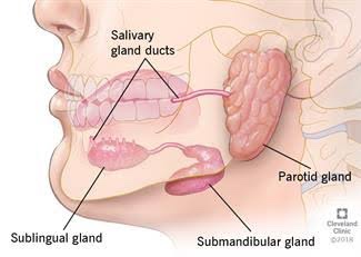
Blood Supply

●Tongue is mainly supplied by lingual artery branch of external carotid artery, tonsilar branch of facial artery and Ascending pharyngeal artery branch of external carotid artery

● Venous drainage : dorsal lingual veins and deep lingual veins

● Sensory supply: lingual nerve supplies anterior 2/3rd of tongue, glossopharngeal nerve supplies posterior 1/3rd of tongue, Vagus nerve supplies posterior most part of tongue

● Motor supply : All extrinsic and intrinsic muscles of the tongue are supplied by hypoglossal nerve except palatoglossus supplied by vagus nerve

Clinical significance

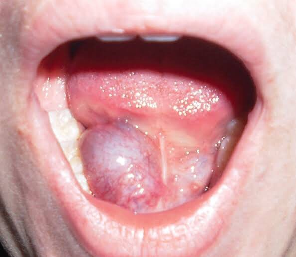
● Ankyloglossia (tongue tie) : Occurs due to abnormal length of frenulum that extends to the tip of the tongue . Ankyloglossia can be corrected surgically.

● Fissured tongue: Occurs when small furrows present on dorsal surface of the tongue. They are generally painless and benign and often associated with other syndromes

● Geographic tongue: asymptomatic and benign characterized by presence of red patches with greyish white border covering dorsum of tongue

Interesting facts

● An average adult has 2000- 4000 taste buds

● You cannot see your taste buds with naked eye. Those tiny pink and white bumps you see are actually papillae

● Tongue muscles are only muscles in the body working independent of the skeleton

● An oversized tongue is indicative of sleep apnea disorder

● Children sense flavours more intensely compared to adults. Umami is new variant of taste , monosodium glutamate is chemical responsible for this taste

● Women have shorter tongue than men.

● Your tongue is germ free only of its pink. If it is white there is a thin film of bacteria on it

● Blue whale has largest tongue in animal kingdom and weighs 5400lbs

Reference: B.D chaurasia and interesting facts from google

Tongue (part-1)

● Tongue is the muscular organ situated in the floor of the mouth

It has 2 surfaces

• Superior surface

• Inferior surface

● Superior surface is divided into 3 parts Anterior 2/3rd or oral part , posterior 1/3rd or pharyngeal part and base or root of the tongue

● Anterior 2/3rd and posterior 1/3rd are divided by V shaped sulcus terminalis, they are structurally and developmentally distinct.

● Inferior surface is covered by smooth mucous membrane. In midline a mucosal fold called frenulum connects tongue with floor of the mouth

● Lateral to frenulum deep lingual veins can be seen through mucosa lateral to lingual vein , mucosal fold called plica fimbriata is present

PAPILLAE OF TONGUE

Superior surface of tongue contains numerous papillae and have taste buds on their surface

Types of papillae

• Circumvalate papillae : present in front of sulcus terminalis , largest among papillae 8 to 12 in number

• Filliform papillae : Thin, long papillae with pointed ends. No taste buds. Identified by keratinization.

• Fungiform papillae: Slightly mushroom shaped , larger than Filliform papillae and present at apex of tongue and margins

• Foliate papillae : short vertical folds , present lateral to terminal sulcus and at margins

TASTE BUDS

Taste buds are sensory receptors for taste. Sensation of taste is called gustation.  Taste buds are located on surface of all papillae except Filliform papillae

Four taste sensation are sour, sweet, salty,  bitter and recently added is umami

MUSCLES OF TONGUE

● Intrinsic muscles: Main function is to alter the shape of the tongue ,they originate and insert within the tongue and no bony attachments.  ( 4) superior longitudinal,  inferior longitudinal,  vertical and transverse muscles

● Extrinsic muscles :  These muscles take origin from outside of the tongue and alter the shape as well.(4) Genioglossus, hyoglossus, styloglossus and palatoglossus

Superior longitudinal muscle: It lies just beneath dorsum of tongue. Action: It curls the tip upward and roll it posteriorly

Inferior longitudinal muscle : lies on each side lateral to the Genioglossus muscle. Action : To curl the tip of the tongue inferiorly


Transverse muscle: lies inferior to the superior longitudinal muscle and run from the septum to margins. Action : They narrow the tongue and increase the height

Vertical muscle: It lies inferolaterally to the Dorsum of tongue. Action : flattens the Dorsum

Genioglossus : origin- from superior mental spine and gets inserted into mucous membrane of tongue. It’s action is to protrude tongue, depress central part of tongue and increase the volume of sucking

Hyoglossus : origin- from greater horn and body of hyped bone and gets inserted to side of the tongue. It’s action is to depress side of the tongue assisting Genioglossus to enlarge the oral cavity

Styloglossus : origin- lower part of styloid process and upper part of stylohyoid ligament and inserts on side of the tongue. Action is to elevate and retract the tongue

Palatoglossus : origin- from soft palate and insertion lateral margin of tongue

♢ Styloglossus and palatoglossus attach the tongue Superiorly ,while Genioglossus and hyoglossus attached to the tongue inferiorly

♢ All the extrinsic and intrinsic muscles of tongue are supplied by hypoglossal nerve except palatoglossus supplied by vagus nerve

Reference: B.D chaurasia

4th ventricle of brain

• Ventricles are the sites for CSF production.

• lateral Ventricles are the largest and most proximal. Interventricular foramen of Monro connects lateral ventricle and third ventricle

• Third ventricle connects fourth ventricle through aqueduct of sylvius.CSF flows through the entire pathway and exits fourth ventricle into surrounding CNS tissue or through spinal cord.

STRUCTURE AND FUNCTION

• As said earlier , Ventricles are the sites for CSF production. Majority of CSF is produced by choroid plexus. Although lateral Ventricles also produce most of the CSF due to their larger surface area

• CSF gets produced in all Ventricles. CSF provides support to brain, absorbs shock during trauma,keeps neurons in homeostasis, provides nutrition and contain immune cells that fight against infections of nervous system

• fourth ventricle is most inferiorly located directly draining into spinal cord.

• Superiorly it connects third ventricle through cerebral aqueduct of sylvius. Anteriorly it’s surrounding by pons and medulla . Posteriorly by cerebellum and inferiorly by spinal cord.

• lateral apertures or foramen of luschka and median aperture or foramen of magendie. These apertures help the spread of CSF around the spinal cord and surrounding neural structures. All the CSF produced does not drain out of these aperture goes into central canal of spinal cord

EMBRYOLOGY

• Neural tube forms around 4th week of gestation. Three dilation that form neural tube are procencephalon, mesencephalon and rhombencephalon.

• Procencephalon or forebrain develops into telencephalon and diencephalon . Mesencephalon becomes the midbrain while rhombencephalon grows into metencephalon ( pons and cerebellum) and melencephalon (medulla)

• Within each of these dilations that develop from neural tube are cavities that become Ventricles

• The cavity located in rhombencephalon becomes fourth ventricle

BLOOD SUPPLY

posterior inferior cerebellar artery , anterior inferior cerebellar artery and superior cerebellar artery supplies majority of blood flow to fourth ventricle

CLINICAL SIGNIFICANCE

• Ependymomas are the third most common pediatric brain tumour. They are CNS malignancies that originate from the ependymal lining of Ventricles. These tumours are often treatable or require surgery to remove these tumors

• Hydrocephalus is presence of too much of CSF, which often results problem with CSF absorption. A condition called fourth ventricle outlet obstruction is a type of non communicating Hydrocephalus where there is disconnection between subarachnoid space and ventricular system

• Dandy walker malformation (DWM)that demonstrates a cystic enlargement of fourth ventricle and agenesis or hypoplasia of cerebellar vermis . CNS disorders associated with DWM include holoprosencephaly, neural tube defects and dysgenesis of corpus callosum

Reference :- B.D chaurasia

Swellings in the angle of Mandible, Floor of Mouth & Palate

Swellings at the angle of Mandible include: ✍🏻👇🏻

🔅Congenital disease

• Branchial Cleft Cyst

🔅Neoplasm

(i) Benign

  • Hemangioma
  • Lymphangioma, Cystic hygroma
  • Pleomorphic adenoma (mixed tumor)
  • Warthin tumor
  • Neurofibroma
  • Angiolipoma
  • Adenoma
  • Hamartoma
  • Lipoma
  • Oncocytoma

(ii) Malignant

  • Mucoepidermoid carcinoma
  • Squamous cell carcinoma
  • Adenoid cystic carcinoma
  • Acinic cell carcinoma
  • Adenocarcinoma
  • Rhabdomyosarcoma
  • Lymphoma, leukemia
  • Metastatic adenopathy

🔅Inflammation/Infection

  1. Parotitis
  2. Parotid Abscess
  3. Tuberculosis
  4. Sarcoidosis
  5. Sjögren disease
  6. HIV

Detailed View🔍

1) Branchial Cleft Cyst:

  • Failure of involution of clefts and pouches lead to cysts, fistulas or sinus tracts.
  • Its a painless fluctuant swelling
  • First branchial cleft cysts are rare usually located at parotid gland or periparotid region.
  • Second branchial cleft cyst – Type II are the most common
  • Typically, second branchial cleft cysts present as a rounded swelling just below the angle of mandible, anterior to the sternocleidomastoid

2) Hemangiomas:

They are the most common benign salivary gland mass. Capillary hemangiomas involve parotids

3) Lymphangiomas

They are congenital malformations of the lymphatic system that may involve the parotid gland (Soft asymptomatic neck mass associated with facial asymmetry)

4) Pleomorphic Adenoma:

Hard painless slow growing mass

5) Warthin Tumor:

Incorporation of heterotopic salivary gland ductal epithelium within intraparotid & periparotid nodes

6) Parotitis & Parotid Abscess:

  • Most common in children
  • Mumps is the most common viral cause of parotitis
  • The condition manifests tender swelling at the angle of Mandible
  • Sialadenitis is most commonly due to bacterial infections caused by Staphylococcus aureus.
  • Premature neonates and immunosuppressed individuals are affected.

Swellings in the floor of Mouth: 👇🏻✍🏻

Ranula presents as a translucent blue, dome-shaped fluctuant swelling & contains viscid, glairy jelly like fluid
  1. Ranula – a type of mucocele found on the floor of the mouth. Present as a swelling of connective tissue consisting of collected Mucin from a ruptured salivary gland by local trauma.
  2. Swellings in the floor of the mouth are more likely to arise from structures above the Mylohyoid muscle. The commonest swellings in the floor of the mouth are denture induced hyperplasia & salivary calculus.
  3. Swellings in the floor of the mouth may inhibit swallowing & speech.
  4. Mandibular tori produce bony hard swelling lingual to the lower premolars.

Differential diagnosis of swellings of the floor of the mouth or neck (Jham et al., 2007): https://www.researchgate.net/figure/Differential-diagnosis-of-swellings-of-the-floor-of-the-mouth-or-neck-Jham-et-al-2007_tbl1_287206404


Swellings on the Palate: 👇🏻✍🏻

  1. Torus palatinus is an intrinsic bone lesion whereas a dental abscess pointing on the palate (usually from the palatal roots of the 1st & 2nd maxillary molars or from upper lateral incisors) is extrinsic.
  2. Salivary neoplasms
  3. Invasive carcinoma from the maxillary sinus may produce a palatal swelling.
  4. Kaposi’s sarcoma, typical of HIV/AIDS may also present as lump on palate.
  5. Paget’s disease.

Differential diagnosis of palatal swellings: https://www.researchgate.net/figure/Differential-diagnosis-of-palatal-swellings_tbl1_221967546

Dentowesome|@drmehnaz🖊


Image source: Google.com

CIRCLE OF WILLIS(Circulus arteriosus cerebri)

The only weapon with which the unconscious patient can immediately retaliate upon the incompetent surgeon is hemorrhage -William S Halsted.

Also known as loop of Willis,Willis polygon and cerebral arterial circle.

Blood vessels supplying the brain consists of 2 separate vascular systems:

  • Internal carotid arteries
  • Vertebral arteries

In the base of the brain both systems are connected bilaterally by posterior communicating arteries and left to right side by anterior communicating arteries forming the “Circle of Willis”(COW).

Other key points:

  • In subclavian steal syndrome ,blood is “stolen” from COW to preserve blood flow to upper limb.
  • This syndrome results from a proximal stenosis of the subclavian artery.
  • Variations in the shape of COW is also seen in mentally ill and migraine patients which include hypoplasia of component vessels,absence of posterior communicating arteries,duplication/triplication of anterior communicating arteries,etc.

Mnemonics:

Cell is Clearly Circulating

C-Cortical branches

C-Central branches

C-Choroidal branches

Sources: BD Chaurasia’s Human anatomy 7th edition-volume 4

Slideshare.net -Circle of Willis by Luiz Roberto Meier Update 2