Burning Palate and Chest Pain: Connecting Oral Health to Systemic Risk

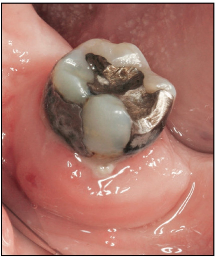
Clinical view of the patient’s palate. Burning Mouth Syndrome or Something More? A Case of Dual Diagnosis

An 80-year-old patient presents to the office with a chief complaint of a continuous burning feeling on his palate. Upon examination you find that the patient is extremely sensitive.When you finish your examination, the patient complains of sudden chest pain. He starts to sweat and has labored breathing.

Q: What is your diagnosis of the patient?

  • Medical: There are a number of possibilities for the patient to have chest pain and labored breathing. The patient might be experiencing an acute myocardial infarction, hyperventilation, or angina pectoris.
  • Dental: The patient has a Candida species infection, which is a gingival disease of fungal origin. It is also known as atrophic (erythematous) candidiasis.

Q: How will you manage the patient if he is having a myo-cardial infarction?

  1. Stop the dental procedure
  2. Administer oxygen to the patient at 4 to 6 liters per minute
  3. Call emergency medical services (EMS) immediately.
  4. Administer nitroglycerin from the emergency kit (if pain continues, most likely not angina)
  5. Administer aspirin (fibrinolytic properties):
    Give the patient 325 mg of non-enteric-coated aspirin to chew if they have no contraindications (e.g., allergies or bleeding disorders)
  6. Monitor vital signs
  7. Keep the patient in a comfortable seated position to minimize strain on the heart.
  8. Manage the patient’s pain with opioids (morphine) or nitrous oxide

Q: What is your approach to treating the dental condition of this patient?

First treat the condition with a topical antifungal (eg, nystatin or clotrimazole troches) applied to the tissue side of the denture four to six times a day for 2 to 3 weeks. If the fungal infection persists,treat the patient with 100 mg fluconazole daily.

@dr.mehnaz


REFERENCESPERIODONTAL REVIEW : A STUDY GUIDE / DEBORAH TERMEIE.

Decoding Oral Malodor: Managing a Mandibular Molar Abscess in a Medically Complex Patient

When Bad Breath Signals Trouble: A Case of Mandibular Molar Abscess

The patient is a 65-year-old man complaining of oral malodor. His dentist referred him to you to access the mandibular right second molar because of swelling, pus, and soreness. When he sits in your chair, he seems disoriented and irritable.When you look in his mouth, you find generalized inflammation of the gingiva and an abscess on the buccal aspect of the mandibular right second molar with suppuration. Charting demonstrates an 8-mm facial pocket and 6-mm palatal and interproximal pockets.

Q: What is your diagnosis of the patient?

Medical:

There are a number of reasons for the patient to appear dazed and irritable:

  • Hypoglycemia or hyperglycemia
  • Alcohol or drug overdose
  • Hyperthyroidism or hypothyroidism
  • Cerebrovascular incident

Dental:

  • The patient may have diabetes mellitus–associated gingivitis related to the endocrine system, under the heading of gingival diseases modified by systemic diseases, which is a subcategory of dental plaque–induced gingival diseases.
  • The patient has a periodontal abscess, which is a subclassification of abscesses of the periodontium.

Q: What could have led to the abscess formation?

  • Diabetes: According to Bjelland et al,18 multiple periodontal abscesses may result from uncontrolled hyperglycemia. Rees19 listed multiple or recurrent periodontal abscesses among the possible indications of undiagnosed or poorly controlled diabetes mellitus.
  • The abscess may also be caused by a preexisting periodontal pocket in association with bacteria at the depth of the pocket.
  • A foreign body can also cause a periodontal abscess.

Q: How will you treat the periodontal abscess?

  • I would ask the patient if he has seen his physician recently and whether he knows his hemoglobin A1c levels to determine if the abscess may be associated with diabetes (only his medical doctor can make that diagnosis).
  • An incision at a 90-degree angle to the long axis of the tooth will drain the exudate.Without removal of the cause (foreign body, bacteria, or calculus), the abscess will recur. If this is not possible, extraction might be necessary.
  • Antibiotics and analgesics should be prescribed. A follow-up with a dentist is also needed.
  • Saline Rinse: Advise warm saline rinses to reduce discomfort and promote healing.

Dentowesome | @dr.mehnaz


References: Periodontal review : a study guide / Deborah Termeie.

Restoring Fractured Teeth in Patients with Parafunctional Habits: Key Considerations

A 43-year-old patient presents with a fractured palatal cusp of an upper five after eating a sandwich. On examination, the following were found.

  • Extraoral: large masseters, bitten fingernails.
  • Intraoral: healthy periodontium (no BPE >1). The palatal cusp of the upper vital and
    unrestored five is fractured subgingivally. Anterior tooth wear, dentine exposure on both upper canine cusp tips.

What is the mechanism leading to this problem?

The fractured palatal cusp of the upper premolar is likely due to excessive occlusal forces exerted over time, especially during parafunctional habits like bruxism (teeth grinding) or clenching. The patient’s large masseters and bitten fingernails suggest bruxism or clenching, which leads to increased stress on the teeth. Over time, this can weaken tooth structure, making it more susceptible to fracture, even during relatively low-stress activities like eating a sandwich.

What is the significance of the anterior wear?

The anterior wear, especially the dentine exposure on the upper canines, indicates a significant amount of tooth surface loss, typically caused by parafunctional habits like bruxism. Canines play a crucial role in guiding the occlusion and protecting the posterior teeth during lateral movements (canine guidance). The loss of this guidance could shift the load to other teeth, like the upper premolars, further contributing to their fracture. The exposed dentine also increases the risk of sensitivity and further wear.

What additional precautions might you take in this case when you plan for a definitive restorative work?

  1. Assessment of Parafunctional Habits: •Address the underlying cause of the excessive forces, likely bruxism or clenching. Consider a thorough evaluation and a potential referral to a specialist to assess for any contributing factors such as stress, anxiety, or sleep disorders. • Fabrication of a night guard (occlusal splint) may be recommended to protect the teeth from further damage.
    1. Occlusal Analysis:
      • Perform a detailed occlusal analysis to identify any interferences, especially in lateral and protrusive movements. Address occlusal discrepancies that could contribute to abnormal forces on teeth.
      • Check for the need to adjust canine guidance, as the wear may have altered the normal function.
    2. Restorative Material Selection:
      • Consider using a durable material such as porcelain, zirconia, or composite for the restoration, especially for posterior teeth under high stress.
      • A crown may be necessary for the fractured tooth to provide full coverage and strength, particularly if the fracture is subgingival.
    3. Subgingival Fracture Considerations:
      • Ensure proper isolation during restorative procedures, as the subgingival fracture may complicate the seating of a restoration.
      • Crown lengthening or orthodontic extrusion may be required to ensure that the margins of the restoration are accessible and that a proper seal can be achieved without violating the biological width.
    4. Patient Education and Follow-up:
      • Educate the patient about the potential for ongoing damage if bruxism is not managed.
      • Regular follow-up is important to monitor the restoration and assess for further wear or damage due to parafunctional habits.

References: Practical Procedures in Dental Occlusion, First Edition. Ziad Al-Ani and Riaz Yar. © 2022 John Wiley & Sons Ltd.

Composite resin strip crowns


Composite is the material of choice for the restoration of primary anterior teeth. An anterior strip crowns with composite resin provides an aesthetic and durable restoration.

Method:

  1. Local anaesthesia and rubber-dam isolation should be used if possible. Alterna- tively, because of age and poor cooperation of younger children, the restorative work may be completed under general anaesthesia.
  2. Select the correct celluloid crown form depending on the mesiodistal width of the teeth.
  3. Remove the caries using a slow-speed round bur.
  4. Using a high-speed tapered diamond or tungsten carbide bur, reduce the incisal
    height by around 2 mm, prepare interproximal slices and place a labial groove at
    the level of gingival and middle thirds of the crown.
  5. Protect the exposed dentine with a glass ionomer lining cement.
  6. Trim the crown form and make two holes in the incisal corners by piercing with
    a sharp explorer.
  7. Etch the enamel for 20 seconds, and wash and dry.
  8. Apply a thin layer of bonding resin and cure for 20 seconds, ensuring all surfaces
    are covered equally.
  9. Fill the crown form with the appropriate shade of composite and seat with gentle,
    even pressure, allowing the excess to exit freely. The use of small wedges may be
    helpful in avoiding interproximal excess.
  10. Light cure each aspect (labially, incisally and palatally) equally.
  11. Remove the celluloid crown gently, and adjust the form and finish with either composite finishing burs or abrasive discs.
  12. Check the occlusion after removing the rubber dam.

Source: Handbook of Pediatric Dentistry, Third Ed


@drmehnaz🖊