WEGNER’S GRANULOMATOSIS

Wegener’s granulomatosis is a disease of unknown aetiology, which basically involves the vascular, renal and respiratory systems. It involves the nose, paranasal air sinuses, lower respiratory tract, gut, joints, nervous system, and kidneys. Involvement of the kidney is the common cause of death.

This disease is caused by an abnormal immune reaction secondary to a nonspecific infection or a hypersensitivity reaction to an unknown antigen.

Clinical Features

>Wegener’s granulomatosis may occur at any age, although the majority of cases are in the fourth and fifth decadesof life.

 >There is a slight predilection for occurrence in males.

 >It is best described as a multisystem disease, which is usually first characterized clinically by the development of rhinitis, sinusitis, and otitis or ocular symptoms.

 >The patient soon develops a cough and hemoptysis as well as fever and joint pain. 

>Hemorrhagic or vesicular skin lesions are also commonly present. 

>Granulomatous lesions of the lungs are found on the chest radiograph, while the glomerulonephritis, which develops ultimately, leads to uraemia and terminal renal failure.

 >In nervous system, sensory neuropathy may be an occasional finding.

>Prognosis—the disease is usually fatal, with mean survival time of 5 months. Death occurs due to involvement of kidney.

Oral Manifestations

Involvement of the gingiva has been the most common and characteristic manifestation, and is termed as strawberry gingivitis.Gingival lesions may manifest as ulcerations, friable granular lesions, or simply enlargements of the gingiva. 

The inflammatory process starts in the interdental papilla and spreads rapidly to the periodontal structure and leads to bone loss and tooth mobility. 

Palate—orallesionstypicallyincludeulcerationofthe palate by extension of nose lesions and destruction of nasal septum. This will lead to perforation of palate.

• Teeth—theremaybelooseningofteethwithinsomecases spontaneous exfoliation. After extraction of teeth patient is usually noticed poor healing.

Laboratory Findings

Laboratory findings include anaemia, leukocytosis, elevated erythrocyte sedimentation rate, and hyperglobulinaemia. Because of kidney involvement, haematuria is common, as well as the finding of albumin, casts, and leukocytes in the urine. Circulating immune complexes have been demonstrated in some patients, but this is not a consistent finding.
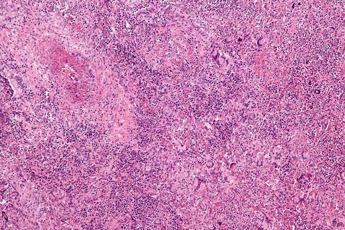
Histologic Findings

Wegener’s granulomatosis presents a pattern of mixed inflammation centred around the blood vessels. 

The lesions in the upper respiratory tract and lungs consist of giant cell necrotizing granulomatous lesions showing vasculitis. 

Oral biopsy specimens show pseudoepitheliomatous hyperplasia and subepithelial abscesses. The gingival and other lesions show a nonspecific granulomatous process with scattered giant cells.

Diagnosis

  • Clinical diagnosis—typical strawberry gingivitis with necrotic ulceration in the oral cavity.
  • Laboratorydiagnosis—cytoplasmiclocalizationispresent with Wegener’s granulomatosis. Histopathologically chronic inflammatory cells and multinucleated giant cells are found.
  • Differential Diagnosis
  • Agranulocytosis, leukemia, lymphoma—diagnosis by blood picture, possibly histology.
  1. Management
    • Cotrimoxazole—it is combination of trimethoprim and sulfamethoxazole. It has proved to be effective as an adju- vant or sole therapy in both localized and generalized forms.
    • Corticosteroids—regimenofcyclophosphamide12mg/ kg body weight/day with prednisolone 1 mg/kg body weight have been utilized to obtain complete remission.
    • Others—other treatment modalities includes cyclo- sporine, intravenous pooled immunoglobulin, and local irradiation.

REFERENCE- SHAFER’S TEXTBOOK OF ORAL PATHOLOGY {8TH ED} AND ANIL GHOM TEXTBOOK OF ORAL MEDICINE

BECHET’S SYNDROME

  • Behçets disease (BD) was initially described by the Turkish dermatologist Hulusi Behçet as a triad of symptoms including 

                        1. Recurring oral ulcers, 

                         2.Recurring genital ulcers, and 

                         3.Eye involvement.

ETIOLOGY

  • The cause of BD is unknown, 
    • but immune dysregulation, 
    • including circulating immune complexes, 
    • autoimmunity, cytokines, and 
    • heat shock proteins, 
    • major factor in the pathogenesis of BD.

CLINICAL MANIFESTATIONS

  • Highest incidence is in young adults between the ages of 25 and 40.
  • The most common site of involvement is oral mucosa. 
  • The genital area is the second most common site of involvement and presents as ulcers
  • The eye lesions consist of uveitis, retinal vasculitis, vascular occlusion, optic atrophy, and conjunctivitis. 
  • Blindness is a common complication of the disease, and periodic evaluation by an ophthalmologist is necessary.

  • Positive pathergy is defined as  an inflammatory reaction  forming within 24 hours of a needle puncture scratch, or saline injection.
  • A positive pathergy test, which is performed by placing a 20 gauge needle 5 mm into the skin of the forearm. The test is positive if an indurated papule or pustule greater than 2 mm in diameter forms within 48 hours.

ORAL MANIFESTATIONS

  • The most common single site of involvement is the oral mucosa. 
  • Recurring oral ulcers appear in over 90% of patients; these lesions cannot be distinguished from RAS .
  • Some patients experience mild recurring oral lesions; others have the deep large scarring lesions characteristic of major RAS.
  • These lesions may appear anywhere on the oral or pharyngeal mucosa.

Histologic Features

The microscopic findings are not diagnostic. They consist of parakeratosis, acanthosis, and polymorphonuclear leukocyte infiltration of epithelium, sometimes with mi- croabscess formation similar to psoriasis. The connective tissue shows a lymphocyte and plasma cell infiltrate.

Laboratory Findings

The patients usually have a mild leukocytosis, an elevated erythrocyte sedimentation rate, and pyuria.

DIFFERENTIAL DIAGNOSIS

  • Sweet Syndrome : oral ulcers, conjunctivitis, episcleritis, Inflamed tender skin papules or nodules.
  • Erythema Multiforme: erosions, target(iris) lesions.
  • Pemphigoid: bullae, erosions.
  • Pemphigus : erosions, flaccid skin bullae.
  • Reiter syndrome: ulcers, conjunctivitis,
  • Herpes simplex virus
  • Lupus erythematosus

TREATMENT

The management of Behçet’s syndrome depends on the severity and the sites of involvement.

  • Azathioprine combined with prednisone has been shown to reduce ocular disease as well as oral and genital involvement.
  • Pentoxifylline, which has fewer side effects than do immunosuppressive drugs or systemic steroids, has also been reperted to be effective in decreasing disease activity, particularly of oral and genital lesions.
  • Dapsone, colchicines and thalidomide have also been reported to be effective to treat mucosal lesions of Behcet’s disease.

REFERENCE- BURKET TEXTBOOK OF ORAL MEDICINE AND SHAFER’S TEXTBOOK OF ORAL PATHOLOGY {8TH EDITION}

Systemic lupus erythematosus{SLE}

  • SLE is a multisystem autoimmune inflammatory disorder of unknown etiology.
  • Main feature is the formation of antibodies to DNA, which may initiate immune complex reactions, in particular a vasculitis. 
  • Female to male ratio of 9:1
  • More common in persons of non-European descent.
  • Etiology
  • Geneticpredisposition—relativeofpatientshavehigher incidences of auto-antibodies, immune deficiency and connective tissue disease. This tendency is greatest among identical twins.
    • Immunological abnormality possibly mediated by viral infection—immune complex consisting chiefly of nucleic acid and antibody account for majority of the tissue changes.
    • Autoimmune disease—as these patients develop antibodies to many of their own cells.
    • Endocrine—thereishighincidenceinfemalesinpreg- nancy. This finding suggestive of increased estrogen level.
    • Biochemicalincreaseinexcretionofmetabolicproducts, particularly tyrosine and phenylalanine, in certain SLEpatient.
  • CLINICAL MANIFESTATIONS
  • Lupus is known as “the great mimic.”
  •  Skin lesions of lupus can be classified 
    • lupus-specific (having diagnostic clinical or histopathologic features) 
    • nonspecific lesions.
  • Three subtypes of lupus-specific 
    • Acute
    • subacute 
    • chronic. 
  • Acute cutaneous lupus occurs in 30 to 50% of patients and is classically represented by the butterfly rash-mask-shaped erythematous eruption involving the malar areas and bridge of the nose
  • Chronic cutaneous lupus occurs in 15 to 20% of cases and affects the skin of the face or scalp in about 80% of cases.
  • The least common subtype, subacute cutaneous lupus, occurs in 10 to 15% of patients and includes papulo­squamous (psoriasiform) and annular-polycystic eruptions, usually on the trunk and arms.
  • Nonspecific but suggestive skin manifestations of lupus are common and include 
    • alopecia (both scarring following discoid lesions and non-scarring)
    • Photosensitivity
    • Raynaud’s phenomenon
    • Urticaria
    • Erythema
    • Telangiectases
    • cutaneous vasculitis.

  • ORAL MANIFESTATIONS
  • Two predominant types of oral lesions are
    •  discoid lesions 
    • ulcerations.
  • Oral ulcerations associated with SLE  they occur with increased frequency on the palate and in the oropharynx and are characteristically painless.
  • Histologically, they are characterized by lymphocytic infiltrate at the base of the ulcer and in the perivascular distribution, which is similar to that observed in discoid lesions.
  • Discoid oral lesions, appear as whitish striae frequently radiating from the central erythematous area, giving a so-called “brush border.”
  • Buccal mucosa, gingiva, and labial mucosa are the most commonly affected intraoral sites.
  • Direct immunofluorescent staining for immunoglobulins and complement C3 factor is a useful aid to diagnosis. Granular deposition of IgM, IgG, and C3 along the basement membrane is characteristic

Diagnosis

• Clinical diagnosis—skin lesion with lesion present on oral mucosa which is atrophic and erythematous will suspect lupus erythematous. Oral and nasopharyngeal ulceration is major diagnostic criteria for SLE.

Laboratory diagnosis—L.E. cell inclusion phenomenon with surrounding pale nuclear mass apparently devoid of lymphocytes. Anemia, leukopenia and thrombocyto- penia, with sedimentation rate increased. Serum gamma globulin increased and Coomb’s test is positive.

Positive lupus band test—it shows deposition of IgG,IgM or complement component in skin.

  1. Differential Diagnosis
    • Lichenplanus—homogenouspicture,nodarkerythema and no telangiectasia. Mucosal changes are usually extensive and symmetrical.
    • Lichenoidreaction—historyofdrugisalwaysthere.
    • Ectopic geographic tongue—systemic manifestation present is lupus erythematous, which is absent in ectopicgeographic tongue.
    • Psoriasis—Auspitz’s’signispositive.
    • Electrogalvanic lesion—dissimilar restorations are seenin oral cavity.
    • Leukoplakiaanderythroplakia—lesionstendtomaintainsame appearance and there are no skin changes.
    • Geographic stomatitis—no skin changes, mucosal lesionschange location rapidly.
    • Benign mucous membrane pemphigoid—no systemiccomplain and serology test to be done.
  • TREATMENT
  • Corticosteriods are the cornerstone of therapy
  • A pulse i.v cyclophosphamide regimen for remission induction followed by quarterly infusions
  • Recently, mycophenolate mofetil and azathioprine
  • NSAIDs for arthritis relief
  • Antimalarial like hydroxychloroquinine – effective in cutaneous lupus 
  • DENTAL MANAGEMENT
  • Recommended prophylactic antibiotics if ANC count falls below 500 – 1000 cells/mm3
  • Adrenal supression –
  • Adenocorticotropic hormone supression test is used to evalute
  • Current guidelines – Replacement therapy with hydrocortisone is unnecessary

REFERENCE- ANIL GHOM TEXTBOOK OF ORAL MEDICINE; BURKIT TEXTBOOK OF ORAL MEDICINE AND GOOGLE[SLIDE SHARE]

Histopathology of dentinal caries

Zone I: Zone of fatty degeneration of odontoblast process
Zone 2: Zone of dentinal sclerosis characterized by deposition of cal- cium salts in dentinal tubules
Zone 3: Zone of decalcification of dentin, a narrow zone, preceding bacterial invasion
Zone 4: Zone of bacterial invasion of decalcified but intact dentin Zone 5: Zone of decomposed dentin

Earldentinal caries

Fatty degeneration oodontob/ast process

>Disposition of fat globules – precedes early sclerotic changes  >Special stains – Sudan red
>Significance-
1.Fat contributes to impermeability 

2.Predisposing factor for dental sclerosis

Sclerotic dentin

>Reaction of vital pulp – calcification of dentinal tubules (DT)

>Seals off DT from further penetration of microorganisms

>Minimal in rapidly advancing caries

>Prominent in slow caries

>Sclerotic dentin – appear white in transmitted light

Decalcification odentinatubules

>Above dentinal sclerosis – zone of decalcification

>Occurs in advance of bacterial invasion of DT 

>Pioneer bacteria
>The initial decalcification – only the walls of DT 

>Study of tubules- pure form of microorganisms

Zone omicrobial invasion

>Proteolytic organisms – predominantly in deeper layers Acidogenic microorganisms – more in early caries
>Supporting the hypothesis that initiation and progression are two distinct processes and must be differentiated

Advanced dentinacaries

>Decalcification of the walls of DT – confluence

>Thickening of sheath of Neumann – along its course • Increase in the diameter of DT – microorganisms

>Focal coalescence of adjacent tubules and ovoid area of destruction- liquefaction foci
>Acidogenic organisms – initial decalcification

>Proteolytic organisms – matrix destruction

>Multiple areas of destruction>Necrotic mass of dentin (leathery consistency)

>Formation of transverse cleftsExtend at right angles to DT and parallel contour line

>Peeling away of carious dentin

REFERENCE- Shafers textbook of oral pathology 8th edition

Histopathology of enamel caries

Four zones are clearly distinguishable, starting from the inner advancing front of the lesion. These are the (1) translucent zone, (2) dark zone, (3) body of the lesion and (4) surface layer.

Zones of enamecarieTranslucenzone {TZ)

First recognizable zone of alteration

Advancing front of the lesion

Half the lesions demonstrate TZ, not always present

Seen in longitudinal ground sections in clearing (quinoline – RI – 1.62)

TZ appears structureless

Pore volume – I% (compared to 0.1 % of sound enamel)

Dark zone

Lies adjacent and superficial to the translucent zone Positive zone

Shows positive birefringence (in contrast to sound enamel.

Pore volume of 2-4% (polarized light)
Presence of small pores; large molecules of quinoline are unable to penetrate
Micropore system – gets filled with air and becomes dark
Medium like water may penetrate

Body othlesion

Between unaffected, surface and dark zone
Area of greatest demineralization
Pore volume – 5% in periphery and 25in centre
Quinoline imbibition – body appears transparent
Water imbibition – positive birefringence compared to sound enamel Striae of Retzius – prominent

Surfaczone

Quantitative studies – partial demineralization of 1-10% • Pore volume – less than 5% of the spaces

Negative birefringence – water imbibition

Positive birefringence – porous subsurface
All the four zones of enamel caries cannot be seen with same immersion medium.

REFERENCE – Shafers textbook of oral pathology 8th edition

GENERALIZED AGGRESSIVE PERIODONTITIS

The term “juvenile periodontitis” was introduced by Chaput and colleagues in 1967 and by Butler in 1969. In 1971, Baer definedit as “a disease of the periodontium occurring in an otherwise healthy adolescent which is characterized by a rapid loss of alveolar bone about more than one tooth of the permanent dentition.

• Usually affecting persons under 30 years of age (however, may be older).

• Generalized proximal attachment loss affecting at least three teeth other than first molars and incisors.

• Pronounced episodic nature of periodontal destruction. • Poor serum antibody response to infecting agents.

Clinical Characteristics

GAP usually affects individuals under age 30, but older patients also may be affected. In contrast to LAP, evidence suggests that individuals affected with GAP produce a poor antibody response to the pathogens present.

Clinically, GAP is characterized by “gen- eralized interproximal attachment loss affecting at least three per- manent teeth other than first molars and incisors.”The destruction appears to occur episodically, with periods of advanced destruction followed by stages of quiescence of variable length (weeks to months or years). 

P. gingivalis, A. actinomycetemcomitans, and Tannerella forsythia (formerly Bacteroides forsythus) frequently are detected in the plaque that is present.

Two gingival tissue responses can be found in cases of GAP. One is a severe, acutely inflamed tissue, often proliferating, ulcerated, and fiery red. One is a severe, acutely inflamed tissue, often proliferating, ulcerated, and fiery red.

Bleeding may occur spontaneously or with slight stimulation. Suppuration may be an important feature. This tissue response is believed to occur in the destructive stage, in which attachment and bone are actively lost.

Some patients with GAP may have systemic manifestations, such as weight loss, mental depression, and general malaise.

Radiographic Findings

No definite pattern of distribution occurs, The radiographic picture in GAP can range from severe bone loss associated with the minimal number of teeth to advanced bone loss affecting the majority of teeth in the dentition.

Prevalence and Distribution by Age
and Gender

A US national survey of adolescents ages 14 to 17 reported that 0.13% had GAP. In addition, blacks were at much higher risk than whites for all forms of aggressive periodontitis, and male teenagers were more likely to have GAP than female adolescents.

Treatment of aggressive periodontitis 

• Successful treatment of EOP depends on early diagnosis use of antibiotics against the infecting micro organisms and provision of an infection free environment for healing 

• EOP both localized and generalized types includes surgery and use of tetracyclines (Lindhe 1982, Christersson and Zambon 1993) 

T/T of GAP – often less predictable – alternative antibiotics directed to specific pathogenic flora require 

– Multidisplinary approach combines clinical labroartory evaluation with conventional periodontal therapeutic methods for diagnosis and t/t of GAP 

Antibiotic Therapy for Aggressive Periodontitis

Associated MicrofloraAntibiotic of Choice
Gram-positive organismsAmoxicillin–clavulanate potassium (Augmentin)12,72
Gram-negative organismsClindamycin22,23,68,72
Nonoral gram-negative, facultative rodsCiprofloxacin41
Pseudomonads, staphylococci
Black-pigmented bacteria and spirochetesMetronidazole22,65
Prevotella intermedia, Porphyromonas gingivalisTetracycline55
Actinobacillus actinomycetemcomitansMetronidazole-amoxicillin22,65 Metronidazole-ciprofloxacin Tetracycline53
P. gingivalisAzithromycin54

REFERENCE- Caranza textbook of periodontology 11edition and Mc donald 9thed

LOCALIZED AGGRESSIVE PERIODONTITIS

The term “juvenile periodontitis” was introduced by Chaput and colleagues in 1967 and by Butler in 1969. In 1971, Baer defined it as “a disease of the periodontium occurring in an otherwise healthy adolescent which is characterized by a rapid loss of alveolar bone about more than one tooth of the permanent dentition.

According to Hart et al. diagnosis of localized early-onset periodontitis is based on 

• attachment loss of > or equal 4 mm on at least two permanent first molars 

and incisors (one of which must be a first permanent molar) 

Not more than two other permanent teeth, which are not first permanent molars or incisors, should be affected 

• Bone loss around primary teeth can be early finding in LAP

Clinical Characteristics

The lack of clinical inflammation despite the presence of deep periodontal pockets and advanced bone loss .

The amount of plaque on the affected teeth is minimal, which seems inconsistent with the amount of periodontal destruction present.The plaque that is present forms a thin biofilm on the teeth and rarely mineralizes to form calculus.

The rate of bone loss is about three to four times faster than in chronic periodontitis.

 Other clinical features of LAP may include (1) distolabial migration of the maxillary incisors with concomitant diastema formation, (2) increasing mobility of the maxillary and mandibular incisors and first molars, (3) sensitivity of denuded root surfaces to thermal and tactile stimuli, and (4) deep, dull, radiating pain during mastication, probably caused by irritation of the supporting structures by mobile teeth and impacted food.

Radiographic Findings

Vertical loss of alveolar bone around the first molars and incisors, beginning around puberty in otherwise healthy teenagers, is a classic diagnostic sign of LAP. Radiographic findings may include an “arcshaped loss of alveolar bone extending from the distal surface of the second premolar to the mesial surface of the second molar”

Prevalence and Distribution by Age
and Gender

LAP affects both males and females and is seen most frequently in the period between puberty and 20 years of age.

RISK FACTORS FOR AGGRESSIVE PERIODONTITIS

Microbiologic Factors

A. actinomycetemcomitans, Capnocytophagaspp., Eikenella corrodens, Prevotella intermedia, and Campylobacter rectus), A. actinomycetemcomitans has been implicated as the primary pathogen associated with LAP.

Immunologic Factors

Some immune defects have been implicated in the pathogenesis of aggressive periodontitis. The human leukocyte antigens (HLAs), which regulate immune responses, have been evaluated as candidate markers for aggressive periodontitis.

Genetic Factors

Familial pattern of alveolar bone loss and have implicated genetic factors in aggressive periodontitis.

Environmental Factors

The amount and duration of smoking are important variables that can influence the extent of destruction seen in young adults.46 Patients with GAP who smoke have more affected teeth and more loss of clinical attachment than nonsmoking patients with GAP.16 However, smoking may not have the same impact on attachment levels in younger patients with LAP.

Treatment

In ps with LAP,

Aa organisms penetrate into crevicular epithelium T/T with antibiotic alone such as 2 week course of doxycycline reduce Aa organisms 

Surgical removal of infected crevicular epithelium and debridement of root surface during surgery while the patient is on a 14 day course of doxycycline hyclate (1gm per day) (Mandell and Sockaransky 1988, Saxen et al 1990). 

Microdentex manufacturers the DMDx(Microdentex, FORT MYERS, Florida) test, a DNA test kit for establisting the risk of aggressive periodontits and confirms whether the child has responded favorably to the use of antimicrobial therapy Retesting after 4-6 weeks after the completion of antibiotic therapy determines the pts response to t/

• Rams and collagues described keyes technique for treating LAP Scaling and root planning of all teeth , with irrigation to probing depth of saturated inorganic salt solutions and 1% chloramine T.((sodium para-toluene sulfonchloramide) In addition recommended administration of systemic tetracycline (18 per day) for 14 days This dose appropriate for pts 12 yrs of age and older 

Home T/T Daily application of paste of sodium bicarbonate and 3% hydrogen peroxide and inorganic salt irrigation.

Associated MicrofloraAntibiotic of Choice
Gram-positive organismsAmoxicillin–clavulanate potassium (Augmentin)12,72
Gram-negative organismsClindamycin22,23,68,72
Nonoral gram-negative, facultative rodsCiprofloxacin41
Pseudomonads, staphylococci
Black-pigmented bacteria and spirochetesMetronidazole22,65
Prevotella intermedia, Porphyromonas gingivalisTetracycline55
Actinobacillus actinomycetemcomitansMetronidazole-amoxicillin22,65 Metronidazole-ciprofloxacin Tetracycline53
P. gingivalisAzithromycin54

REFERENCE-

Caranza textbook of periodontology 11edition and Mc Donalds 9thedition

RUBBER DAM

Rubber dam was introduced byBarnum, a New York dentist in 1863

Advantages of using a rubber dam

• It is raincoat for the teeth
• It helps in improving accessibility and visibility of the working area
• It gives a clean and dry aseptic field while working
• It protects the lips, cheeks and tongue by keeping them out of the way
• It helps to avoid unnecessary contamination through infection control
• It protects the patient from inhalation or ingestion of instruments and medicaments
• It helps in keeping teeth saliva free while performing a root canal so that tooth does not get decontaminated by bacteria present in saliva
• It improves the efficiency of the treatment
• It limits bacterial laden splash and splatter of saliva and blood

• It potentially improves the properties of dental material.
• It provides protection of patient and dentist.

Disadvantages of using a rubber dam

• Takes time to apply 
• Communication with patient can be difficult 
• Incorrect use may damage porcelain crowns/crown margins/ traumatize gingival tissues 
• Insecure clamps can be swallowed or aspirated.Contraindications of use of rubber dam

• Asthmatic patients
• Allergy to latex
• Mouth breathers
• Extremely malpositioned tooth • Third molar (in some cases).

Rubber dam equipment

• Rubber dam sheet• Rubber dam clamp • Rubber dam forceps• Rubber dam frameRubber dam accessories•Lubricant/petroleum jelly• Dental floss• Rubber dam napkin.

Rubber Dam Sheet

  • The rubber dam sheet is normally available in size 5 × 5 or 6 × 6 squares in green or black color
  • It is available in three thicknesses, i.e. light, medium and heavy
  • The middle grade is usually preferred as thin is more prone to tearing and heavier one is more difficult to apply
  • Latex-free dam is necessary as number of patients are increasing with latex allergy
  • Flexi dam is latex-free dam of standard thickness with no rubber smell.

Rubber Dam Clamps

  • Rubber dam clamps, to hold the rubber dam onto the tooth are available in different shapes and sizes.
  • Clamps mainly serve two functions:
    1. They anchor the rubber dam to the tooth.
    2. Help in retracting the gingiva.

Rubber Dam Forceps

  • Rubber dam forceps are used to carry the clamp to the tooth.
  • They are designed to spread the two working ends of the forceps apart when the handles are squeezed together.
  • The working ends have small projections that fit into two corresponding holes on the rubber dam clamps.
  • The area between the working end and the handle has a sliding lock device which locks the handles in positions while the clinician moves the clamp around the tooth.
  • It should be taken care that forceps do not have deep grooves at their tips or they become very difficult to remove once the clamp is in place.

Rubber Dam Frame

Rubber dam frame supports the edges of rubber dam .Frames have been improved dramatically since their old style with the huge ‘butterflies’.Modern frames have sharp pins which easily grip the dam. These are mainly designed with the pins that slope backwards.

• Rubber dam frames are available in either metal or plastic. 

• Plastic frames have advantage of being radiolucent.
• When taut, rubber dam sheet exerts too much pull on the rubber dam clamps, causing them to come loose,especially clamps attached to molars.
• To overcome this problem, a new easy-to-use rubber dam frame (Safe-T-Frame) has been developed that offers a secure fit without stretching the rubber dam sheet. Instead, its “snap-shut” design takes advantage of the clamping effect on the sheet, which is caused when its two mated frame members are firmly pressed together. In this way, the sheet is securely attached, but without being stretched. Held in this manner, the dam sheet is under less tension, and hence, exerts less tugging on clamps—especially on those attached to molars.

SAFE T FRAME

Rubber Dam Punch

  • Rubber dam punch is used to make the holes in the rubber sheet through which the teeth can be isolated.
  • The working end is designed with a plunger on one side and a wheel on the other side.
  • This wheel has different sized holes on the flat surface facing the plunger.
  • The punch must produce a clean cut hole every time.
  • Two types of holes are made, single and multihole.
  • Single holes are used in endodontics mainly.
  • If rubber dam punch is not cutting cleanly and leavingbehind a tag of rubber, the dam will often split as it is stretched out.
  1. Rubber Dam Template
    • It is an inked rubber stamp which helps in marking the dots on the sheet according to position of the tooth.
    • Holes should be punched according to arch and missing teeth.
  1. Rubber Dam Accessories
  2. Lubricant or Petroleum Jelly
  3. It is usually applied on the undersurface of the dam.
  4. It is helpful when the rubber sheet is being applied to theteeth.
  5. dental floss
  6. It is used as flossing agent for rubber dam in tight contact areas.
  7. It is usually required for testing interdental contacts.

Rubber Dam Napkin

• This is a sheet of absorbent materials usually placed between the rubber sheet and soft tissues.
• It is generally not recommended for isolation of single tooth.

REFERENCE – NISHA GARG TEXTBOOK OF ENDODONTICS AND GROSSMAN’S TEXTBOOK OF ENDODONTICS

RUBBER DAM PLACEMENT

  1. Placement of Rubber Dam
  2. Before placement of rubber dam, following procedures should be done:
    • Thorough prophylaxis of the oral cavity.
    • Check contacts with dental floss.
    • Check for any rough contact areas.
    • Anesthetize the gingiva if required.
    • Rinse and dry the operated field.
  • Methods of Rubber Dam Placement.
  • Method I: Clamp placed before rubber dam
  • Select an appropriate clamp according to the tooth size.
  • Tie a floss to clamp bow and place clamp onto the tooth
  • Larger holes are required in this technique as rubber dam has to be stretched over the clamp. Usually two or three overlapping holes are made.
  • Stretching of the rubber dam over the clamps can be done in the following sequence:
  • – Stretch the rubber dam sheet over the clamp
  • – Then stretch the sheet over the buccal jaw and allow tosettle into place beneath that jaw
  • – Finally, the sheet is carried to palatal/lingual side andreleased.
    This method is mainly used in posterior teeth in both adults and children except third molar.

Method II: Placement of rubber dam and clamp together

  • Select an appropriate clamp according to tooth anatomy.
  • Tie a floss around the clamp and check the stability.
  • Punch the hole in rubber dam sheet.
  • Clamp is held with clamp forceps and its wings are insertedinto punched hole.
  • Both clamp and rubber dam are carried to the oral cavityand clamp is tensed to stretch the hole.
  • Both clamp and rubber dam is advanced over the crown.First, jaw of clamp is tilted to the lingual side to lie on thegingival margin of lingual side.
  • After this, jaw of the clamp is positioned on buccal side.
  • After seating the clamp, again check stability of clamp.
  • Remove the forceps from the clamp.
  • Now, release the rubber sheet from wings to lie around thecervical margin of the tooth.
  • Method III: Split dam technique: This method is split dam technique in which rubber dam is placed to isolate the tooth without the use of rubber dam clamp. In this technique, two overlapping holes are punched in the dam. The dam is stretched over the tooth to be treated and over the adjacent tooth on each side. Edge of rubber dam is carefully teased through the contacts of distal side of adjacent teeth.

Split dam technique is indicated:

• To isolate anterior teeth
• When there is insufficient crown structure
• When isolation of teeth with porcelain crown is required. In such cases placement of rubber dam clamp over the crown margins can damage the cervical porcelain.
• Dam is placed without using clamp.
• Here two overlapping holes are punched and dam is stretched over the tooth to be treated and adjacent tooth on each side.

REFERENCE- NISHA GARG TEXTBOOK OF ENDODONTICS

ABFRACTION

  1. It is also called as ‘stress lesion’. It is the loss of tooth structure that results from flexure which is caused by occlusal stresses. The magnitude of tooth tissue loss depends on the size, duration, direction, frequency and location of the forces.
  1. Causes and mechanism

• Occlusal restoration—some suggested that occlusal restoration may lead to weakening of tooth ability to resist the stresses of occlusion leading to abfraction.

• Predisposing factors—factors, such as erosion and abrasion may play a significant role in tooth tissue loss.

Clinical features

  • Location—itusuallyaffectsbuccal/labialcervicalareas of teeth. Commonly affects single teeth with excursive interferences or eccentric occlusal loads.
  • Appearance—itappearsasdeep,narrowV-shapednotch. The lesion is typically wedge shaped with sharp line angles, but occlusal abfraction may present as circular invaginations.

REFERENCE- ANIL GHOM TEXTBOOK OF ORAL MEDICINE [2nd ed]