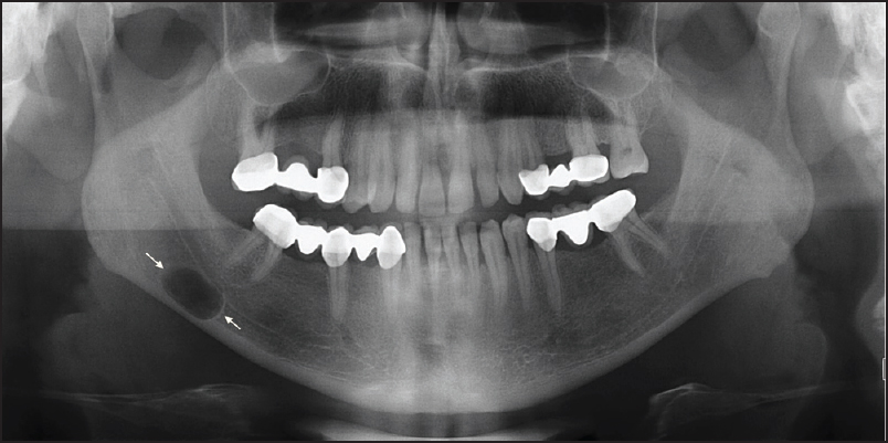
A radiolucent lesion was found incidentally on a dental panoramic radiograph in a 30-year-old man. The cyst was located in the mandible below the inferior alveolar canal. It was roughly oval in outline.
A Stafne cavity is a developmental depression or concavity that occurs in the border of the mandible, which is the lower jaw. It is important to note that Stafne cavities are not true cysts but can sometimes be mistaken for cysts on imaging studies like dental X-rays.
Stafne cavities are typically found in the posterior region of the mandible, near the angle of the jaw. They are more commonly seen in adult males and are considered to be a normal anatomical variation rather than a pathological condition.
These cavities are usually asymptomatic, which means they don’t cause any symptoms or problems for the patient. They are often discovered accidentally during routine dental X-rays or radiographic examinations.
The appearance of a Stafne cavity on an X-ray can resemble that of a cyst, but there are some differences. Unlike cysts, Stafne cavities do not cause expansion or erosion of the surrounding bone. They usually have a well-defined and smooth appearance. It’s also important to note that Stafne cavities are typically found on one side of the mandible and are usually symmetrical.
In most cases, treatment is not necessary for Stafne cavities because they are harmless and don’t cause any issues. However, if there is uncertainty about the diagnosis or if the lesion shows unusual features, further evaluation may be recommended. This can include additional imaging studies like a CT scan or even a biopsy to rule out any other potential pathological conditions.
It’s essential for dental professionals to be aware of Stafne cavities and their characteristic appearance on X-rays. This knowledge helps prevent confusion with other conditions and ensures appropriate management for the patient.

