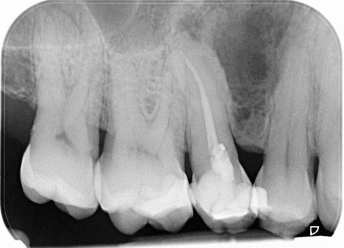
A multilocular radiolucent lesion was found in the interdental bone between the upper first and second premolars. The teeth were vital and after enucleation, the pathologist reported that the cyst had features of a developmental periodontal cyst lined by squamous epithelium with focal thickened areas.
As a dental student, it’s essential to learn about various dental conditions. One specific condition you may come across is called a botryoid cyst or a lateral periodontal cyst.
Botryoid cysts develop from small pieces of tissue called odontogenic epithelial remnants, which are found in the periodontal ligament. These remnants are leftover tissue from tooth development. The term “botryoid” refers to the appearance of these cysts under a microscope, which resembles a cluster of grapes.
Unlike some other cysts caused by inflammation or infection, botryoid cysts are not primarily driven by inflammation. Instead, they are considered a developmental abnormality originating from these remaining epithelial tissues.
When examining a botryoid cyst under a microscope, you may observe focal areas where the cyst lining appears thicker or denser compared to the surrounding epithelium. These areas are known as focally thickened epithelial plaques. The presence of these plaques is a characteristic feature that helps identify botryoid cysts microscopically.
As a dental student, it’s important to keep in mind that this information is based on knowledge available until September 2021. Stay engaged in your studies, seek guidance from your professors, and consult trusted dental resources for the most up-to-date and comprehensive information on dental conditions like botryoid cysts.

