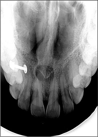
A pear-shaped and well-circumscribed radiolucent lesion with a corticated outline was found on a radiograph related to the root of an upper central incisor. The tooth was not restored and proved vital on testing.
A nasopalatine duct cyst is a specific type of cyst that develops from remnants of the nasopalatine duct. During embryonic development, this duct is involved in the formation of the nasal and oral cavities. Sometimes, remnants of this duct can persist and give rise to a cystic lesion later in life.
The nasopalatine duct extends from a structure called the incisive canal, which is located in the midline of the maxillary bone (the bone that comprises the upper jaw). The cyst can develop anywhere along this tract.
When we examine the lining of a nasopalatine duct cyst under a microscope, we typically observe the presence of both respiratory and squamous epithelium. This finding is significant because it indicates the origin of the cyst from the nasopalatine duct. The respiratory epithelium represents the lining of the nasal portion of the duct, while the squamous epithelium represents the lining of the oral portion.
Furthermore, a nasopalatine duct cyst often contains a neurovascular bundle within its capsule. This bundle consists of nerve fibers and blood vessels that supply the nasopalatine area. Its presence within the cyst is a characteristic feature.
In clinical practice, nasopalatine duct cysts are usually asymptomatic and are often discovered incidentally during routine dental examinations. However, if the cyst becomes large or infected, it can cause pain, swelling, and discomfort in the affected area.
As a dental student, it’s important to be familiar with the clinical presentation, radiographic appearance, and management of nasopalatine duct cysts. Treatment typically involves surgical removal of the cyst to relieve symptoms and prevent potential complications.

