A 58-year-old man presented with a brown–red granular epulis. A periapical radiograph showed underlying bone destruction and a biopsy was reported as showing osteoclast-like giant cells in a spindle-cell background with numerous thin- walled vessels. Haemosiderin and extravasated red cells were abundant.
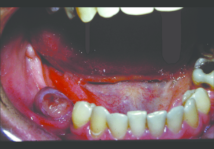

The clinical and radiographic findings suggest that the 58-year-old man may have a peripheral giant-cell granuloma (PGCG). This is a benign, non-cancerous tumor that often arises from the gum tissue and can cause bone destruction in the underlying jawbone.
The biopsy findings support the diagnosis of PGCG, as the presence of osteoclast-like giant cells and spindle cells in a background of abundant thin-walled vessels is characteristic of this condition. The haemosiderin and extravasated red cells seen in the biopsy are likely a result of bleeding within the lesion, which is common in PGCG.
These same features may also be observed in hyperparathyroidism. However, in hyperparathyroidism, serum calcium levels are typically elevated, while this is not the case in giant-cell granuloma. Therefore, measuring serum calcium levels can be a useful diagnostic tool in differentiating between these two conditions, especially when giant-cell granuloma features are observed.
Treatment for PGCG typically involves surgical removal of the lesion, along with the underlying affected bone. Recurrence is possible, so close follow-up and monitoring is important.

