➡️ A acute self limiting Dermatitis
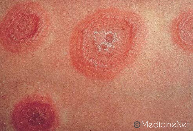
➡️ Clinical eruptions – Iris/target lesions
🔹Etiology:
- Drug usage (Sulfa Drugs)
- Infectious agents
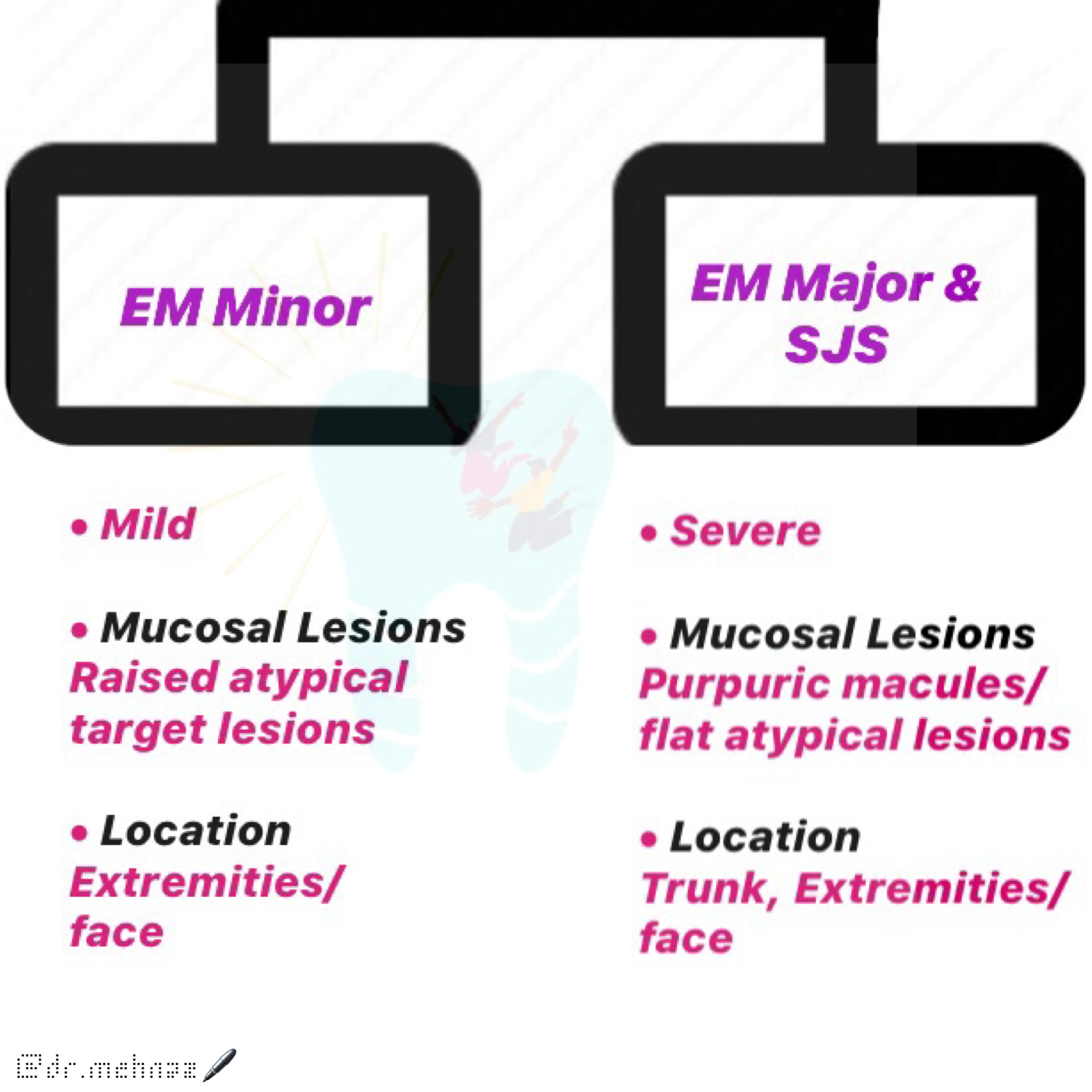
- Herpetic Etiology – Erythema multiforme major (55%); Erythema multiforme minor (100%)
- Mycoplasma infection
🔹Clinical Features:
- Prevalance: 2-4th decade of life
- M>F
- Occurrence of asymptomatic, vividly erythematous discrete macules, papules or vesicles & bullae in symmetrical pattern over hands, arms, foot, legs, face, Neck.
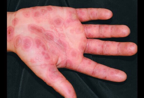
- Concentric ring like appearance of lesions in hands, wrist, ankles 👇🏻
- Target
- Iris
- Bull’s eye
- Lesions appear rapidly in a day/two. Persist for days to weeks & then fade & clear off. Recur after years.
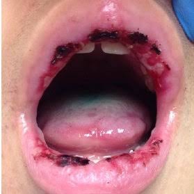
- Oral lesions – Hyperemic macules, papules/vesicles become eroded/ulcerated & bleed freely.
🔹STEVEN’S JOHNSON’S SYNDROME (Muco-cutaneous ocular disease)
- Bullous form of Erythema Multiforme
- Involves skin, oral cavity, eyes & genitalia
- Fever, malaise, photophobia
- Cutaneous lesions – Haemorrhagic vesicles/bullae
- Oral lesions – painful, mastication impossible
- Vesicles/bullae rupture and leave surface with thick white/yellow exudate
- Erosions on pharynx
- Lips – painful, upceration with bloody crusting..👇🏻
- Eye Lesions:
- Photophobia
- Conjunctivitis
- Corneal ulceration
- Panopthalmitis
- Keratoconjunctivitis Sicca
- Blindness due to bacterial infection
- Genital: Non-specific urethritis, balanitis, vaginal ulcers
- Other: Tracheobronchial ulceration, Pneumonia
🔹Histopathological Features:
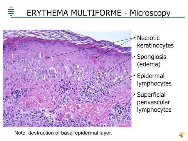
- Lesions exhibit intracellular edema of spinous layer of epithelium & edema of superficial connective tissue – subepidermal vesicle.
- Zone of severe liquefaction degeneration in upper layers of epithelium, intraepithelial vesicle formation & absence of basal membrane.
- Dilatation of superficial capillaries & lymphatic vessels in the connective tissue.
- Lymphocytes, Neutrophils & Eosinophilic infilteration.
🔹Differential Diagnosis:
- Aphthous stomatitis
- Contact stomatitis
- ANUG
- Pemphigus
- Bullous lichen planus
- Dermatitis herpetiformis
- Herpes Zoster
- Chicken pox
- Toxic epidermal Necrolysis
⬇️
Scalding burn (bullous drug eruption)
🔹Treatment:
➡️ Eliminate the cause, drug withdrawal & treat infections after culture tests.
Symptomatic –
- Antihistamins
- Analgesics
- Mouthwash
- Oral antacids
- 0.05% chlorhexidine – bathing
- Corticosteroids therapy: Patients with infection-induced erythema multiforme do worse when steroids are given.
Dr. Mehnaz Memon🖊
References: Shafer’sTextbook Of Oral Pathology

