Muhad Noorman P- Dentowesome 2020
1.DENTURE STOMATITIS
Denture-induced stomatitis is a common inflammatory reaction related to the wearing of dentures usually involving Candida (yeast) species. Less common forms of denture stomatitis may be due to mechanical trauma or a contact reaction.
Predisposing factors
Denture-related stomatitis is very common, with over 50% of denture wearers affected in some populations. It is the most common clinically important condition developing in the mouth
- Complete upper (maxillary) denture – probably due to the large contact area between denture and oral mucosa
- Acrylic dentures – Candida species seem to have a particular binding affinity for acrylic resin
- Poor dental hygiene – Candida species and Lactobacillus bacteria stick to denture surfaces and should be removed chemically and/or mechanically at least daily
- Poorly fitting dentures – mechanical trauma damages the mucosa, making it more prone to infection
- Denture age – old dentures are commonly associated with denture stomatitis probably due to poor fitting and rough surface in which Candida can hide
- Continuous wearing of denture – failure to remove at night increases the risk
- Men – are twice as likely to develop denture stomatitis than women
- Diabetes mellitus – diabetics are more prone to developing yeast infections
- Dry mouth (xerostomia) – saliva normally helps flush the mouth and clean the denture surface
CLINICAL FEATURE , Denture stomatitis usually does not cause any symptoms ,Some complain of burning sensations in mouth . But on examination the mouth lining in contact with the denture will be red and swollen sometimes with small red dots (petechial haemorrhages).
Treatment: regular oral hygiene, oral nystatin gargle, cleansing denture in nystatin solution, Not wearing denture at night.
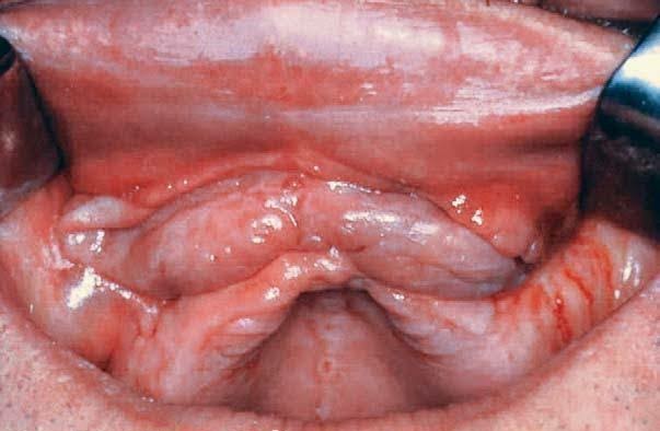
2.DENTURE INDUCED FIBROUS HYPERPLASIA aka EPULIS FISSURATUM
Denture-induced fibrous hyperplasia (DIFH) is a persistent lesion caused mostly by the prolonged wear of an ill-fitting, over-extended denture. Although the condition frequently coexists with denture stomatitis, it is a distinct entity .Develops as a reactive lesion to chronic mechanical irritation produced by the flange of a poorly fitting denture.More simply, epulis fissuratum is where excess folds of firm tissue form inside the mouth, as a result of rubbing on the edge of dentures that do not fit well. It is a benign entity.
CLINICAL FEATURE: The lesion is usually painless.The usual appearance is of two excess tissue folds in alveolar vestibule/buccal sulcus, with the flange of the denture fitting in between the two folds. It may occur in either the maxillary or mandibular sulci,although the latter is more usual.Anterior locations are more common than posterior. Less commonly there may be a single fold, and the lesion may appear on the lingual surface of the mandibular alveolar ridge.
Surgically excised, reconstruction of denture, denture rebasing can be done
3. DENTURE INDUCED ULCER
Traumatic ulcers caused by dentures with overextended or unbalanced occlusion are seen in about 5% of denture wearers.Overextention of flanges,sequestration of bony spicules, illfitting, sharp boders are cause of Traumatic ulcer.
CLINICAL FEATURE: Superficial, Painful, shallow ulceration often with erythematous halo and greyish membrane covering the ulcer.
Symptomatic relief by topical analgesic,steroid oinments and repair of denture.
4. INFLAMMATORY PAPILLARY HYPERPLASIA OF PALATE
Inflammatory papillary hyperplasia (IPH) is a benign lesion of the palatal mucosa. It is usually found in denture-wearers but also has been reported in patients without a history of use of a maxillary prosthesis use.The lesion almost exclusively involves the hard palate
CLINICAL FEATURES:
Inflammatory papillary hyperplasia is usually asymptomatic. It presents as a cluster of individual papules or nodules that may be erythematous, somewhat translucent, or normal in surface coloration. Mucosa is erythematous and has a pebbly or papillary surface. Many cases are associated with denture stomatitis.
Often the entire vault of the hard palate is involved .Has a female prediliction.
This may be aided by use of topical antibiotic or antifungal therapies. Small lesions are also typically treated with mouthrinses such as chlorhexidine mouthrinse at 0.12% or antifungal mouthrinse/ gels. Larger lesions recquired surgical excision.
References: Neville Textbook of Oral pathology.
Shafers textbook of pathology. Cover photo image: Internet

