11) CARDIOMEGALY
▪️Presentation: Poor exercise tolerance.
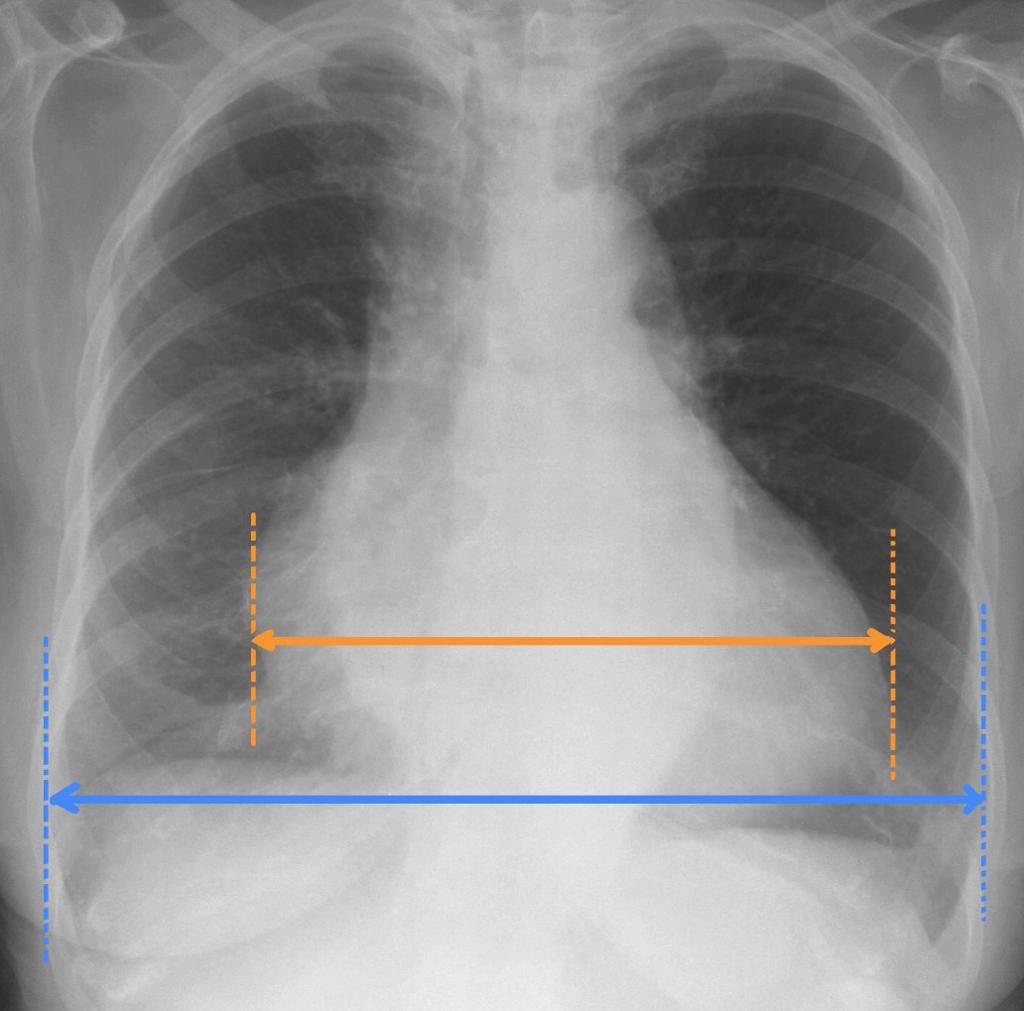
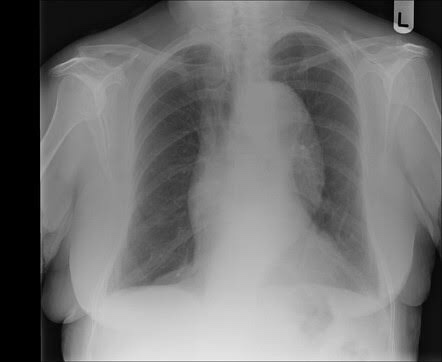
• PA and lateral chest x-rays demonstrate markedly enlarged cardiac silhouette.
• Cardiothoracic ratio is measured on a PA chest x-ray, and is the ratio of:
- maximal horizontal cardiac diameter
- maximal horizontal thoracic diameter (inner edge of ribs / edge of pleura)
• A normal measurement should be less than 0.5.
▪️Imaging differential diagnosis
Pericardial Effusion
Page 11 of 14
12) THORACIC AORTIC ANEURYSM
➡️ Abnormal dilatation of aorta, elastic fibers in the media are replaced by fibrosis.
▪️Etiology:
- Atherosclerosis
- Syphilis
- Marfan’s syndrome
- Lupus erythematosis
- Ehlers-Danlos syndrome
- Turner’s and Noonan’s syndrome
• The thoracic aorta can usually be seen on both frontal and lateral chest radiographs, and aneurysms are often obvious. However, it is difficult to assess size accurately.
• An aortic aneurysm, as aneurysm elsewhere, can be described as saccular or fusiform. Aortic root aneurysms can cause your aortic valve to work incorrectly, which can cause a heart murmur.
▪️Treatment and prognosis:
➡️ Mild to moderate aneurysmal dilatation can usually be treated conservatively and monitored. When the diameter reaches 5-6 cm intervention is usually considered as the risk of rupture is significantly elevated. Treatment options include:
- Open Repair:
- Elephant graft repair
- Endovascular repair
➡️ In general, when possible, endovascular repair is the treatment of choice, with reduced morbidity and mortality.
➡️ The majority of patients with thoracic aortic aneurysms either die of a direct complication of the aneurysm (rupture most frequently) or other cardiovascular complications.
Complications
- rupture
- distal embolization
- fistula formation
Page 12 of 14

