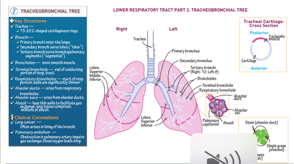
Key structures:
Trachea:
- Cartilaginous “trunk” of the tree.
- Comprises 15 – 20 C-shaped cartilaginous rings, are stacked vertically and connected via anular rings.
- Trachealis posterior forms posterior wall of trachea; moves to accommodate foods passing posteriorly through the esophagus.
Bronchi:
- Primary bronchi enter the lungs.
- Secondary bronchi serve lobes of lung (“lobar” bronchi).
- Tertiary bronchi serve bronchopulmonary segments (“segmental” bronchi); 10 on the right, 8-10 on the left.
Bronchioles:
- Numerous, and narrow as they branch.
- Have more smooth muscles in their walls, but still have cartilage in their walls.
- Terminal bronchiole is the final passageway of the conduction portion of the respiratory system.
Respiratory bronchioles:
- Demarcate the respiratory portion of the respiratory tract.
- Thin walls allow some gas exchange.
Alveolar ducts:
- Arise from respiratory bronchioles.
Alveolar sacs:
- Terminal ends of the alveolar ducts.
Alveoli:
- Thin-walled out-pockets of the alveolar sacs.
- Surrounded by pulmonary capillaries.
- Facilitate gas exchange between the respiratory and cardiovascular systems.
Lungs:
- Hundreds of millions of alveoli.
- Left lung = superior and inferior lobes; heart nestles into medial left lobe.
- Right lung = superior, middle, and inferior lobes.
Clinical Correlations:
Lung cancer often originates in the bronchi.
Pulmonary embolism (aka PE) obstructs arterial supply. In a PE, gas exchange is reduced, and blood oxygen levels drop.