1) PRIMARY STAGE
Development of chancre at site of inoculation. Usually Solitary in nature.
Intra-orally,
➡️ The chancre is an ulcerated lesion covered by grayish white membrane, painful due to secondary infection.
➡️ Sites:
- Lips (Brownish crusted appearance)
- Tongue
- Gingiva
- Palate
- Tonsils
- Fresh Extraction Wound

Microscopically:-
- Superficial Ulcer
- Intense inflammatory infiltrate – Plasma Cells
- Silver Stain – Demonstration of Micro-organisms
Heals: 3 weeks – 2 months
2) SECONDARY/METASTATIC STAGE
- 6 weeks after primary lesion
- Diffuse eruptions on mucous membrane.
- Multiple, painless, grayish white plaques overlying an ulcerated surface.
- Mucous Patches:
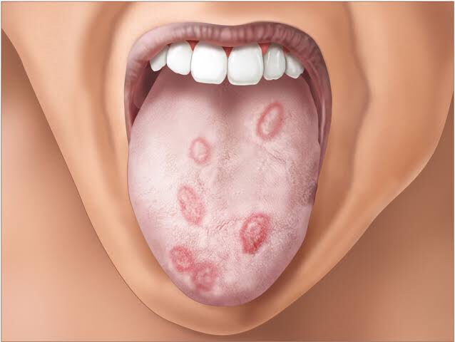
- Site: Tongue, Gingiva, Buccal Mucosa
- Highly infectious
- Appearance: Ovoid, irregular & surrounded by erythematous zone.
3) TERTIARY/LATE SYPHILIS:
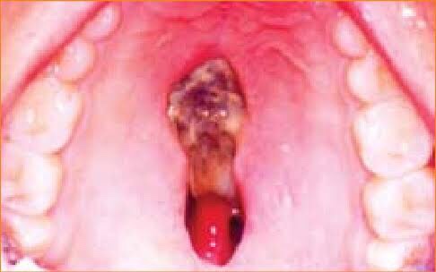
- Characterized by formation of GUMMA
- Site: Tongue, Palate
- Appearance: Firm, Nodular mass in the tissue; Ulcerates➡️ Deep painless ulcer
- Lesions of Palate cause perforation by sloughing of Necrotic Mass of tissue.
ATROPHIC/INTERSTITIAL GLOSSITIS
➡️ Most characteristic & important lesion of syphilis due to endarteritis obliterans.

➡️ In syphilitic glossitis, the surface of the tongue gets broken up by fissures due to atrophy.
- Males affected
- Fibrosis
- Hyperkeratosis
- Carcinomatous transformation – Epidermoid Ca.
LEUKOPLAKIC INVOLVEMENT
White patches on tongue – Tertiary Syphilis
Due to vasculitis & endarteritis, there’s circulatory deficiency to the lingual papillae
⬇️
Atrophy of filiform & fungiform papillae
⬇️
Bald, smooth, lingual surface
⬇️
Leukoplakic involvement (Dysplastic type)
References: Shafer’sTextbook Of Oral Pathology, Image source: Google

